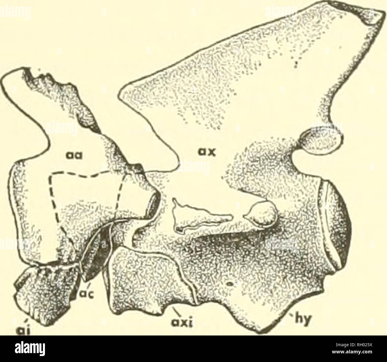
. Bulletin. Natural history. Text-fig. 39. Atlas-axis vertebrae in Clidastes propython (YPM 1100. X li). A. Anterior view of atlas. B. Lateral view of atlas-axis. Abbreviations: aa, atlas neural arch; ai, atlas intercentrum; ac, atlas centrum (odontoid); ax, axis; axi, axis intercentrum; hy, facet for liypapophysis (intercentrum) of third cervical vertebra. (Clidastes, Ectenosaurns, Mosasauriis). Spinal nerves I and II exited in front of and behind the base of the spinous portion of the atlantal arch, respectively. The spinous process expands somewhat distally and has a slightly ventromedially
Image details
Contributor:
Library Book Collection / Alamy Stock PhotoImage ID:
RH025XFile size:
7.2 MB (247.3 KB Compressed download)Releases:
Model - no | Property - noDo I need a release?Dimensions:
1699 x 1471 px | 28.8 x 24.9 cm | 11.3 x 9.8 inches | 150dpiMore information:
This image is a public domain image, which means either that copyright has expired in the image or the copyright holder has waived their copyright. Alamy charges you a fee for access to the high resolution copy of the image.
This image could have imperfections as it’s either historical or reportage.
. Bulletin. Natural history. Text-fig. 39. Atlas-axis vertebrae in Clidastes propython (YPM 1100. X li). A. Anterior view of atlas. B. Lateral view of atlas-axis. Abbreviations: aa, atlas neural arch; ai, atlas intercentrum; ac, atlas centrum (odontoid); ax, axis; axi, axis intercentrum; hy, facet for liypapophysis (intercentrum) of third cervical vertebra. (Clidastes, Ectenosaurns, Mosasauriis). Spinal nerves I and II exited in front of and behind the base of the spinous portion of the atlantal arch, respectively. The spinous process expands somewhat distally and has a slightly ventromedially incurved dorsoposterior corner. The tips of these processes are usually longi- tudinally ribbed and contact each other in Clidastes and Plotosaiirus, are sepa- rate in Platecarpus, and widely separate in Tylosaurus and Mosasaurus where the spinous processes are relatively short. Thus a true atlas neural spine is absent in mosasaurs. Midway between the tips of the spinous and synapophyseal processes on the dorsal margin of the atlas there is a longitudinally giooved region in the center of which is a slight excavation. This pit probably marks the tendonous insertion of the M. longissimus cervicis and anterior attachment of a tendonous sheet that extends dorsoposteriorly through the muscle masses of the Mm. transversalis capitis and articidoparietalis in Varanus (Nishi, 1916, p. 233). The site of inser- tion of the M. obliquus capitis inferior is probably located immediately ante- rior to the above-mentioned pit. According to Nishi (1916, p. 243) a small group of fibers from the M. obliquus capitis magnus (M. obliquus capitis superior) arises on the surface of the atlas just lateral to the longissimus tendons. In some mosasaurs (Plotosaurs, Platecarpus, Ectenosauriis, Tylosaurus) a prominent posterodorsally directed tuberosity is developed at this point, in others (Clidastes, Mosasaurus) only a small ridge is present. Each neural arch meets the atlas intercentrum ventromedially.