1-800-457-3801

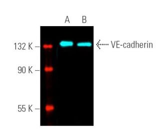
VE-cadherin Antibody (F-8): sc-9989

4.6(54)
Write a reviewAsk a question

Datasheets
  • VE-cadherin Antibody (F-8) is a mouse monoclonal IgG1 κ VE-cadherin antibody, cited in 383 publications, provided at 200 µg/ml
  • specific for an epitope mapping between amino acids 768-784 at the C-terminus of VE-cadherin of human origin
  • VE-cadherin Antibody (F-8) is recommended for detection of VE-cadherin of mouse, rat and human origin by WB, IP, IF, FCM and ELISA; also reactive with additional species, including porcine
  • Anti-VE-cadherin Antibody (F-8) is available conjugated to agarose for IP; HRP for WB, IHC(P) and ELISA; and to either phycoerythrin or FITC for IF, IHC(P) and FCM
  • also available conjugated to Alexa Fluor® 488, Alexa Fluor® 546, Alexa Fluor® 594 or Alexa Fluor® 647 for WB (RGB), IF, IHC(P) and FCM, and for use with RGB fluorescent imaging systems, such as iBright™ FL1000, FluorChem™, Typhoon, Azure and other comparable systems
  • also available conjugated to Alexa Fluor® 680 or Alexa Fluor® 790 for WB (NIR), IF and FCM; for use with Near-Infrared (NIR) detection systems, such as LI-COR®Odyssey®, iBright™ FL1000, FluorChem™, Typhoon, Azure and other comparable systems
  • Contact our Technical Service Department (or your local Distributor) for more information on how to receive a FREE 10 µg sample of VE-cadherin (F-8): sc-9989.
  • m-IgG Fc BP-HRP is the preferred secondary detection reagent for VE-cadherin Antibody (F-8) for WB applications. This reagent is now offered in a bundle with VE-cadherin Antibody (F-8) (see ordering information below).

QUICK LINKS

SEE ALSO...

loader

VE-cadherin Antibody (F-8) is an IgG1 κ mouse monoclonal VE-cadherin antibody (also designated CDH5 antibody, CD144 antibody or Cadherin 5 antibody) that detects the VE-cadherin protein of mouse, rat and human origin by WB, IP, IF, FCM and ELISA. VE-cadherin Antibody (F-8) is available as both the non-conjugated anti-VE-cadherin antibody form, as well as multiple conjugated forms of anti-VE-cadherin antibody, including agarose, HRP, PE, FITC and multiple Alexa Fluor® conjugates. The cadherins are a family of Ca2+-dependent adhesion molecules that function to mediate cell-cell binding critical to the maintenance of tissue structure and morphogenesis. Cadherins each contain a large extracellular domain at the amino-terminus, which is characterized by a series of five homologous repeats, the most distal of which is thought to be responsible for binding specificity. The relatively short carboxy-terminal, intracellular domain interacts with a variety of cytoplasmic proteins, including β-catenin, to regulate cadherin function. VE-cadherin (for vascular endothelial cadherin, also designated cadherin-5) is localized at intercellular junctions of endothelial cells, where it is thought to play a role in the cohesion and organization of intercellular junctions.

For Research Use Only. Not Intended for Diagnostic or Therapeutic Use.

Alexa Fluor® is a trademark of Molecular Probes Inc., OR., USA

LI-COR® and Odyssey® are registered trademarks of LI-COR Biosciences

References:

  1. Genomic organization and chromosomal mapping of the mouse P-cadherin gene.  |  Hatta, M., et al. 1991. Nucleic Acids Res. 19: 4437-41. PMID: 1886768
  2. The cadherins: cell-cell adhesion molecules controlling animal morphogenesis.  |  Takeichi, M. 1988. Development. 102: 639-55. PMID: 3048970
  3. Functional properties of human vascular endothelial cadherin (7B4/cadherin-5), an endothelium-specific cadherin.  |  Breviario, F., et al. 1995. Arterioscler Thromb Vasc Biol. 15: 1229-39. PMID: 7627717
  4. Desmosomal cadherins: another growing multigene family of adhesion molecules.  |  Koch, PJ. and Franke, WW. 1994. Curr Opin Cell Biol. 6: 682-7. PMID: 7833048
  5. Cadherins and catenins: interactions and functions in embryonic development.  |  Ranscht, B. 1994. Curr Opin Cell Biol. 6: 740-6. PMID: 7833053
  6. Spatial and temporal relationships between cadherins and PECAM-1 in cell-cell junctions of human endothelial cells.  |  Ayalon, O., et al. 1994. J Cell Biol. 126: 247-58. PMID: 8027182
  7. Dynamics of cadherin/catenin complex formation: novel protein interactions and pathways of complex assembly.  |  Hinck, L., et al. 1994. J Cell Biol. 125: 1327-40. PMID: 8207061
  8. Morphogenetic roles of classic cadherins.  |  Takeichi, M. 1995. Curr Opin Cell Biol. 7: 619-27. PMID: 8573335

I use this antibody long time ago and it was perfect. Unfortunately, Months ago I bought same antibody but I had nonspecific binding with high background and faint band. I did not change anything in the my protocol. So what do you think the problem?

Asked by: fawza
Thank you for contacting Santa Cruz Biotechnology . Please contact technical service by email [email protected] or call (800)-457-3801.
Answered by: BlakeJ
Date published: 2023-05-01

I am wondering this antibody sc9989, when it bind with ve cadherin, it will disrupt the adhersion junction between ve cadherin? Thank you so much!

Asked by: HX906
Thank you for your question. Anti-VE-cadherin Antibody (F-8) is only recommended for detection of VE-cadherin of mouse, rat and human origin by WB, IP, IF, FCM and ELISA. Its interference with biological functions has not determined in any way.
Answered by: Tech Support Europe
Date published: 2022-03-09

Is VE- Caherin sc9989 antibody work for Immunohistochemistry on paraffin section

Asked by: gjoseph
Thank you for your question. It would be helpful if you could call us, allowing for a more interactive discussion of this and other related questions.
Answered by: Technical Support
Date published: 2021-09-01

Hello, We have used the polyclonal version of this antibody to stain HMVEC vascular permeability. However, I have not been able to see staining using this version of the antibody. Is there a recommended staining method for this version?

Asked by: Abigail
Thank you for your question. You can find all recommended protocols under this link: https://www.scbt.com/resources/protocols Please reach out to our technical service team if problems with the antibody persist for troubleshooting advice.
Answered by: Tech Support Europe
Date published: 2021-06-25

Can I use this antibody for immunofluorescense staining of bovine cells? Which species can recognize?

Asked by: Alberto302
thank you for your question. Anti-VE-cadherin Antibody (F-8) is recommended for detection of VE-cadherin of mouse, rat and human origin by WB, IP, IF, FCM and ELISA; also reactive with additional species, including porcine. We have not tested the antibody in bovine samples, but PMID 28743502 cites such a use of this antibody.
Answered by: Tech Support Europe
Date published: 2021-01-14

Hello, I'm interested in doing FACS. However, I wanted to know how this stains the VE-cadherin even when the cells are in suspension(after trypsin) and there is no cell-cell adhesion. Does this antibody stains even when there is no cell-cell adhesion?

Asked by: SaraBS
Thank you for your question. Yes, it should stain either way. We permeabilize the cells for FACS for this protein. You can find our recommended protocol here: https://www.scbt.com/resources/protocols/flow-cytometry
Answered by: Tech Service
Date published: 2019-12-12

I am currently attempting to use this antibody for IHC, but I'm having some trouble. I'm using an in vitro model of rat primary lymphatic endothelial cells, and I typically use a 1:200 dilution of AB. Do you happen to have any recommended protocols?

Asked by: MH13
Thank you for your question. We recommend a starting dilution of 1:50 for IHC for this antibody. You can find our recommended protocol here: https://www.scbt.com/resources/protocols/immunoperoxidase-staining Please reach out to Technical Service for any further assistance with this antibody.
Answered by: Tech Service
Date published: 2019-12-03

Hi, I was wondering if this antibody binds the intra- or extracellular domain of the protein?

Asked by: MSuessmilch
Thank you for your question. This antibody is specific for an epitope mapping between amino acids 768-784 at the C-terminus of VE-cadherin of human origin. According to Uniprot, this corresponds to a cytoplasmic domain. https://www.uniprot.org/uniprot/P33151#subcellular_location
Answered by: Technical Support
Date published: 2019-12-03
  • y_2024, m_5, d_2, h_3CST
  • bvseo_bulk, prod_bvqa, vn_bulk_3.0.42
  • cp_1, bvpage1
  • co_hasquestionsanswers, tq_17
  • loc_en_US, sid_9989, prod, sort_[SortEntry(order=LAST_APPROVED_ANSWER_SUBMISSION_TIME, direction=DESCENDING)]
  • clientName_scbt
  • bvseo_sdk, java_sdk, bvseo-4.0.0
  • CLOUD, getContent, 106ms
  • QUESTIONS, PRODUCT
Rated 5 out of 5 by from Excellent signal on IF stainsWe used this antibody to stain human lung endothelial cells and it work perfectly for us. Good junctional labeling.
Date published: 2024-03-21
Rated 5 out of 5 by from Super reliableWe've been using this antibody for years and it works perfectly in WB and IHC
Date published: 2022-02-24
Rated 5 out of 5 by from goodIt is appropriate to examine expression of target peotein by WB.
Date published: 2020-10-29
Rated 5 out of 5 by from Clear band in WBWe did WB experiment with this antibody in HUVEC, and the band is clear.
Date published: 2020-09-01
Rated 5 out of 5 by from WB效果很棒学校里有很多相关课题组用这个抗体,做出来效果果然不错,终于解决了我的一个大烦恼,可以愉快的继续做实验了
Date published: 2020-07-01
Rated 5 out of 5 by from Very good efficiency for immunofluorescence assayGreat signal revealed by confocal analysis of VE-cadherin on HUVEC cells.
Date published: 2019-07-25
Rated 5 out of 5 by from lab TechThis antibody was used for immunofluorescence-staining Ve-Cadherin in human lung endothelial cells. the signal produced by this antibody is very clear and very strong.
Date published: 2019-04-18
Rated 1 out of 5 by from 蛍光免疫染色4%PFA固定したラット組織の凍結切片と、賦活化処理したパラフィン切片に対して蛍光免疫染色を行いましたが特異的なシグナルは得られませんでした。
Date published: 2018-11-26
  • y_2024, m_5, d_2, h_3
  • bvseo_bulk, prod_bvrr, vn_bulk_3.0.42
  • cp_1, bvpage1
  • co_hasreviews, tv_0, tr_54
  • loc_en_US, sid_9989, prod, sort_[SortEntry(order=SUBMISSION_TIME, direction=DESCENDING)]
  • clientName_scbt
  • bvseo_sdk, java_sdk, bvseo-4.0.0
  • CLOUD, getReviews, 15ms
  • REVIEWS, PRODUCT
VE-cadherin Antibody (F-8) is rated 4.6 out of 5 by 54.
  • y_2024, m_5, d_2, h_3
  • bvseo_bulk, prod_bvrr, vn_bulk_3.0.42
  • cp_1, bvpage1
  • co_hasreviews, tv_0, tr_54
  • loc_en_US, sid_9989, prod, sort_[SortEntry(order=SUBMISSION_TIME, direction=DESCENDING)]
  • clientName_scbt
  • bvseo_sdk, java_sdk, bvseo-4.0.0
  • CLOUD, getAggregateRating, 105ms
  • REVIEWS, PRODUCT