Structural basis of ligand recognition and transport by Sfh2, a yeast phosphatidylinositol transfer protein of the Sec14 superfamily.
Chen, L., Tan, L., Im, Y.J.(2022) Acta Crystallogr D Struct Biol 78: 853-864
- PubMed: 35775985
- DOI: https://doi.org/10.1107/S2059798322005666
- Primary Citation of Related Structures:
7WVT, 7WWD, 7WWE, 7WWG - PubMed Abstract:
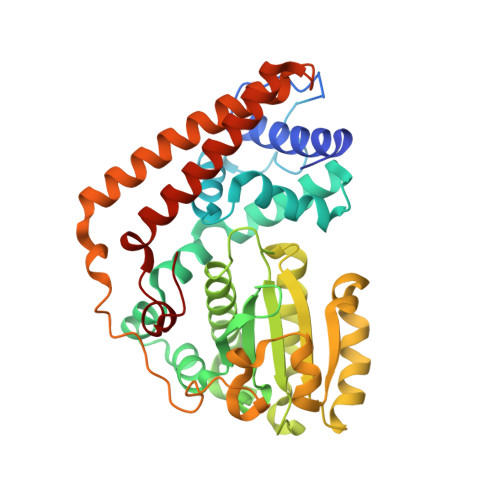
Sec14-like phosphatidylinositol transfer proteins (PITPs) are involved in lipid metabolism and phosphatidylinositol 4-phosphate signaling by transporting phosphatidylinositol (PI) and a secondary ligand between the organellar membranes in eukaryotes. Yeast Sfh2 is a PITP that transfers PI and squalene without phosphatidylcholine transfer activity. To investigate the structural determinants for ligand specificity and transport in Sfh2, crystal structures of Sfh2 in complex with PI and squalene were determined at 1.5 and 2.4 Å resolution, respectively. The inositol head group of PI is recognized by highly conserved residues around the pocket entrance. The acyl chains of PI bind into a large hydrophobic cavity. Squalene is accommodated in the bottom of the cavity entirely by hydrophobic interactions. The binding of PI and squalene are mutually exclusive due to their overlapping binding sites, correlating with the role in lipid exchange. The binding mode of PI is well conserved in Sfh family proteins. However, squalene binding is unique to the Sfh2 homolog due to the specific hydrophobic residues forming a shape-complementary binding pocket. Recombinant apo Sfh2 forms a homodimer in vitro by the hydrophobic interaction of the gating α10-α11 helices in an open conformation. Ligand binding closes the lid and dissociates the dimer into monomers. This study reveals the structural determinants for the recognition of the conserved PI and a secondary ligand, squalene, and provides implications for the lipid-transfer function of Sfh2.
Organizational Affiliation:
College of Pharmacy, Chonnam National University, Gwangju 61186, Republic of Korea.