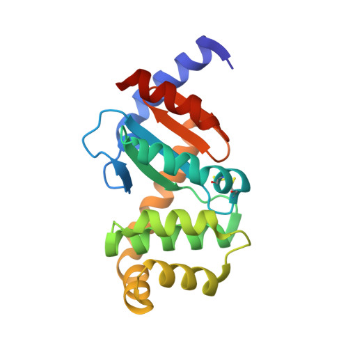
The atypical thiol-disulfide exchange protein alpha-DsbA2 from Wolbachia pipientis is a homotrimeric disulfide isomerase.
Walden, P.M., Whitten, A.E., Premkumar, L., Halili, M.A., Heras, B., King, G.J., Martin, J.L.(2019) Acta Crystallogr D Struct Biol 75: 283-295
- PubMed: 30950399
- DOI: https://doi.org/10.1107/S2059798318018442
- Primary Citation of Related Structures:
6EEZ - PubMed Abstract:
Disulfide-bond-forming (DSB) oxidative folding enzymes are master regulators of virulence that are localized to the periplasm of many Gram-negative bacteria. The archetypal DSB machinery from Escherichia coli K-12 consists of a dithiol-oxidizing redox-relay pair (DsbA/B), a disulfide-isomerizing redox-relay pair (DsbC/D) and the specialist reducing enzymes DsbE and DsbG that also interact with DsbD. By contrast, the Gram-negative bacterium Wolbachia pipientis encodes just three DSB enzymes. Two of these, α-DsbA1 and α-DsbB, form a redox-relay pair analogous to DsbA/B from E. coli. The third enzyme, α-DsbA2, incorporates a DsbA-like sequence but does not interact with α-DsbB. In comparison to other DsbA enzymes, α-DsbA2 has ∼50 extra N-terminal residues (excluding the signal peptide). The crystal structure of α-DsbA2ΔN, an N-terminally truncated form in which these ∼50 residues are removed, confirms the DsbA-like nature of this domain. However, α-DsbA2 does not have DsbA-like activity: it is structurally and functionally different as a consequence of its N-terminal residues. Firstly, α-DsbA2 is a powerful disulfide isomerase and a poor dithiol oxidase: i.e. its role is to shuffle rather than to introduce disulfide bonds. Moreover, small-angle X-ray scattering (SAXS) of α-DsbA2 reveals a homotrimeric arrangement that differs from those of the other characterized bacterial disulfide isomerases DsbC from Escherichia coli (homodimeric) and ScsC from Proteus mirabilis (PmScsC; homotrimeric with a shape-shifter peptide). α-DsbA2 lacks the shape-shifter motif and SAXS data suggest that it is less flexible than PmScsC. These results allow conclusions to be drawn about the factors that are required for functionally equivalent disulfide isomerase enzymatic activity across structurally diverse protein architectures.
Organizational Affiliation:
Institute for Molecular Bioscience, University of Queensland, Brisbane, QLD 4072, Australia.