A receptor-binding site as revealed by the crystal structure of CfaE, the colonization factor antigen I fimbrial adhesin of enterotoxigenic Escherichia coli.
Li, Y.F., Poole, S., Rasulova, F., McVeigh, A.L., Savarino, S.J., Xia, D.(2007) J Biol Chem 282: 23970-23980
- PubMed: 17569668
- DOI: https://doi.org/10.1074/jbc.M700921200
- Primary Citation of Related Structures:
2HB0 - PubMed Abstract:
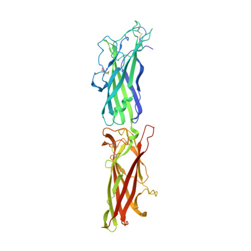
CfaE is the minor, tip-localized adhesive subunit of colonization factor antigen I fimbriae (CFA/I) of enterotoxigenic Escherichia coli and is thought to be essential for the attachment of enterotoxigenic E. coli to the human small intestine early in diarrhea pathogenesis. The crystal structure of an in cis donor strand complemented CfaE was determined, providing the first atomic view of a fimbrial subunit assembled by the alternate chaperone pathway. The in cis donor strand complemented variant of CfaE structure consists of an N-terminal adhesin domain and a C-terminal pilin domain of similar size, each featuring a variable immunoglobulin-like fold. Extensive interactions exist between the two domains and appear to rigidify the molecule. The upper surface of the adhesin domain distal to the pilin domain reveals a depression consisting of conserved residues including Arg(181), previously shown to be necessary for erythrocyte adhesion. Mutational analysis revealed a cluster of conserved, positively charged residues that are required for CFA/I-mediated hemagglutination, implicating this as the receptor-binding pocket. Mutations in a few subclass-specific residues that surround the cluster displayed differential effects on the two red cell species used in hemagglutination, suggesting that these residues play a role in host or cell specificity. The C-terminal donor strand derived from the major subunit CfaB is folded as a beta-strand and fits into a hydrophobic groove in the pilin domain to complete the immunoglobulin fold. The location of this well ordered donor strand suggests the positioning and orientation of the subjacent major fimbrial subunit CfaB in the native assembly of CFA/I fimbriae.
Organizational Affiliation:
Laboratory of Cell Biology, Center for Cancer Research, NCI, National Institutes of Health, Bethesda, Maryland 20892-4256, USA.