Review

The Role of Structural Bioinformatics in Understanding Tumor Necrosis Factor α-Interacting Protein Mechanisms in Chronic Inflammatory Diseases: A Review

by
Luana Luiza Bastos
1,*,
Diego Mariano
1,
Rafael Pereira Lemos
1,
Tatiane Senna Bialves
1,
Carlo Jose Freire Oliveira
2 and
Raquel C. de Melo-Minardi
1,*
1
Laboratory of Bioinformatics and Systems, Department of Computer Science, Institute of Exact Sciences, Universidade Federal de Minas Gerais, Belo Horizonte 31270-901, Brazil
2
Laboratory of Immunology and Bioinformatics, Institute of Natural and Biological Sciences, Federal University of Triângulo Mineiro, Uberaba 38025-180, Brazil
*
Authors to whom correspondence should be addressed.
Immuno 2024, 4(1), 14-42; https://doi.org/10.3390/immuno4010002
Submission received: 21 November 2023 / Revised: 21 December 2023 / Accepted: 12 January 2024 / Published: 15 January 2024
(This article belongs to the Section Structural Immunology)

Abstract

:
Tumor necrosis factor α (TNF-α) is a multifunctional cytokine protein acknowledged as a vital mediator in cell differentiation, proliferation, and survival. Additionally, TNF-α is a crucial component of the host’s defense by mediating inflammatory and immune responses against various aggressive agents, including viruses, bacteria parasites, and tumors. However, excessive production can be detrimental to the body and is also implicated in developing several inflammatory and immune-mediated disorders. Therefore, there is great interest in studying its role and its modulation, in various diseases, both in in vitro, in vivo, and in silico experiments. In this review, we evaluated the structures of proteins related to TNF-α available in public databases. In addition, we described the main antibodies blocking this cytokine and its applications and commented on the potential of naturally produced binding molecules, such as TNF-α-binding proteins produced by ticks. We also discuss the role of structural bioinformatics techniques in understanding the mechanisms of chronic inflammatory diseases related to TNF-α. We hope that the data presented in this review will be useful for studies that aim to better understand the mechanisms of the interactions of TNF-α with other proteins and will lead to new drugs or treatments.

1. Introduction

Tumor necrosis factor α (TNF-α) is a cytokine protein described as a mediator of inflammatory and immune responses in mammals. TNF-α production is performed predominantly by activated macrophages and lymphocytes [1,2,3]. Nevertheless, under inflammatory conditions, it can be generated by various cell types, encompassing T and B lymphocytes, mast cells, endothelial cells, neutrophils, cardiac cells, muscle cells, fibroblasts, osteoblasts, and natural killer cells (NK cells) [1,2,3,4].
The cytokine was identified in 1975 by Carswell and colleagues [1] during the study of hemorrhagic necrosis of tumors produced by endotoxins. In 1985, TNF-α was cloned for the first time by D. Pennica and colleagues [5]. They demonstrated that cytokines induce hemorrhagic necrosis in mice [1,2,3,4]. TNF-α is generated as a precursor form called transmembrane TNF-α, which is expressed as a type II cell surface polypeptide of 233 amino acid residues (26 kDa). After being processed by metalloproteinases such as the TNF-converting enzyme (TACE), which cleaves the TNF-α backbone between the residues A76 (alanine 76) and V77 (valine 77), the soluble form of TNF-α of 157 amino acid residues (17 kDa) is released. Moreover, it mediates its biological activities through binding with tumor necrosis factor α (TNF-α) 1 receptors (TNF-R1) and TNF-α 2 receptors (TNF-R2). Both TNF-α in its soluble form and its transmembrane form are biologically active. Transmembrane TNF-α exerts its biological function in cell-to-cell contacts, while soluble TNF-α acts at sites distant from cytokine-producing cells. Transmembrane TNF-α also binds to both receptors, but its biological activities are mainly mediated by TNF-R2 [2,6].
Chronic inflammatory and autoimmune diseases represent a group of long-lasting pathological conditions, characterized by persistent and often unregulated inflammation in the body. The tumor necrosis factor alpha (TNF-α) plays a crucial role in this context, being a central pro-inflammatory cytokine involved in the pathological process that determines tissue damage. It is implicated in various diseases, including rheumatoid arthritis, Crohn’s disease, ankylosing spondylitis, and psoriasis, among others. TNF-α triggers exacerbated inflammatory responses, leading to tissue destruction and the chronic symptoms of these diseases. The modulation of immune mechanisms triggered by TNF-α is so important that a significant number of medications and immunobiologicals act strategically by down-regulating the production or action of this cytokine. A better understanding of its functioning and interaction with other proteins involved in its participation in the inflammatory process opens up the possibility for the study and development of new treatments for chronic inflammatory and autoimmune diseases [7,8,9,10,11].

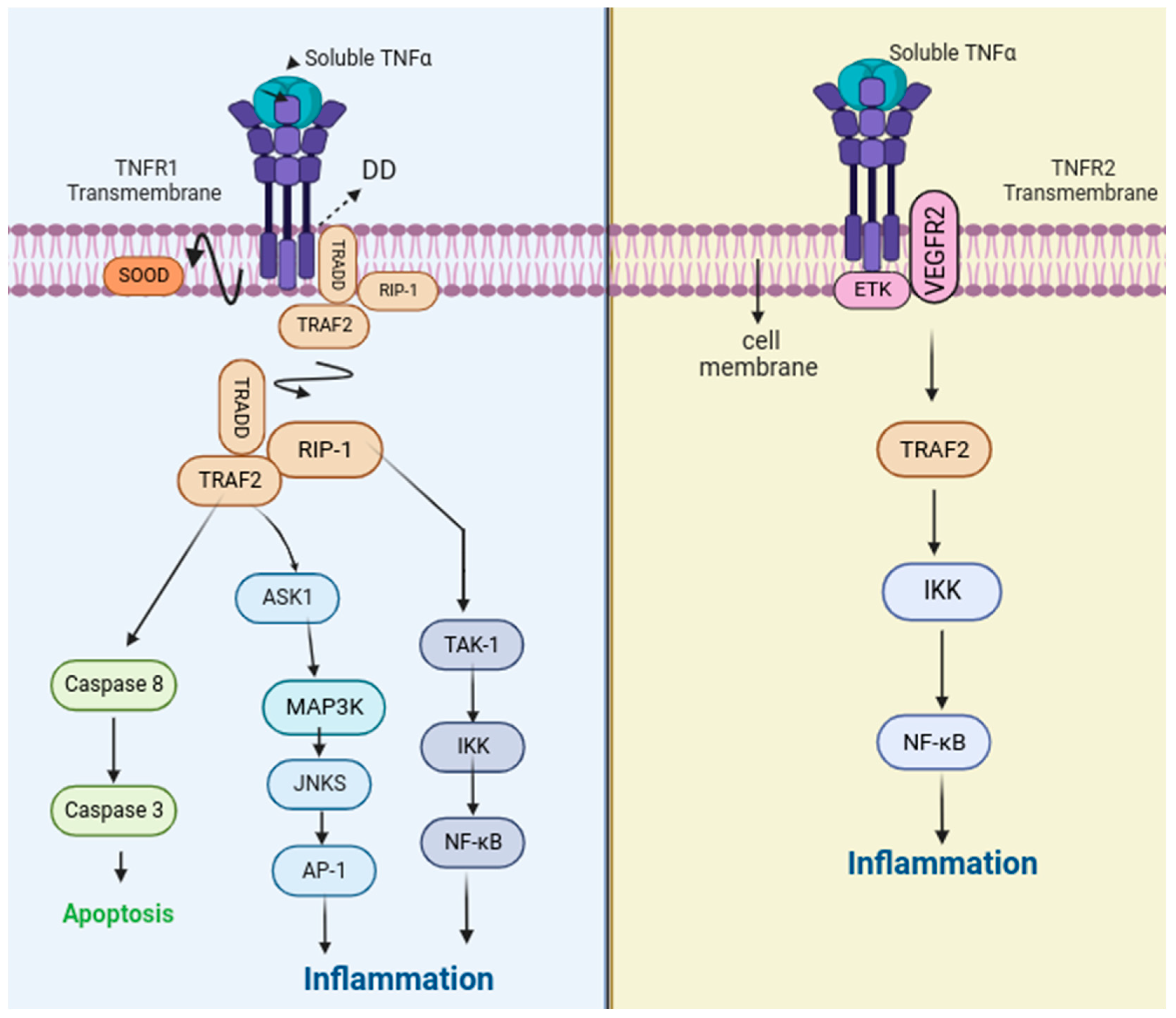
2. Function and Signaling
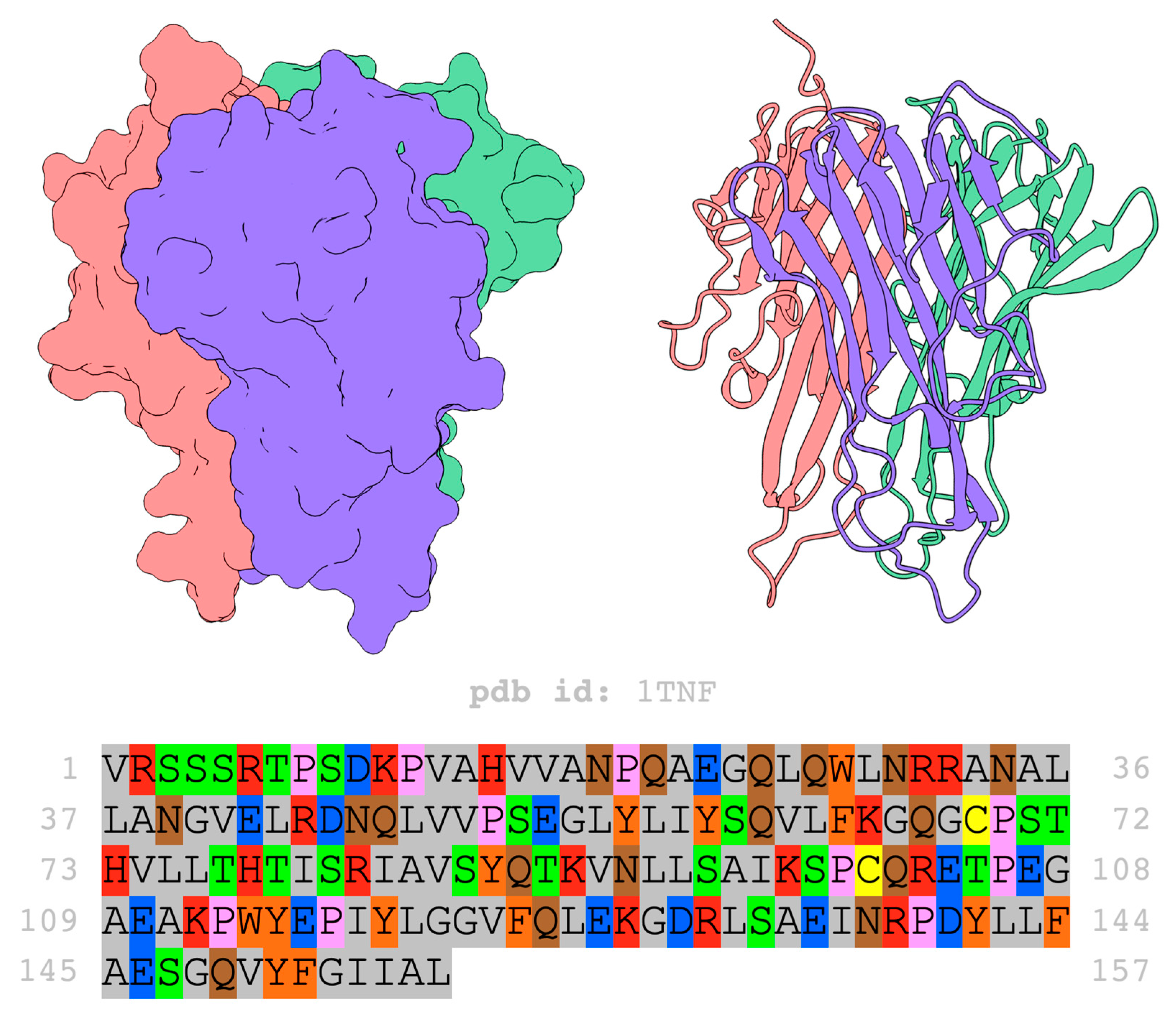
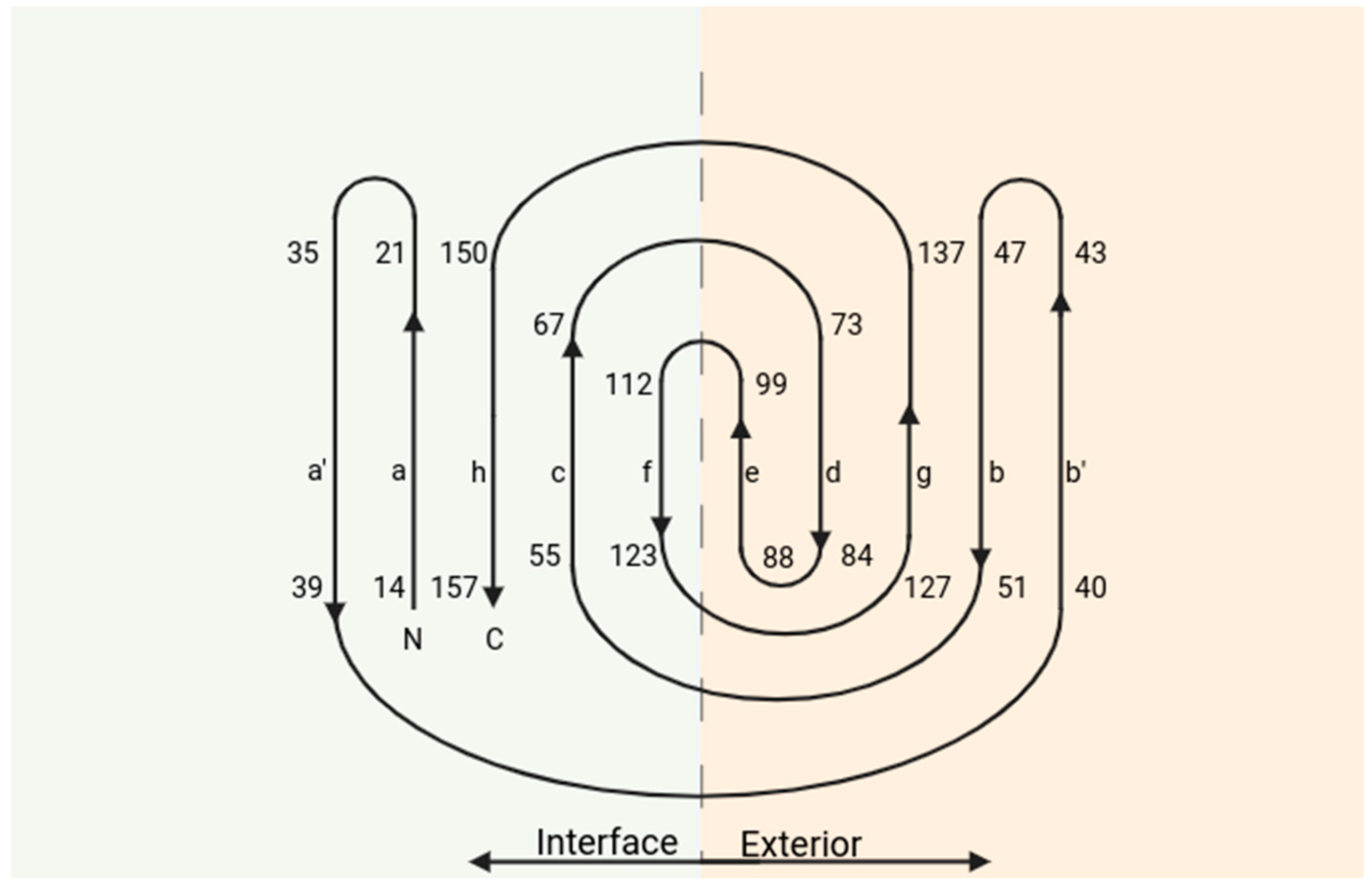
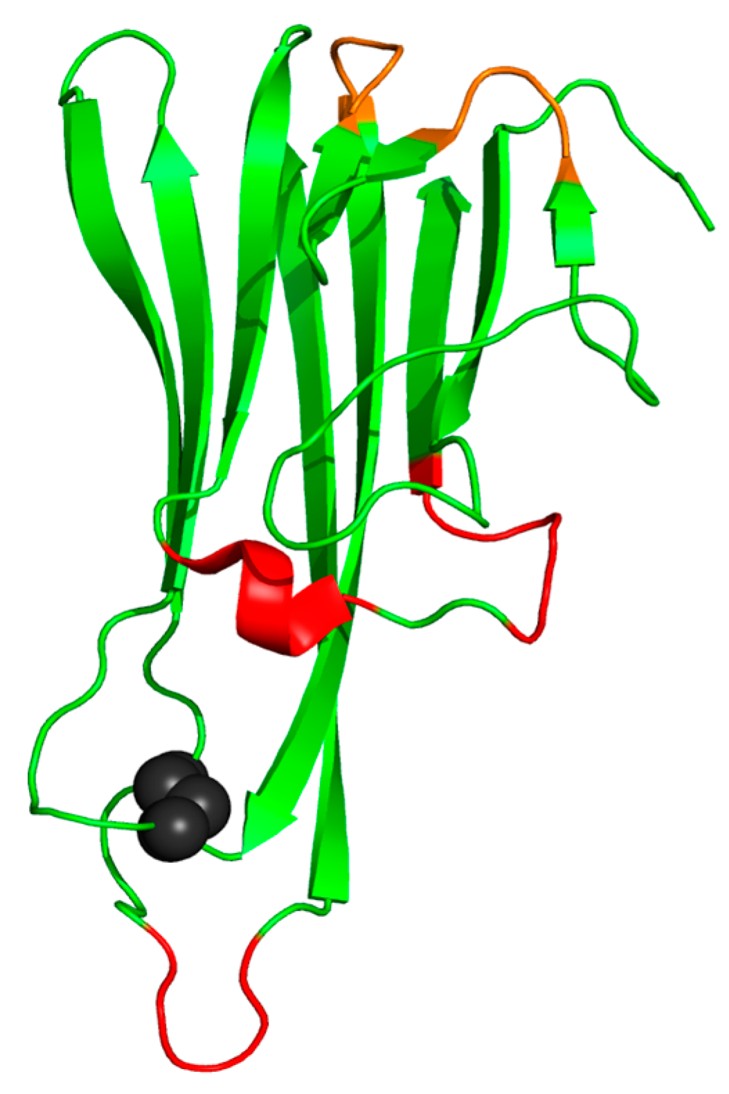
TNF-α-mediated signaling is dependent on binding to the TNF-R1 and TNF-R2 receptors. Receptors interact with the same and different molecules, which makes them capable of activating common and distinct pathways. TNF-R1 is more associated with the activation of pro-inflammatory and cell death pathways, while TNF-R2 is more associated with tissue repair and angiogenesis pathways [2,3,6,12,13].
Both receptors are transmembrane proteins and members of the tumor necrosis factor receptor (TNFR) superfamily. Like the other members, they have cysteine-rich domains (CRDs) and are characterized by having two to four CRDs, which form three disulfide bonds each. The receptors have four CRDs, of which CRD1 and CRD2 have approximately 30% identity, while the other domains have no identity [14,15,16,17]. TNF-R1 has a death domain (DD), a region of around 80 residues located in the intracellular part of the receptor, close to the C-terminus, which is responsible for its cytotoxicity (Figure 1). Its signaling pathway begins with binding to TNF-α, which causes the silencer of the death domain (SOOD) to dissociate from binding to the DD region of the receptor. The DD region binds to the TNFR1-associated death domain protein (TRADD) [2,18].
TRADD is a protein possessing a death domain (DD) that engages with TNFR1, facilitating programmed cell death signaling and triggering the activation of nuclear factor kappa B (NF-kB). NF-kB is a protein complex responsible for regulating transcription, cytokine production, and cell survival. Signaling proceeds with the recruitment carried out by TRADD of two proteins: the Receptor-interacting Protein 1 (RIP-1), a serine-threonine kinase that binds TRADD through its DD region, and TNF receptor-associated factor 2 (TRAF-2), an E3 ubiquitin ligase that binds a protein that recruits ubiquitin-conjugating enzymes. This TRADD–RIP-1–TRAF-2 protein conjugation is released from TNF-R1 after its junction [18,19,20].
Then, signaling involves the recruitment and activation of different mitogen-activated kinase (MAP3K) proteins. RIP-1 mediates the recruitment of growth factor β-activated kinase 1 (TAK), which promotes the activation of the IkB (inhibitor of nuclear factor kappa B) kinase complex called IKK. IKK generates the phosphorylation of IkB proteins, signaling their ubiquitination and proteasome-mediated degradation, allowing the NF-kB factor to enter the nucleus and initiate gene transcription [2,19,21,22].
The TRADD–RIP-1–TRAF-2 complex can also recruit apoptosis signal-regulating kinase 1 (ASK-1). The factor activates MAP3K phosphorylates and c-Jun N-terminal kinases (JNKs) mitogen-activated protein kinases (p38 MAPKs), a class of MAPKs that respond to stress with cytokines. Activated JNKs phosphorylate the C-jun region, a subunit of the transcription factor activating protein (AP-1), which allows the cAMP response element binding protein (CBP/p300) reaction [2].
In addition to mediating cell survival and pro-inflammatory signals through NF-kB and AP-1, TNF-R1 can initiate cell death signaling pathways. This signaling involves the binding of the Fas-associated DD protein (FADD), which associates with the TRADD protein, forming the TRADD-FADD complex, recruiting pro-caspases 8, which releases activated caspase 8 and initiates apoptosis through the cleavage and activation of pro-caspase 3 [2,18,19].
The TNF-R2 signaling pathways are not yet clearly defined. TNF-R2 does not have the DD; however, it can still interact with TRAF-2, which binds directly to the receptor. TNF-R2 can also activate endothelial/epithelial tyrosine kinase (ETK), implicating TRAF-2-independent cell adhesion, migration, proliferation, and survival. ETK is a regulator of epithelial cell junctions and participates in the mediation of angiogenesis through TNF-induced Phosphatidylinositol 3-kinase (PI3K), which is mediated by ETK and binds to vascular endothelial growth factor receptor 2 (VEGFR2) [2,12,14,15,16,23].
The receptor associates with an inactive form of ETK independently, through the 16 amino acid sequence at the end of TNF-R2. It is believed that TNF-α induces a conformational change in TNF-R2 that triggers the unfolding of the closed and inactive form of ETK. In endothelial cells, TNF-α induces the assembly of a trimolecular complex containing TNFR2-ETK-VEGFR2, which results in the activation of PI3K (Figure 2) [2,12,14,15,16,23].
Both TNF-R1 and TNF-R2 are expressed in most cell lines and primary tissues. The expression of receptors is regulated by the presence of pro-inflammatory cytokines such as TNF-α itself, interleukin 1 (IL-1), and interleukin 2 (IL-2) [2,12,14,15,16,23].
Many of the pro-inflammatory effects of TNF-α can be explained based on the cytokine’s effects on the vascular endothelium and interactions with endothelial leukocytes. In response to TNF-α during inflammation, endothelial cells exhibit a distinct temporal, spatial, and anatomical model; this response generates the recruitment of different populations of leukocytes. Furthermore, the TNF-α-induced expression of cyclooxygenase 2 (COX-2) can increase the production of prostacyclins, resulting in vasodilation, causing flushing and heat through increased local blood flow, which are classic signs of inflammation [2,12,14,15,16,23].
The receptors connect to the cytokine by binding between two TNF-α chains (Figure 2); thus, a trimer, which is the active form of TNF-α, was able to interact with three receptor molecules. The receptors interact with the following residues of the TNF-α interface: Gln-21, Glu-23, Arg-31, Arg-32, Ala-33, Asp-143, Phe-144, Ala-145, Glu-146, Gln-149, Ser-86, His-73, and Tyr-87. The Arg-31 residue stands out, and when mutated, it conferred a lower binding affinity of cytokine with the receptors [12].
In the next section, we will discuss the TNF-α three-dimensional structure in more detail.

3. Structure

The structure of the soluble human tumor necrosis factor α (TNF-α) was identified in 1989 by Eck and Sprang and their results published the following year [3], recorded in the Protein Data Bank (PDB) as 1TNF (Figure 3). The structure was identified via X-ray diffraction at a resolution of 2.6 Å and an R-value of 0.23, and its refinement was carried out using X-PLOR. The protein is a homotrimer with a total of 52,106 kDa, 3552 atoms, and 471 residues distributed in three identical chains of 157 amino acids [3,14,15,16,25,26,27].
Each monomer of TNF-α forms an elongated and antiparallel β-sandwich, corresponding to a structure composed of two antiparallel β-sheets. In this case, these are pleated β-sheets, in which the amino groups (NH) of a fully extended β-sheet interacted by forming hydrogen bonds with the carbonyl groups (C=O) of the adjacent strand. The monomers have a jelly roll topology (Figure 4), which is defined as a supersecondary protein fold composed of eight β-strands (named from “a” to “h”) composed of two sheets of four strands [3,14,15,16,25,26,27].
The inner sheet comprises ribbons (a, c, f, and h); they are involved in the internal contacts that form the trimer, while the strands (b, g, d, and e) form the outer surface. Both the inner and outer sheets are made up of five β-sheets. The inner sheets facing the trimer axis are essentially flat, while the outer sheets are very curved. It is observed that the leaves are twisted at around 60° clockwise [3,14,15,16,25,26,27].
There are three helical segments in TNF-α, none extending more than one turn. The segments are formed by residues 106–110, 138–142, and 145–150 (Figure 5). There is a disulfide bond between residues 69–101, which connect the e and f strands with the c and d strands. Loops that connect the strands are also found in the structure, formed by residues 37–42, connecting strand a to b, and the loop formed by residues 49–57 connecting strand b to c [3,14,15,16,25,26,27].
The spatial distribution of side chain residues in TNF-α is typical of β-sandwich proteins; the interior of the subunit is packed largely with hydrophobic amino acid side chains. There are short sequences of hydrophobic and hydrophilic residues that alternate in the outer leaves [3,14,15,16,25,26,27].

4. TNF-α Role in the Body’s Inflammatory Process

As observed earlier, tumor necrosis factor α (TNF-α) plays a pivotal role in the body’s inflammatory response and serves as a critical component in the host’s defense against various harmful agents, such as viruses, bacteria, and parasites. Nevertheless, excessive production can be detrimental to the body and is also implicated in developing several immune-related disorders [12,28]. Conversely, reduced plasma concentrations of this cytokine, whether occurring naturally or due to particular disease treatments, are linked to an elevated risk of bacterial and fungal infections or the reactivation of latent tuberculosis [12,28].
Chronic inflammatory and autoimmune diseases constitute a category of health conditions distinguished by enduring and persistent inflammation in various body regions. Chronic inflammation may arise from an immune response that is dysregulated, causing the immune system to erroneously target the body’s healthy tissues. This results in continual inflammation and frequently leads to the progressive degeneration of the organs affected. These conditions can have a significant impact on patients’ quality of life and often require long-term treatment to control symptoms and prevent complications [29]. Below, we present some examples of chronic inflammatory diseases in which TNF-α plays a fundamental role in the inflammatory process.

4.1. Rheumatoid Arthritis

Rheumatoid arthritis is characterized as a persistent autoimmune disorder primarily impacting the joints, causing inflammation, pain, stiffness, and, in more advanced stages, structural damage to the involved joints. This ailment manifests when the immune system erroneously targets its own healthy cells and tissues, initiating an inflammatory response within the synovial membranes that line the joints [30,31].
The pathogenesis of rheumatoid arthritis involves the activation of pro-inflammatory cytokines, with TNF-α playing a central role. This cytokine is found at elevated levels in patients with the disease and exerts a significant influence on the progression of inflammation. The inflammatory process is associated with the accumulation of various immune cells, including type 1 helper T cells (Th1), macrophages, B cells, plasma cells, and dendritic cells (DCs) [30,31].
TNF-α, secreted by Th1 cells and macrophages, participates in several events that lead to the activation of synovial fibroblasts, epidermal hyperplasia, and recruitment of further inflammatory cells. Thus, in response to stimulation by cytokines such as IL-1, IL-6, and TNF-α, synovial fibroblasts increase the expression of cathepsins and matrix metalloproteinases (MMPs), promoting the degradation of collagen and proteoglycans. This process culminates in the destruction of cartilage and bones, ultimately leading to joint erosion. Additionally, osteoclasts play a significant role in the progression of rheumatoid arthritis pathology, being activated by TNF-α and inducing synovial hyperplasia and angiogenesis [30,31].

4.2. Psoriasis

Psoriasis is a persistent skin condition identified by red, scaly lesions, typically appearing on the scalp, elbows, knees, and lower back. This condition arises from an abnormal acceleration of the skin cell life cycle, resulting in the rapid generation of new cells and the buildup of dead tissue on the surface of the skin [32].
The pathogenesis of psoriasis involves an overactive immune response, in which TNF-α plays a significant role. This pro-inflammatory cytokine is found at high levels in patients with psoriasis and is associated with the activation of several cells of the immune system, including T cells, dendritic cells, and macrophages. Elevated TNF-α activity in psoriasis contributes to the chronic inflammation and rapid cell turnover observed in the condition. Furthermore, TNF-α triggers the excessive production of keratinocytes in the epidermis, leading to the formation of the characteristic psoriatic plaques. Blocking TNF-α activity through targeted therapies may be effective in treating psoriasis, providing symptom relief, and reducing disease progression [33].

4.3. Crohn’s Disease

Crohn’s disease is a chronic and inflammatory condition that impacts the gastrointestinal tract and can manifest in any segment of the digestive system, ranging from the mouth to the anus. It is characterized by chronic inflammation of the intestinal wall, resulting in symptoms such as abdominal pain, diarrhea, weight loss, and fatigue. Additionally, Crohn’s disease can lead to complications, such as strictures, fistulas, and abscesses [8].
TNF-α is one of the principal inflammatory mediators associated with the pathogenesis of Crohn’s disease. Elevated levels of TNF-α have been identified in patients with Crohn’s disease, indicating the crucial role of this cytokine in the progression and maintenance of intestinal inflammation [8]. TNF-α plays a multifaceted role in Crohn’s disease, as it stimulates the activation of inflammatory cells such as macrophages and T lymphocytes, resulting in the release of other pro-inflammatory cytokines and the formation of granulomas, a hallmark of this disease. Moreover, TNF-α can contribute to the impairment of the intestinal barrier function and alterations in the regulation of the immune response within the intestinal mucosa [10,34].
Anti-TNF-α therapy, which involves inhibiting the activity of TNF-α, has been shown to be effective in treating Crohn’s disease, providing symptom relief, and promoting the healing of intestinal lesions. This therapeutic approach has revolutionized the treatment of Crohn’s disease and is extensively employed in patients exhibiting moderate to severe forms of the condition [10,34].

4.4. Ankylosing Spondylitis

Ankylosing spondylitis is a form of chronic inflammatory arthritis primarily impacting the spine and sacroiliac joints situated in the lower back and pelvis. This progressive condition can lead to the fusion of the vertebrae, resulting in stiffness and limited spine mobility. Furthermore, ankylosing spondylitis can affect other joints, such as the shoulders, hips, and knees, and also present extra-articular manifestations, such as inflammation in the eyes, skin, and intestines. Although the exact cause of the disease is not completely understood, genetic factors play an important role in its predisposition. Ankylosing spondylitis can significantly impact the quality of life of affected individuals, and early diagnosis and appropriate treatment are essential to minimize symptoms and prevent long-term complications [35].
In ankylosing spondylitis, the heightened expression and activity of TNF-α are linked to the persistent inflammation of the sacroiliac joints and spine. This inflammation results in the formation of scar tissue and eventual ankylosis, characterized by the fusion of the vertebrae. Beyond its direct pro-inflammatory impact, TNF-α plays a role in activating immune system cells, including T lymphocytes, and stimulating the production of other inflammatory cytokines. This cascade of events leads to the perpetuation of chronic inflammation in the affected joints, resulting in the characteristic symptoms of the disease, such as pain, stiffness, and progressive limitation of joint mobility [35,36].

4.5. Systemic Sclerosis

Systemic sclerosis (SSc), also referred to as systemic scleroderma, is a rare, chronic autoimmune disease that impacts the body’s connective tissues. Characterized by excessive collagen production, SSc results in fibrosis, i.e., the thickening of the skin, often involving the internal organs. This complex condition can manifest itself heterogeneously, presenting a variety of symptoms that range from localized cutaneous sclerosis to more severe forms that affect multiple organ systems. The precise etiology of systemic sclerosis is not yet fully understood, but it is thought to encompass a combination of genetic and environmental triggers. Dysregulated immune response plays a key role, resulting in chronic inflammation and abnormal collagen deposition in affected tissues. In addition to cutaneous symptoms, SSc can cause systemic complications, including pulmonary, cardiac, renal, and gastrointestinal dysfunction [37,38].
TNF-α plays a pivotal role in the initial host response to infections and is involved in the pathogenesis of various immune-mediated systemic diseases. Patients with systemic sclerosis (SSc) often exhibit elevated serum levels of TNF-α, which contribute to the development of pulmonary fibrosis and pulmonary arterial hypertension. Additionally, inflammatory arthritis can manifest in SSc patients. The use of infliximab and etanercept may provide improvements in inflammatory arthritis and disability in individuals with SSc.TNF-α inhibitors reduce systemic inflammation and improve endothelial function, thus reducing the risk of the progression of pulmonary arterial hypertension and acute cardiovascular and/or cerebrovascular events. Clinicians need to be aware of the potential risks of tuberculosis reactivation and opportunistic infections. Randomized clinical trials with TNF-α inhibitors in patients with SSc are needed to confirm the potential role of these agents in the treatment of SSc.
The management of systemic sclerosis involves a multidisciplinary approach, with treatment targeting specific symptoms and regular monitoring to detect possible complications. Research continues to explore new insights into the underlying mechanisms of the disease, as well as identifying more effective therapies. Although there is no cure for SSc, advances in understanding pathogenesis and the development of targeted therapeutic strategies offer hope for improving the quality of life for patients affected by this debilitating condition [37,38].

4.6. Hidradenitis Suppurativa

Hidradenitis suppurativa (HS) is a chronic, inflammatory, debilitating skin disease that affects the apocrine sweat glands, resulting in painful nodules, recurrent abscesses, and subcutaneous tunnel formation. This dermatological condition has a significant impact on patient’s quality of life, not only due to physical pain but also due to the possible formation of scars, deformities, and psychosocial complications. Although the exact etiology of HS is not completely understood, genetic factors, chronic inflammation, and immune system dysfunctions are among the contributing elements. HS usually manifests itself in areas of skin folds, such as armpits, groin, and perianal region. Symptoms can range from mild to severe, and the disease often progresses over time. Patients with HS face significant challenges in managing the condition, and treatment may involve a multimodal approach that includes measures such as antibiotics, anti-inflammatories, corticosteroids, and in some cases, surgical interventions. The appropriate management of hidradenitis suppurativa requires an individualized approach, considering the extent of the disease, the patient’s response to treatment, and the specific needs of each case. Continued research is critical to improving therapeutic options and providing a better understanding of the underlying mechanisms of this complex condition [39,40,41].
The exact pathophysiology of HS is unclear, although current theory involves follicular obstruction, rupture, and subsequent inflammation, leading to the development of fistulas and abscesses in the intertriginous skin. Several inflammatory modulators have been implicated in the development of HS, including tumor necrosis factor α (TNF-α), as well as interleukin (IL)-1β, IL-10, and IL-17. Initial evidence for the use of TNF-α inhibitors in HS resulted from the recognition that patients with inflammatory bowel disease treated with these medications saw a simultaneous improvement in their HS symptoms. Early case reports and case series have illustrated the value of TNF-α inhibitors in the treatment of HS. Despite advances in understanding the relationship between hidradenitis suppurativa and TNF-α, it is important to highlight that each patient responds uniquely to treatments. The choice of appropriate therapy must be individualized, taking into account the severity of symptoms, the patient’s medical history, and other relevant factors. Treatment of HS remains challenging, and continued research is essential to develop more effective and personalized approaches to improve the quality of life for individuals affected by this complex dermatological condition [39,40,41].

4.7. Vasculitis

Vasculitis is a medical condition characterized by the inflammation of blood vessels. This inflammation can lead to damage to the vessels, resulting in narrowing, weakening, or even blocking blood flow. There are several types of vasculitis, classified based on the size of the affected blood vessels and the extent of inflammation. Symptoms can vary widely depending on the organs affected but often include fever, fatigue, joint pain, rash, and, in more serious cases, damage to vital organs. The exact etiology of vasculitis is not always clear, but in many cases, it is an autoimmune response, where the immune system mistakenly attacks its blood vessels. Other causes may include infections, reactions to certain medications, or genetic disorders. Diagnosis usually involves a combination of blood tests, imaging tests such as angiography, and, in some cases, a biopsy of affected tissue [42,43,44].
Vasculitis treatment aims to control inflammation and may involve the use of immunosuppressive medications, corticosteroids, and in some cases, biological therapies. Long-term management requires a multidisciplinary approach with regular follow-up care from a rheumatologist or autoimmune disease specialist. The prognosis may vary depending on the type and severity of vasculitis, but with appropriate treatment, many people can manage the condition and maintain a good quality of life [42,43,44].
The relationship between vasculitis and tumor necrosis factor α (TNF-α) is complex and may vary depending on the specific type of vasculitis. In some forms of vasculitis, especially Takayasu arteritis and granulomatosis with polyangiitis (GPA), there is evidence suggesting that TNF-α plays a role in the pathogenesis and maintenance of vascular inflammation. In cases of vasculitis associated with rheumatic diseases, such as Takayasu arteritis, some clinical studies have explored the use of anti-TNF medications, such as infliximab and etanercept, as part of treatment. These medications are designed to inhibit the action of TNF-α and thus modulate the inflammatory response. However, the effectiveness and safety of these treatments vary, and the decision to use them will depend on the specific characteristics of each patient and the assessment of a healthcare professional. It is essential to emphasize that the decision to use TNF-α inhibitors in the treatment of vasculitis must be carefully considered and individualized. The management of vasculitis is complex and often requires a multidisciplinary approach, involving rheumatologists and other specialists, to optimize treatment results and minimize risks. As medical research is constantly evolving, it is always advisable to consult a healthcare professional who is up to date on the latest and most appropriate therapeutic options for each clinical situation [42,43,44].

5. TNF-α Inhibitors as Therapeutic Drugs

The excessive expression of tumor necrosis factor α (TNF-α) is associated with chronic inflammatory diseases. It is also known that to carry out its signaling and function, it is necessary for the cytokine to be in its trimer conformation to interact with its receptors. Thus, an alternative treatment for diseases with chronic inflammatory characteristics is the blockage of the interaction between the cytokine and its receptors, which is carried out using anti-TNF-α antibodies [6].
Antibodies are glycoproteins whose structure is formed by two identical light chains plus two other identical heavy chains. The lower region of the antibody, where the “tail” is formed, is called the Fc domain. It is responsible for interacting with surface receptors. The amino-terminal ends of light and heavy chains are called Fab fragments (fragment antigen-binding). This is where the antigen binds to the antibody [45].
The FDA (Food and Drug Administration) and EMA (European Medicines Agency) have approved four antibodies and a fusion protein that interacts with TNF-α. Etanercept, trade name Embrel, is a fusion protein formed by the dimeric fusion consisting of the extracellular ligand-binding portion of the TNF-R2 receptor, linked to the Fc portion of human IgG1. This fusion protein was approved by the FDA in 1998 and by the EMA in 2000 [46].
Etanercept binds to both forms of the cytokine, covering the binding site and preventing it from interacting with its receptors [46]. Etanercept binds to soluble TNF-α and transmembrane TNF-α, inactivating them by blocking their interactions with receptors. It binds exclusively to active trimeric TNF-α, positioning itself in the cleft between the subunits. With a half-life of 3–3.5 days after subcutaneous administration, etanercept also demonstrates the ability to modulate pro-inflammatory genes, such as NF-κB, in plaque psoriasis (PS), resulting in a significant reduction in production of TNF-α. Additionally, it promotes the apoptosis of dendritic cells (DCs) in substantial quantities in the psoriasis plaque, interrupting the positive feedback associated with TNF-α through early apoptotic cell death, before DC activation and maturation [47].
Infliximab, commercially known as Remicade, was developed in 1993 and is the first anti-TNF-α antibody to be approved for use by the FDA in 1998 and by the EMA in 2000. It was initially approved for treating Crohn’s disease and later extended to other diseases. with characteristic chronic inflammatory conditions. The antibody neutralizes the biological activity of the cytokine, binding to TNF-α in its soluble and transmembrane form. Infliximab is a chimeric antibody composed of a human 1gG1 constant region (75%) linked to a murine-derived antigen-binding variable region (25%). Each antibody binds to one chain of the trimer. The EF loop of TNF-α plays a crucial role in antigen–antibody interaction. The interface is highly complementary and corresponds to a large region. The total surface area is complementary and has a buried interface between infliximab and TNF-α of 1977 Å2, corresponding to a larger area than typical protein–protein interfaces ranging between 1560 and 1700 Å2 [6,28].
The contact interface corresponds to the CD and EF loop residues, as well as the GH loop residues. It comprises 12 residues, within which Arg-32 is found, which is also part of the binding site for TNF-α with its receptors. By carrying out mutations in the residues of the site, it was identified that residues Gln-67, Arg-138, and Tyr-141, when mutated, significantly decrease the affinity of the complex. The antibody binds to residues Gln-67, Pro-70, Ser-71, Arg-32, Thr-105, Glu-107, Ala-109, Glu-110, Asn-137, Arg-138, Asp-140, and Tyr-141 of the TNF-α interface [6,22].
Infliximab was specifically designed to target all forms of TNF-α in humans, effectively preventing the binding of TNF-α to its soluble and transmembrane receptors. Following infliximab treatment in patients with inflammatory bowel disease (IBD), there is a promotion of lysis of cell lines expressing TNF-α through complement-dependent and antibody-dependent cytotoxicity, leading to a reduction in inflamed tissue. Furthermore, infliximab induces apoptosis, inhibits the production of IFN-γ in colonic and stimulated T cells, and contributes to an anti-inflammatory effect. Moreover, it exerts a negative regulatory effect on intracellular adhesion molecule 1 (ICAM-1) and vascular cell adhesion molecule 1 (VCAM-1), while also modulating the balance of matrix metalloproteinases (MMPs) and tissue inhibitor of metalloproteinases (TIMPs). Infliximab has an approximate half-life of 8 to 10 days, and its efficacy can be maintained by administering doses every eight weeks [6,28].
Adalimumab, commercially named Humira, is a fully human antibody approved in 2005 by the FDA and in 2008 by the EMA. The antibody binds to the cytokine via a highly complementary surface, with a total buried surface area of approximately 2540 Å2. The antibody binds to two monomers of the trimer. The Adalimumab epitope comprises a series of discontinuous fragments formed by 19 residues, including Gln-21, Glu-23, Ala-145, and Glu-146, which are part of the receptor binding site. It was observed that making mutations in the residues Glu-23, Asn-91, Lys-65, Gln-67, Glu-135, and Glu-146 decreases the binding affinity between the antibody and the cytokine. Antibody binds to residues Pro-19, Gln-20, Glu-23, Lys-65, Gln-67, Glu-10, Pro-113, Tyr-141, Ala-145, Glu-146, Thr-71, Gln-21, Thr-77, Thr-79, Ser-81, Lys-89, Asn-91, Glu-135, and Asn-13 at the interface of TNF-α [6,48].
Adalimumab requires less frequent subcutaneous administration due to its comparatively prolonged half-life, which ranges from 10 to 13 days. Furthermore, it has a lower propensity for immunogenicity compared to infliximab. Due to its better tolerance and lower incidence of adverse immunological reactions, adalimumab is effective in the treatment of Crohn’s disease (CD) and may be an option for patients who have had allergic reactions to infliximab. Psoriasis (PS) patients treated with adalimumab demonstrated reductions in TNF-α and IL-6 levels, as well as markers of the acute phase of inflammation. Furthermore, the treatment of rheumatoid arthritis (RA) with adalimumab has shown the ability to inhibit IL-17, overproduced by Th17 cells, while increasing the number of regulatory T cells (Treg) compared to untreated patients [6,48].
Certolizumab-pegol, brand name Cimzia, was approved by the FDA in 2008 and in 2009 by the EMA. The antibody has a different structure from other antibodies, being formed by a monovalent Fab fragment of a humanized anti-TNF-α antibody without an Fc region. The Fab region is linked to two cross-linked chains of a 20 kDA polyethylene glycol. The antibody binds to a single cytokine monomer, and the binding comprises a total accessible area of 1887 Å2, corresponding to a larger area than that of common protein–protein complexes. The binding region is formed by 16 residues, including Ser-86 and Thr-87, which are part of the TNF-α receptor sites. It can be noted that when residues Gln-88 and Arg-138 are mutated, the binding affinity of the complex decreases. The antibody site at the TNF-α interface is formed by residues Gly-24, Asp-45, Gln-47, Thr-77, Ile-83, Val-85, Ser-86, Gln-88, Thr-89, Lys-90, Arg-131, Glu-135, Asn-137, Arg-138, Pro-139, Asp-140, and Thr-87 [49].
Certolizumab pegol, as it does not have an Fc region, does not trigger complement or antibody-dependent cytotoxicity. This TNF-α inhibitor has a distinct mechanism of action compared to others in the same class. Due to PEGylation, Certolizumab pegol can be more efficiently distributed into inflamed tissues than Infliximab and Adalimumab. Its unique structure is suggested as the reason behind the greater efficacy, and PEGylation increases its half-life to two weeks, favoring the concentration of the inhibitor in inflamed tissues [47].
Golimumab, commercially named Simponi, was approved by the FDA and EMA in 2009. It is a fully human antibody that binds to just one monomer of the cytokine. The complex has a buried region of 1902 Å2 and its epitope in TNF-α involves 13 residues. Golimumab binds to residues Gly-24, Lys-65, Gln-67, Ser-71, Glu-104, Thr-105, Pro-106, Glu-107, Gly-108, Ala-111, Arg-138, Asp-140, and Tyr-141 at the TNF-α interface [50,51].
Golimumab, with superior affinity compared to infliximab and adalimumab, effectively neutralizes both soluble and transmembrane TNF-α, inhibiting its biological activity. It further hinders leukocyte infiltration by blocking cell adhesion proteins (E-selectin, ICAM-1, and VCAM-1) and suppressing the secretion of pro-inflammatory cytokines. Golimumab boasts a half-life ranging from 7 to 20 days [47].
Infliximab binds to both monomeric and trimeric forms of soluble TNF-α, while etanercept binds only to trimeric forms of soluble TNF-α. Each infliximab molecule can bind to two molecules of soluble TNF-α, allowing for up to three infliximab molecules per TNF-α homotrimer. In contrast, etanercept forms a one-to-one complex with a TNF-α homotrimer. Monoclonal antibodies (mAbs), but not etanercept, form large protein complexes in vitro. Infliximab, adalimumab, golimumab, and etanercept show similar binding activities to soluble TNF-α and almost identical neutralization capabilities against TNF-α receptor signaling [44,47,52].
Golimumab has higher affinity for soluble TNF-α compared to infliximab and adalimumab, being conformationally more stable. Its inhibitory capacity against TNF-α-induced cytotoxicity and the activation of human endothelial cells is superior to that of infliximab and adalimumab. In antibody-dependent cell activity (ADCC), all agents show similar activities on specific target cells, except etanercept, which shows discrepancies in different experimental conditions [44,47,52].
Infliximab, adalimumab, golimumab, Certolizumab pegol, and etanercept share the human IgG1 hinge region. Proteases such as MMP-3 and MMP-12 can cleave infliximab and adalimumab, preserving the neutralization capacity of soluble TNF-α. However, the cleavage of etanercept results in the loss of soluble TNF-α-neutralizing activity. Mucosal metalloproteinases and anti-hinge autoantibodies may contribute to the lack of response to anti-TNF-α agents in inflammatory bowel disease (IBD) [44,47,52].
Distribution studies in animal models show that Certolizumab pegol has more effective distribution to inflamed joints compared to infliximab and adalimumab. Prolonged exposure to inflamed tissue is more significant for Certolizumab pegol than for the other agents. Additional studies in mouse models of IBD for a more comprehensive understanding are awaited [44,47,52].
In conclusion, the variety in mechanisms of action and pharmacokinetic properties among tumor necrosis factor α (TNF-α) inhibitors offers a spectrum of therapeutic choices for inflammatory diseases like rheumatoid arthritis, Crohn’s disease, and psoriasis. Infliximab, adalimumab, golimumab, certolizumab pegol, and etanercept exhibit distinct affinities, binding capacities, and immune response modulations. Grasping these nuances is essential for tailoring personalized treatment strategies for individual patients. Additionally, ongoing research into the distribution characteristics and impacts of pharmaceutical modifications, such as PEGylation, holds promising prospects for enhancing the efficacy and safety of these agents. Although challenges such as immunogenicity and variable response persist, the constant evolution in anti-TNF-α therapies highlights the importance of a holistic and evolving approach to the management of inflammatory diseases [44,47,52].

6. New Anti-TNF-α Agents

Tumor necrosis factor alpha (TNF-α) inhibitor antibodies are widely employed in the treatment of autoimmune and inflammatory diseases, but they pose certain challenges and potential issues, including: (i) Immunogenic response: The development of antibodies against the TNF-α inhibitor itself can diminish the effectiveness of the treatment over time. (ii) Side effects: Some patients may experience side effects such as injection site reactions, opportunistic infections, and cardiovascular risks associated with the use of these medications. (iii) Cost: TNF-α inhibitory antibodies can be expensive, potentially limiting access for some patients due to financial constraints.
In addition, aspects such as the possibility of therapeutic failure, since patients may not respond adequately to treatment with TNF-α inhibitor antibodies, and aspects such as long-term safety, since the long-term safety of continuous use of these antibodies medications is still being studied, must also be considered. Currently, new anti-TNF-α agents are being developed to overcome shortcomings such as the non-response of existing TNF-α inhibitors [53,54,55]. Below, we present some examples.

6.1. Ozoralizumab (TS-152)

Ozoralizumab, also known as Nanozora, was developed by Taisho Pharmaceutical Co., Ltd. (Tóquio, Japan), under license from Ablynx, an affiliate of Sanofi, for the treatment of rheumatoid arthritis (RA). The approval for this novel tumor necrosis factor (TNF-α) inhibitor was granted in Japan in September 2022, targeting patients with rheumatoid arthritis (RA) who have not responded adequately to conventional treatments. Administration is carried out subcutaneously every 4 weeks, with a dose of 30 mg. Ozoralizumab acts potently by inhibiting TNF-α through two human TNF-α-binding domains, in addition to a human serum albumin-binding domain that prolongs its plasma half-life, allowing for longer intervals of administration. Its molecular weight is 38 kDa, approximately a quarter of the molecular weight of conventional immunoglobulin G [56,57].
We highlight that, although there is a consolidated experience in using TNF-α inhibitors to treat RA, Ozoralizumab presents a completely new structure. For this reason, it is essential to gather long-term efficacy and safety data after its introduction into clinical practice. A long-term extension study (NCT04077567; JapicCTI-194932) is currently underway in patients who have demonstrated a positive clinical response [56,57].

6.2. ZINC09609430

In the 2019 study conducted by Saddala and Huang [58], the primary objective was to identify novel small molecules capable of directly binding to TNF-α and/or TNFR1. This aimed to inhibit the interaction between these proteins and regulate the subsequent signaling pathways. The authors employed a range of cheminformatics techniques, including pharmacophore modeling, virtual screening, molecular docking, and in silico ADMET analysis, to explore the Zinc database [59] database for new TNF-α and TNFR1 inhibitors. Pharmacophore models were used to select the most promising compounds in the Zinc database, similar to existing drugs [58].
The most successful molecules were then mapped to the key features of the TNF-α pharmacophore, TNFR1, and the TNF-α–TNFR1 complex. They were subjected to additional evaluations, such as molecular docking, the analysis of protein–ligand interactions, as well as in silico ADMET studies. Molecular coupling analysis revealed the binding energies of TNFα, TNFR1, and the TNF-α–TNFR1 complex, serving as a basis for selecting the five best compounds regarding binding energy. Moreover, in silico ADMET studies revealed that all 15 compounds (ZINC09609430, ZINC49467549, ZINC13113075, ZINC39907639, ZINC25251930, ZINC02968981, ZINC09544246, ZINC58047088, ZINC72021182, ZINC08704414, ZINC05462670, ZINC35681945, ZINC23553920, ZINC05328058, and ZINC17206695) met the Lipinski criteria and exhibited no toxicity. These new selective inhibitors of the TNF-α, TNFR1, and the TNF-α–TNFR1 complex have the potential to be used as anti-inflammatory agents and represent promising candidates for future investigations and experimental trials [58].

6.3. CP-690334-01

In 2020, Kwak and colleagues [60] conducted a study that used a computational approach to find new therapies for Crohn’s disease in patients who were resistant to anti-TNF-α treatment, characterized by alternating periods of remission and deterioration. They used a transcriptomic dataset (GSE100833) of patients with this refractory form of Crohn’s, available at NCBI GEO [61]. After a thorough co-expression analysis, they investigated the extent of protein–protein interactions between genes grouped into clusters based on data from the STRING database [62]. Pathway analysis was conducted using the clEnrich function, which is based on KEGG gene sets. Co-expressed genes in clusters 1, 2, 3, and 4, as well as up- or down-regulated genes and all differentially expressed genes, showed high connectivity. Among them, cluster 1, notably enriched in chemokine signaling, also demonstrated enrichment in cytokine–cytokine receptor interactions, identifying several drugs, including cyclosporine, that are known for their efficacy in the disease. Furthermore, Vorinostat, histone deacetylase inhibitors, and piperlongumine, known for its inhibitory effect on NF-κB activity, were identified. Some alkaloids have also been highlighted as potential candidates for therapeutic drugs. These findings indicate that they may represent a new therapeutic option for antiTNF-α–refractory Crohn’s disease, corroborating the importance of using public molecular data and computational methods in identifying new therapeutic alternatives for the condition [60].

6.4. TNF-α Inhibitory Activity Detected in Ticks

In a study published in 2006, Konik et al. [39] detected an anti-tumor necrosis factor α activity in the saliva of the tick Ixodes ricinus. The experiment used ELISA (Enzyme-Linked Immunosorbent Assay), which consists of an enzymatic test that identifies specific antibodies. Specific antibodies for human and mouse TNF-α were used [63].
As is widely known, ticks need to feed on blood to complete their development and maintain their life cycle. During an invasion process after an animal bite (such as a tick), the host organism tends to activate the defense system with pro-inflammatory and immunomodulatory pathways, the formation of a hemostatic plug generating vasoconstriction, and tissue remodeling. If successful, these processes would lead to the rejection of the tick, preventing attachment and feeding from being completed. To circumvent the host’s defense system, a fundamental mechanism is the injection of saliva, which contains anti-hemostatic, anti-inflammatory, and immunomodulatory molecules that facilitate a satisfactory diet [64,65].
In a previous study, the authors demonstrated that saliva, as well as Ixodes ricinus salivary gland extract, significantly reduced the level of the cytokine [53]. Carrying out a digestion experiment with trypsin (a protein that participates in the digestion process, promoting the breakdown of proteins into peptides), in which saliva compounds were exposed to trypsin, it was noted that the inhibitory activity of TNF-α was lost, demonstrating that the factor that generates the inhibitory activity is a protein [53].
Next, a rapid fractionation experiment was carried out using liquid chromatin in proteins from saliva and salivary gland extract, where a peak of TNF-α inhibition was identified, corresponding to a protein with a molar mass of 23 kDa. It has been suggested that the mechanism involved in cytokine inhibition is direct binding to the protein [53].
Identifying a TNF-α inhibitory activity sparked interest in identifying such a protein and identifying the existence of the same activity in other species. Years later, in 2017, Kezková and Kopecky compared the presence of TNF-α inhibition in 11 species of ticks from the Ixodidae family, Amblyomma americanum, Dermacentor marginatus, D. reticulatus, Haemaphysalis concinna, Ixodes ricinus, I. persulcatus, I. hexagonus, I. scapularis, Rhipicephalus appendiculatus, R. pulchellus, and R. sanguineus. Partially fed females were used in the experiment over 6–7 days [66]. The test was performed using ELISA to estimate the effect of TNF-α inhibition on tick saliva and its salivary gland. As a result, inhibition for the cytokine was found in ticks of the genus Ixodes and Haemaphysalis, while ticks of the genus Rhipicephalus, Dermacentor, and Amblyomma do not show TNF-α inhibitory activity [66].
Taking into account that the active form of TNF-α is in the trimer form and that the cleavage of the structure by a protease would inhibit its function, it was tested whether inhibiting the activity of metalloproteases and proteases in saliva contents the inhibitory activity of TNF-α would be lost, thus changing the shape of the trimer. The results demonstrated that the inhibitory activity of proteases and metalloproteases did not reduce the inhibition of TNF-α. This suggests that the inhibition mechanism is directly linked to the cytokine and not the cleavage of its structure [56].

7. Limitations in TNF-α Research

Research on tumor necrosis factor α (TNF-α) faces several limitations that influence the complete understanding of its complex biological functions. One of the notable limitations is the diversity of cellular and tissue responses induced by TNF-α, which makes it challenging to discern its specific effects in different physiological and pathological contexts. Furthermore, the temporal and spatial regulation of TNF-α expression presents a complexity that often complicates the interpretation of experimental results. Genetic heterogeneity between individuals also contributes to variability in responses to TNF-α, impacting the generalizability of findings in clinical studies. Furthermore, research faces significant challenges when transitioning results obtained in vitro and animal models to human physiology. Experimental models often simplify the complexity of the biological environment, not fully capturing the diversity of cellular responses and the interaction of systems in human organisms. Furthermore, differences between species may influence the interpretation of results, as TNF-α signaling pathways may vary between animals and humans. Establishing more precise correlations between results obtained in experimental models and clinical response in patients is crucial for validating and effectively applying therapies that aim to modulate TNF-α [67,68,69,70,71,72,73].
To overcome limitations in TNF-α research, innovative strategies can be adopted. One promising approach is to utilize advanced imaging and unique cell analysis techniques to map tissue-specific TNF-α expression at the cellular level. This would allow a more detailed understanding of the cellular interactions and spatial dynamics of TNF-α. Furthermore, an integration of genomic and proteomic data can provide comprehensive insights into signaling pathways mediated by TNF-α. In the clinical context, personalizing therapy with TNF-α inhibitors, taking into account genetic factors and molecular profiles, may represent an advance in treatment efficacy. Future perspectives in research on TNF-α must involve a multidisciplinary approach, integrating biological, clinical, and genetic data. The identification of specific biomarkers associated with TNF-α activation may improve patient stratification and facilitate the development of more targeted therapies. Furthermore, studies exploring interactions between TNF-α and other affected mediators may provide a more holistic view of the signaling networks involved. The advancement initiated in research on TNF-α is crucial not only for understanding its role in pathological conditions but also for informing innovative and more effective therapeutic strategies in a variety of inflammatory diseases [67,68,69,70,71,72,73].

8. Bioinformatics Approaches

Recently, bioinformatics has been revolutionizing several areas of life sciences through new algorithms, methods, and tools with a user-friendly interface. New technologies such as next-generation sequencers or cryogenic transmission electron microscopy have enabled the construction of large biological databases, which have become easily accessible today via the internet. With bioinformatics methodologies, new paths have been traced to accelerate knowledge discovery through high-throughput analysis. In this section, we summarize some of the computational strategies previously cited here to understand the mechanisms of interaction of proteins such as TNF-α and their role in several chronic inflammatory diseases and present others that could be applied in future studies.

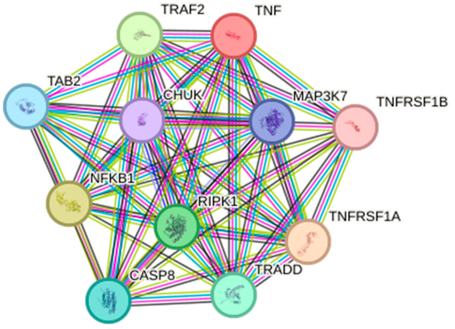
8.1. Biological Databases

Biological databases provide access to data obtained through in vitro and in vivo experiments. Although data are stored in several formats and structures, most of these databases have APIs (application programming interfaces) or even user-friendly interfaces and can generally be accessed through internet sites. Some examples of biological databases are Uniprot [74], PDB [75], and KEGG [76].

8.1.1. Sequences

UniProt (Universal Protein Resource) [42] is an openly accessible database offering a diverse range of information on protein sequences, functions, and structures. Additionally, it provides biological annotations from various services such as Pfam [77], ChEMBL [78], and PDB [75]. The UniProt web service features a user-friendly interface with robust search mechanisms for exploring protein annotations and sequences based on similarities. Access UniProt at <https://www.uniprot.org/> (accessed on 21 December 2023).
To date, there are 9738 entries under the query “TNF-α” in the Uniprot, with 1163 being high-quality reviewed and curated proteins and 8575 unreviewed automatically translated entries. Of those, 383 are human proteins (322 reviewed and 61 unreviewed), with most being components of the TNF family. However, some entries interact directly with TNF-α (Uniprot ID P01375). Some examples are the MAP kinase-activating death domain protein (Uniprot ID Q8WXG6), which may play a role in MAPK activation and TNF-R1 linkage with the pathway [79,80]; splicing factor Cactin (Uniprot ID Q8WUQ7), which is upregulated by TNF-α [81]; and Interleukin-32 (IL-32, Uniprot ID P24001), which induces TNF-α and NF-kB and p38 MAPK pathways [82].

8.1.2. D-Structures

PDB (Protein Data Bank) [75] is a public database that collects and provides access to the 3D structures of biological macromolecules, such as proteins, peptides, nucleic acids, and small ligands. Proteins are complex molecules composed of chains of amino acids, and their 3D structure is crucial to their function [58]. Understanding the 3D structure of proteins is important in various fields, including biology, medicine, and drug discovery. PDB contains detailed information on the three-dimensional structures of these molecules, obtained by experimental techniques such as nuclear magnetic resonance (NMR), X-ray crystallography, cryogenic electron microscopy (Cryo-EM), and so on. PDB is available at <https://www.rcsb.org/> (accessed on 21 December 2023) [75].
Regarding TNF-α 3D structures deposited in the PDB, there are entries without ligands (PDB ID 1TNF [3]) and with various molecules bound to the protein, like inhibitors (PDB IDs 2AZ5, 3L9J, 3WD5) [25,83,84] and other complexes (PDB IDs 3ALQ, 3IT8 5M2I) [14,85,86]. There are also residue-specific mutated entries, with some examples being: the R31D mutant, with a higher affinity for TNF-R1 than TNF-R2 (PDB ID 1A8M [17]); M3S (L29S, S52I, Y56F, and deletion of N-terminal seven amino acids), with low systemic toxicity in vivo (PDB IDs 5TSW and 4TSV [87]); and R1antTNF (A84S, V85T, S86T, Y87H, Q88N, T89Q), which is a TNF-R1-selective antagonistic TNF-α mutant (PDB ID 2E7A [88]).
In addition to the PDB, there are several other databases focused on storing 3D macromolecule structures, as in the case of peptides. Similar to proteins, peptides are molecules composed of sets of amino acids connected by peptide bonds, with sizes ranging from 2 to 50 amino acids. Owing to their increased flexibility and low toxicity, peptides have become a focal point in medical research, particularly for pharmaceutical applications aimed at treating various diseases, including chronic inflammatory conditions. Notable peptide databases include PeptiDB [89], PepX [90], and Propedia [91,92].

8.1.3. Pathways

The KEGG (Kyoto Encyclopedia of Genes and Genomes) database offers a comprehensive compilation of biological pathways, complemented by its integration with other databases such as UniProt and Ensembl. Various features are available within the KEGG (Kyoto Encyclopedia of Genes and Genomes) database, including the analysis of protein–protein interactions, the enrichment of gene ontology (GO) terms, network analysis of biologically relevant pathways, literature exploration, and the integration of genomics, transcriptomics, and proteomics approaches. These features collectively aid in identifying differentially expressed genes and proteins. KEGG is available at <https://www.genome.jp/kegg/> (accessed on 21 December 2023) [93,94].
Regarding the TNF-α, KEGG can be used to better understand mechanisms in chronic inflammatory diseases since it also includes pathway information regarding inflammatory and immune responses. Several studies incorporated KEGG-enriched information on pathways and GO terms to understand TNF-α response in inflammation, including Crohn’s disease [95], Arteriovenous Fistula [96], NF-kB transcriptional targets [22], and inflammatory bowel disease [97]. Under the orthology entry “K03156”, one can access most of the information related to this protein in the database, including a full visualization of the TNF-α (map04668), MAPK (map04010), NF-kB (map04064), and rheumatoid arthritis (map05323) pathway maps, among others.
This information, coupled with the data available from other databases and bioinformatics resources, can be extremely helpful in better understanding the role of TNF-α and its interacting proteins in the context of chronic inflammatory diseases.
The STRING tool [62,98] is a valuable and comprehensive bioinformatics platform designed to analyze and visualize interactions between proteins. It integrates information from various sources, such as protein interaction experiments, computational analyses, and the scientific literature. Through an intuitive interface, researchers can explore network interactions between proteins, identify protein complexes, and gain insights into associated biological functions and pathways. Additionally, STRING offers a variety of confidence metrics to assess the strength of interactions, aiding in the interpretation of results. This tool plays a crucial role in advancing research in molecular and cellular biology, providing a powerful platform for understanding the complex networks of interactions that underpin biological processes. The tool can be useful for analyzing proteins that interact with TNF-α (Figure 6), such as the interaction network of TRAF2, CASP8, NFKB1, MAP3K7, and RIP-1 proteins that are part of the TNF-α signaling pathway [2] and its receptors TNF-R1 and TNFR2 [12]. The interaction network also includes the protein CHUK inhibitor of the alpha subunit of nuclear factor kappa-B kinase [99], and TAB2 includes the TGF-beta-activated kinase 1 and MAP3K7-binding protein 2 [100,101].
The Zinc database [59] is a valuable source of information on bioactive ligands, including small molecules that interact with proteins. It offers an extensive collection of three-dimensional structures of ligands and target proteins, making it an essential tool for research in medicinal chemistry and molecular biology. When it comes to studying tumor necrosis factor alpha (TNF-α) and its relationship to chronic inflammatory diseases, the Zinc database can play a crucial role in providing access to potential chemical compounds that can modulate the activity of this pro-inflammatory cytokine. Researchers can carry out virtual screenings, identifying candidate molecules that could serve as therapeutic agents in treating diseases such as rheumatoid arthritis and Crohn’s disease, which are associated with high levels of TNF-α. The use of the database as a resource for drug discovery is supported by a series of studies demonstrating its effectiveness in identifying molecules with therapeutic potential (references available upon request). Therefore, this tool plays a crucial role in advancing research into chronic inflammatory diseases by offering a valuable platform for the discovery and development of novel therapeutic agents targeting TNF-α [84].
The Gene Expression Omnibus (GEO) [102] is a public repository maintained by the National Center for Biotechnology Information (NCBI), which stores gene expression data from a wide variety of biological experiments. It is an essential tool for the scientific community, enabling access to high-quality gene expression datasets. For studying tumor necrosis factor alpha (TNF-α) and its relationship with chronic inflammatory diseases, GEO offers a wealth of information. Researchers can explore gene expression datasets to identify TNF-α expression patterns in different pathological conditions and biological contexts. This provides valuable insights into how TNF-α is involved in chronic inflammatory processes and provides a solid basis for more detailed studies on its function and regulation in different physiological and pathological contexts. For specific references related to the use of GEO in the study of TNF-α and chronic inflammatory diseases, we recommended consulting specialized scientific literature and recent reviews on the subject [103].

8.2. Protein 3D-Modeling

As previously discussed, protein structures are traditionally obtained by experimental approaches. Although these approaches are well established, they can be costly to implement. Hence, computational techniques can represent a cost reduction for obtaining three-dimensional structures. Traditionally, modeling techniques for proteins and other macromolecules are based on two approaches: (i) comparative modeling and (ii) ab initio modeling (also known as de novo modeling) [104,105,106,107,108].
Comparative modeling is based on similarities between sequences of unknown protein structures and known 3D structures. Although obtaining protein sequences also depends on experimental bench approaches, they can be obtained via low cost through NGS technologies. For instance, a single NGS sequencing run can obtain millions of gene sequences, which can be used to infer protein structure. Thus, we can compare them with known 3D structures of homolog proteins (called “templates”) and obtain their approximate structure of the target sequence [104,105,106,107,108].
On the other hand, the ab initio approach is based on trying to obtain the final 3D structure without a reference template. Recently, artificial intelligence approaches based on neural networks have obtained success in this challenge. An example is the algorithm Alphafold [105].
AlphaFold is a deep learning model developed for predicting the three-dimensional structure of proteins and other macromolecules. Its algorithm uses a neural network-based approach to predict structures by analyzing the amino acid sequence of a protein to reconstruct the 3D arrangement of atoms within the protein. AlphaFold can potentially accelerate drug discovery, enhancing our comprehension of diseases at a molecular level and helping the development of novel treatments and therapies [105,109,110].
Obtaining 3D structures based on an in silico approach has been considered a 50-year challenge [111]. Indeed, Alphafold marked a substantial advancement in computational biology, earning recognition for its potential to revolutionize our comprehension of biology and contribute to the development of new medical treatments [110].
AlphaFold has stood out as a revolutionary tool in the study of biomolecules, including its crucial application in understanding tumor necrosis factor alpha (TNF-α). TNF-α stands as a multifunctional cytokine intricately involved in various immunological and inflammatory responses, playing a pivotal role in pathological conditions like autoimmune diseases and cancer. Leveraging AlphaFold for three-dimensional protein modeling empowers researchers to garner valuable insights into the intricate structure of TNF-α, facilitating a deeper understanding of its molecular interactions and functional mechanisms [112,113].
The precision and efficiency exhibited by AlphaFold in predicting protein structures have ushered in notable advancements in molecular biology. This capability has streamlined the identification of binding sites, functional domains, and specific interactions of TNF-α. This innovative approach holds the potential to expedite biomedical research, fostering the development of more targeted and effective therapies. Additionally, it facilitates the rational design of drugs specifically targeting TNF-α. The use of AlphaFold to explore the structural complexity of TNF-α promises to open new perspectives for understanding diseases associated with this cytokine and for developing more advanced and personalized therapeutic strategies [112,113].

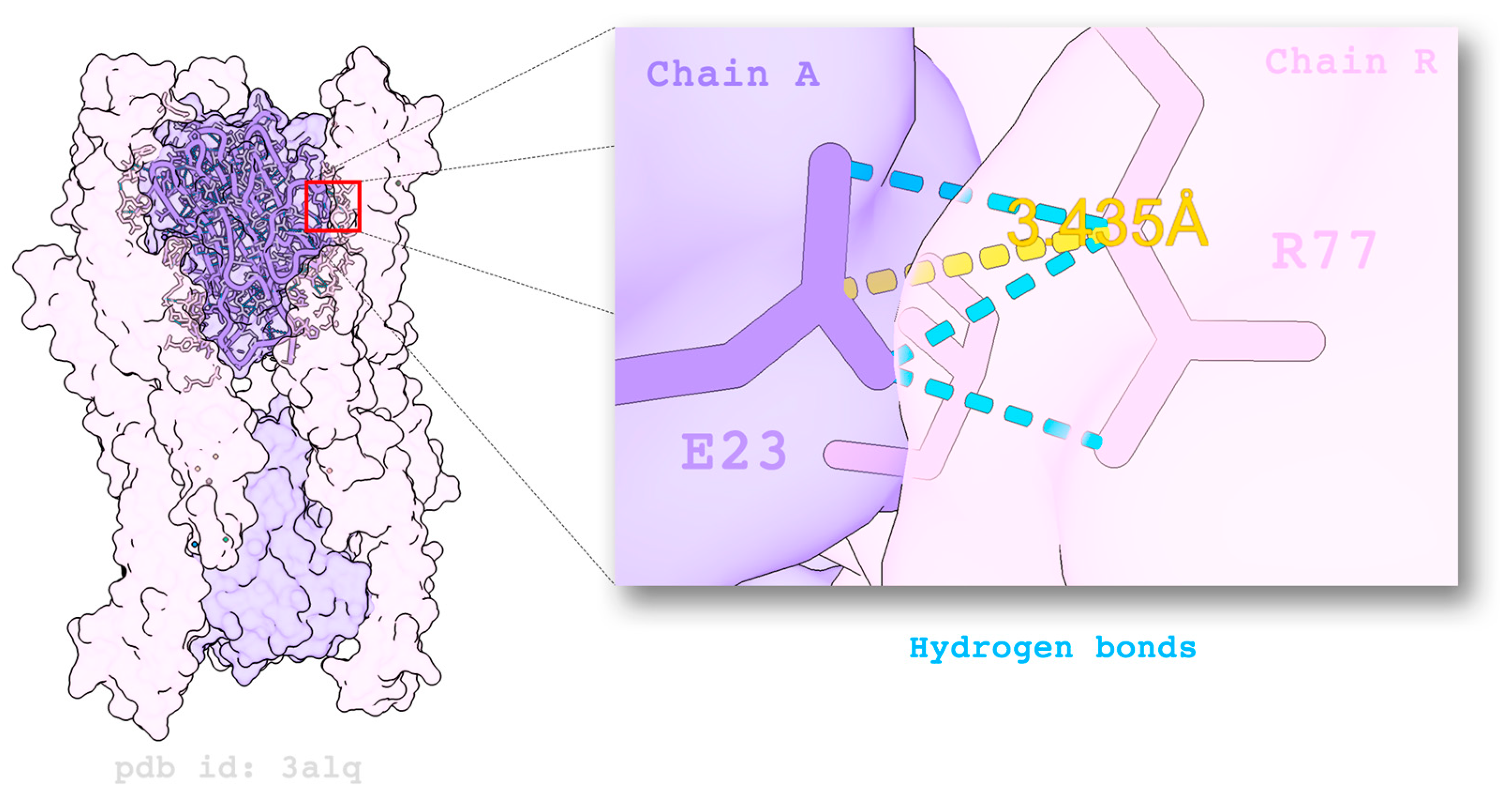
8.3. Assessing Intra- and Inter-Molecular Interactions

Contacts are weak interactions performed by amino acid residues with other amino acids or with other small molecules. Weak interactions shape protein structure and are responsible for much of their role in biological processes [114]. Hence, the evaluation of contacts, i.e., intra- and inter-molecular interactions, through in silico experiments and understanding the mechanisms related to diseases are important, as they allow the proposal of new medicines. Figure 7 illustrates the inter-molecular interactions performed between two residues (E23 and R77) of TNF-α homotrimers (purple) and TNF-R2 receptors (pink). Calculating contacts is usually carried out using distances between specific atoms [114].
There are several tools for evaluating contacts in different contexts. For example, Arpeggio [115] is a web server for evaluating different types of contacts, such as hydrogen bonds, ionic interactions, aromatic stacking, and hydrophobic interactions, among others. Likewise, VTR [116] is a web tool with a similar objective, but which performs structural alignments to evaluate the conservation of these types of contacts in pairs of proteins. Moreover, the E-volve [117] tool also performs structural alignments to evaluate contacts while considering possible mutations and their evolutionary impact. Another tool that considers evolutionary impact is Vermont [118,119], which focuses its analysis on sequence conservation. Furthermore, the nApoli [120] tool systematically evaluates involved contacts between protein and ligand. On the other hand, the LUNA [121] library presents a set of functionalities for contact analysis using Python version 3
Techniques based on structural signatures can also be used for computational modeling of biological problems, with diverse applications, such as graph-based protein modeling [122,123], mutation analysis [124,125], and the evaluation of the binding between protein–ligand [126], small-molecule pharmacokinetic and toxicity properties, or antibodies.

8.4. Molecular Docking and Molecular Dynamics Simulation

Molecular docking is a computational approach of the structural biology field commonly used in drug discovery, for example, in predicting the binding interactions between molecules. Docking aims to determine how and where a ligand binds to a molecule target, estimating the binding affinity or energy of the interactions by score functions. These interactions can be (i) protein–ligand, when the binding occurs between a target protein and small molecules (also called ligand); (ii) protein–protein, when the interactions predicted are between two protein molecules; (iii) protein–peptide, when the interactions between proteins and peptides are estimated; (iv) protein–acid nucleic, when interactions between proteins and acid nucleic molecules, such as DNA and RNA, are detected; and (v) other types of interactions among molecules [127,128,129].
Molecular dynamics (MDs) simulation is a computational approach used to evaluate the conformational behavior of molecules and their interactions in certain conditions and a determined period. In this simulation, the molecules are allocated in a system exposed to field forces: mathematical models that simulate particle interactions [130,131].

9. Bioinformatics Applied to Research on TNF-α Associated with Inflammatory Diseases

In the literature, we can find several examples of works in which bioinformatics tools were applied to research related to TNF-α associated with inflammatory diseases and the search for new potential pharmaceuticals for the treatment of these diseases. For example, we can cite the MDs study carried out by Abdullah-Al-Kamran Khan and colleagues [132] and published in 2021, in which the authors proposed a systematic approach in silico to find variants of the Adalimumab antibody with improved properties. In this study, bioinformatics tools were used to identify significant amino acid residues in the antibody. Then, the authors proposed the adaptation of the remaining residues to mutate the significant residues, and from the combinations of appropriate mutations, 143 variants were designed using comparative modeling methods. To find the most significant ones, the binding properties of the variants were compared with wild-type adalimumab using molecular docking and molecular dynamics simulation. From several docking analyses, the authors selected five significant variants, and after molecular dynamics simulation, a more significant variant with improved binding affinity was identified, whose structural properties are similar to those of wild-type Adalimumab. The engineered variant from this study may provide newer insights into the structure-based affinity improvements of monoclonal antibodies [132].
In the 2021 publication by Mustafá and colleagues [133], the authors explored the role of both TNF-α and IL-6 in promoting the proliferation of synovial membrane cells, thereby triggering the production of matrix metalloproteinases and other cytotoxins. This process contributes to bone erosion and cartilage destruction in rheumatoid arthritis. The study also delved into the potential of growth differentiation factor 11 (GDF11) and growth differentiation factor 8 (GDF8), also known as myostatin, as antagonists for inflammatory responses associated with rheumatoid arthritis. To elucidate the evolutionary relationships of GDF11 with its homologs from closely related organisms, the authors conducted a comprehensive phylogenetic analysis. The resulting phylogram revealed close evolutionary ties between the primate clade within the superorder Euarchontoglires and the order Cetartiodactyla of the superorder Laurasiatheria. Fifty tetrapeptides were developed from conserved regions of GDF11 that served as ligands in protein–ligand coupling against TNF-α and IL-6, followed by drug screening and ADMET profiling of the best-selected ligands. SAGP peptides showed strong interactions with IL-6, and AFDP and AGPC peptides showed strong interactions with TNF-α, and all three peptides met all pharmacokinetic parameters that are important for bioavailability. The potential of GDF8 as an antagonist to TNF-α and IL-6 was investigated through a protein–protein coupling approach. The findings revealed that the binding patterns of GDF8 with TNF-α and IL-6 indicated its potential use as an inhibitor for treating rheumatoid arthritis [133].
In turn, Hong and colleagues [134] carried out an analysis in 2021 using Adalimumab, commercially known as Humira. Taking into account that neonatal Fc receptors can mediate the transcytosis of the Humira–TNF-α complex structures and process them in degradation pathways, which reduces the therapeutic effect of Humira and allows the Humira–TNF-α complex structures to dissociate into Humira and soluble TNF-α in the early endosome to allow recycling of Humira. In the study, the authors used the cytoplasmic pH (7.4), the initial endosomal pH (6.0), and the pK a of the histidine side chains (6.0–6.4) to mutate the residues of the determining regions of complementarity with histidine. Humira (W1-Humira), developed by the authors, has been shown to bind to tumor necrosis factor α (TNF-α) in plasma at neutral pH and dissociate from TNF-α in the endosome at acidic pH. In the study, constant pH molecular dynamics, Gaussian accelerated molecular dynamics, two-dimensional mean potential force profiles, and in vitro methods were used to investigate the characteristics of W1-Humira. The results presented by the authors revealed that the proposed Humira is able to bind to TNF-α with pH-dependent affinity in vitro. W1-Humira was weaker than wild-type Humira at a neutral pH in vitro, and the prediction results were close to the in vitro results. Furthermore, the presented approach demonstrated high accuracy in predicting pH-dependent antibody binding characteristics, which could facilitate the design of antibody drugs. The authors highlighted that advances in computational methods and computational power could further help address challenges in antibody drug design [134].
In Abraham’s study, published in 2003 [135], the author computationally designed variant TNF-α molecules to inhibit the pro-inflammatory cascade. The author employed bioinformatics tools to model mutant structures and assessed their interaction with receptors and cellular activation. The author suggests the potential use of inserting mutations as a tool to investigate ligand–receptor interactions and their significance in signaling processes [135].
As mentioned earlier, Saddala and Huang [58], in their 2021 publication, employed a chemoinformatics pipeline consisting of pharmacophore modeling, virtual screening, molecular docking, and in silico ADMET analysis. This approach was used to screen for new inhibitors of TNF-α and TNFR1 in the Zinc database. Pharmacophore-based models were generated to select the best drug-like compounds from the Zinc database. As a result, the authors found new selective inhibitors of the TNFα, TNFR1, and TNFα–TNFR1 complex that can serve as anti-inflammatory agents and are promising candidates for future research [58].
Halder and colleagues in 2022 [101] published a study that used TNF-α as the main target for the virtual screening of drugs approved by the US-FDA for reuse using the in silico method using molecular docking, ADMET analysis, and prime MMGBSA. After that, drugs were selected according to dock score, ADMET parameters, and MM GBSA dG binding score. Following the initial screening, the selected drug molecules underwent induced docking analysis. Among them, two highly promising molecules, namely ZINC000003830957 (Iopromide) and ZINC000003830635 (Deferoxamine), were selected for molecular dynamics simulation. The authors concluded the study by employing bioisosteric substitution to enhance the ADMET properties of these molecules. Through this research, the authors offered valuable insights into drug exploration and computational tools for drug discovery in the treatment of inflammatory bowel diseases [101].
Agnihotri and colleagues, in 2023 [136], used structural bioinformatics tools to carry out studies targeting TNF-α and nuclear factor kappa B (NF-kB) through the metabolites of rheumatoid arthritis (RA), to inhibit the activity of TNF-α and prevent the NF-kB signaling pathways, thus mitigating the severity of RA disease. The structure of TNF-α and NF-kB was obtained from the PDB database, and the AR metabolites were selected from a literature survey. In silico studies were carried out via molecular docking using the AutoDock Vina software version 1.2.0. The known inhibitors of TNF-α and NF-kB were compared and revealed the metabolite’s ability to target the respective proteins. The most suitable metabolite was then validated via molecular dynamics simulation to verify its efficiency against TNF-α. A total of 56 known differential metabolites of RA were coupled to TNF-α and NF-κB compared to their corresponding inhibitory compounds. Four metabolites, namely chenodeoxycholic acid, 2-hydroxyestrone, 2-hydroxyestradiol (2-OHE2), and 16-hydroxyestradiol have been identified as the common inhibitors of TNF-α with binding energies ranging from −8.6 to −8.3 kcal/mol, followed by docking with NF-kB. Furthermore, 2-OHE2 was selected because it has a binding energy of −8.5 kcal/mol, which inhibits inflammation, and the effectiveness was validated by mean square fluctuation, the radius of gyration, and molecular mechanics with generalized birth solvation and area of surface against TNF-α. Thus, 2-OHE2, an estrogen metabolite, was identified as a potential inhibitor, attenuated inflammatory activation, and can be used as a therapeutic target to disseminate the severity of RA [136].
When it comes to molecular modeling, we have the example of the study carried out by Pierri and colleagues in 2016 [137], entitled “Molecular modeling of antibodies for the treatment of immunological diseases related to TNF-α”, where the authors used bioinformatics tools to understand the interaction of TNF-α inhibitory antibodies better taking into account that therapeutic monoclonal antibodies (mAbs) are highly effective in treating immunological diseases related to TNF-α. In addition to neutralizing TNF-α, these IgG1 antibodies exert Fc receptor-mediated effector functions, such as complement-dependent cytotoxicity (CDC) and antibody-dependent cellular cytotoxicity (ADCC). The crystallizable fragment (Fc) of these IgG1 contains a single glycosylation site at Asn 297/300 that is essential for CDC and ADCC. Glycosylated antibodies without core fucosylation showed improved ADCC. However, structural data regarding the ligand-binding interaction of these mAbs used in TNF-α-related diseases and the role of fucosylation are not available. Therefore, in this study, the authors performed comparative modeling to generate complete 3D mAb models that included the antigen-binding fragment (Fab) portions of infliximab, complexed with TNF-α (4G3Y.pdb), the Fc region of fucosylated human IGHG1 (3SGJ), and afucosylated (3SGK) complexed with the Fc receptor Fc subtype γ RIIIA and the Fc region of a murine immunoglobulin (1IGT). After a few thousand energy minimization steps on the resulting 3D mAb models, final minimized models were used to quantify the interactions occurring between Fc γ RIIIA and the fucosylated/afucosylated Fc fragments. Although fucosylation does not affect Fab-TNF-α interactions, it was found that in the absence of fucosylation, the Fc-mAb and Fcγ RIIIA domain are closer together and new strong interactions are established between G129 of the receptor and S301 of the Chimera 2 Fc mAb; novel polar interactions are also established between the chimera 2 Fc residues Y299, N300, and S301 and the γ RIIIA Fc residues K128, G129, R130, and R155. These data help explain the reduced ADCC observed in fucosylated mAbs, suggesting specific AA residues involved in binding interactions [137].
In 2023, Abechi and colleagues [138] conducted an in silico screening study to identify potential inhibitors of tumor necrosis factor α (TNF-α) using molecular modeling, molecular docking, and pharmacokinetic evaluations. In this study, a set of molecular modeling techniques were applied, including the QSAR model, docking, and pharmacokinetic prediction to identify and optimize novel TNF-α inhibitors. The results showed that the function of these discovered compounds was not linked to lipophilicity, while less long N N bonds and long substituents could lead to very bioactive molecules. The discovered results indicate a promising inhibition against TNF-α and show no harmful effects. Most of the discovered molecules had a higher binding affinity to TNF than the reference substance. Moreover, in comparison to the reference drug rating (ds) of 0.38, molecule 74 with the PubChem ID 2998055 demonstrates enhanced properties, achieving a drug rating (ds) of 0.76. Collectively, the identified molecules showcase favorable pharmacokinetic, pharmacodynamic, and drug interaction properties, indicating promising TNF-α inhibition and suggesting their potential as drug candidates [138].
In 2023, Erba and colleagues conducted a study titled “Head or tail? A molecular dynamics approach to the complex structure of TNF-associated factor TRAF2” [112]. The researchers utilized bioinformatics tools to gain a deeper understanding of the dynamics of TRAF2, a pivotal protein in the TNF-α signaling cascade. The study focused on analyzing the in silico dependence of TRAF2 dynamics on the length of its tail. The authors employed the crystallographic structure of a C-terminal fragment of TRAF2 (168 of 501 aa), denoted as TRAF2-C, and that of a longer construct referred to as TRAF2-plus. The TRAF2-plus structure was reconstructed using the AlphaFold2 code [112].
The results of the study revealed that the longer N-terminal tail of TRAF2-plus significantly influenced the dynamics of the globular regions in the C-terminal head of the protein. The quaternary interactions between the TRAF2-C subunits asymmetrically changed over time, whereas the movements of the TRAF2-plus monomers were comparatively limited and more ordered than those of the shorter construct. These findings provide new insights into the dynamics of TRAF subunits and the protein’s mechanism in vivo. The balance between TRAF monomer-trimer interactions is crucial for various functions, such as receptor recognition, membrane binding, and hetero-oligomerization, emphasizing the protein’s significant role in the inflammation signaling cascade [112].

10. Conclusions

In this presentation, we underscored the significance of TNF-α in chronic inflammatory diseases, examining it through the lens of structural biology. Additionally, we showcased examples of bioinformatics techniques, databases, tools, and strategies that can be employed to unravel insights into the biological complexities associated with TNF-α. We anticipate that the information provided in this review will prove valuable for studies seeking a deeper comprehension of the interactions between TNF-α and other proteins, potentially paving the way for the development of novel drugs or treatments.

Author Contributions

Conceptualization, L.L.B., C.J.F.O. and R.C.d.M.-M.; formal analysis, R.C.d.M.-M. and C.J.F.O.; writing—original draft preparation, L.L.B.; writing—review and editing, D.M., T.S.B. and R.P.L.; visualization, D.M., T.S.B. and L.L.B.; supervision, R.C.d.M.-M.; project administration, R.C.d.M.-M.; funding acquisition, R.C.d.M.-M. All authors have read and agreed to the published version of the manuscript.

Funding

This research was partly funded by CAPES (funding code 001), CNPq, and FAPEMIG.

Conflicts of Interest

The authors declare no conflicts of interest.

References

  1. Carswell, E.A.; Old, L.J.; Kassel, R.L.; Green, S.; Fiore, N.; Williamson, B. An Endotoxin-Induced Serum Factor That Causes Necrosis of Tumors. Proc. Natl. Acad. Sci. USA 1975, 72, 3666–3670. [Google Scholar] [CrossRef]
  2. Bradley, J. TNF-Mediated Inflammatory Disease. J. Pathol. 2008, 214, 149–160. [Google Scholar] [CrossRef]
  3. Eck, M.J.; Sprang, S.R. The Structure of Tumor Necrosis Factor-Alpha at 2.6 A Resolution. Implications for Receptor Binding. J. Biol. Chem. 1989, 264, 17595–17605. [Google Scholar] [CrossRef]
  4. Ruddle, N.H. Tumor Necrosis Factor (TNF-α) and Lymphotoxin (TNF-β). Curr. Opin. Immunol. 1992, 4, 327–332. [Google Scholar] [CrossRef]
  5. Pennica, D.; Hayflick, J.S.; Bringman, T.S.; Palladino, M.A.; Goeddel, D.V. Cloning and Expression in Escherichia Coli of the cDNA for Murine Tumor Necrosis Factor. Proc. Natl. Acad. Sci. USA 1985, 82, 6060–6064. [Google Scholar] [CrossRef]
  6. Hu, S.; Liang, S.; Guo, H.; Zhang, D.; Li, H.; Wang, X.; Yang, W.; Qian, W.; Hou, S.; Wang, H.; et al. Comparison of the Inhibition Mechanisms of Adalimumab and Infliximab in Treating Tumor Necrosis Factor α-Associated Diseases from a Molecular View. J. Biol. Chem. 2013, 288, 27059–27067. [Google Scholar] [CrossRef]
  7. Tracey, K.J.; Cerami, A. Tumor Necrosis Factor: A Pleiotropic Cytokine and Therapeutic Target. Annu. Rev. Med. 1994, 45, 491–503. [Google Scholar] [CrossRef]
  8. Torres, J.; Mehandru, S.; Colombel, J.-F.; Peyrin-Biroulet, L. Crohn’s Disease. Lancet 2017, 389, 1741–1755. [Google Scholar] [CrossRef]
  9. Smolen, J.S.; Aletaha, D.; McInnes, I.B. Rheumatoid Arthritis. Lancet 2016, 388, 2023–2038. [Google Scholar] [CrossRef]
  10. Papadakis, K.A.; Targan, S.R. Role of Cytokines in the Pathogenesis of Inflammatory Bowel Disease. Annu. Rev. Med. 2000, 51, 289–298. [Google Scholar] [CrossRef]
  11. Neurath, M.F. Cytokines in Inflammatory Bowel Disease. Nat. Rev. Immunol. 2014, 14, 329–342. [Google Scholar] [CrossRef]
  12. Idriss, H.T.; Naismith, J.H. TNF Alpha and the TNF Receptor Superfamily: Structure-Function Relationship(s). Microsc. Res. Tech. 2000, 50, 184–195. [Google Scholar] [CrossRef]
  13. Wajant, H.; Pfizenmaier, K.; Scheurich, P. Tumor Necrosis Factor Signaling. Cell Death Differ. 2003, 10, 45–65. [Google Scholar] [CrossRef]
  14. Mukai, Y.; Nakamura, T.; Yoshikawa, M.; Yoshioka, Y.; Tsunoda, S.; Nakagawa, S.; Yamagata, Y.; Tsutsumi, Y. Solution of the Structure of the TNF-TNFR2 Complex. Sci. Signal. 2010, 3, ra83. [Google Scholar] [CrossRef]
  15. Mukai, Y.; Nakamura, T.; Yoshioka, Y.; Shibata, H.; Abe, Y.; Nomura, T.; Taniai, M.; Ohta, T.; Nakagawa, S.; Tsunoda, S.; et al. Fast Binding Kinetics and Conserved 3D Structure Underlie the Antagonistic Activity of Mutant TNF: Useful Information for Designing Artificial Proteo-Antagonists. J. Biochem. 2009, 146, 167–172. [Google Scholar] [CrossRef]
  16. Zhao, L.; Fu, Q.; Pan, L.; Piai, A.; Chou, J.J. The Diversity and Similarity of Transmembrane Trimerization of TNF Receptors. Front. Cell Dev. Biol. 2020, 8, 569684. [Google Scholar] [CrossRef]
  17. Reed, C.; Fu, Z.Q.; Wu, J.; Xue, Y.N.; Harrison, R.W.; Chen, M.J.; Weber, I.T. Crystal Structure of TNF-Alpha Mutant R31D with Greater Affinity for Receptor R1 Compared with R2. Protein Eng. 1997, 10, 1101–1107. [Google Scholar] [CrossRef]
  18. Pobezinskaya, Y.L.; Liu, Z. The Role of TRADD in Death Receptor Signaling. Cell Cycle 2012, 11, 871–876. [Google Scholar] [CrossRef]
  19. Gaeta, M.L.; Johnson, D.R.; Kluger, M.S.; Pober, J.S. The Death Domain of Tumor Necrosis Factor Receptor 1 Is Necessary but Not Sufficient for Golgi Retention of the Receptor and Mediates Receptor Desensitization. Lab. Investig. 2000, 80, 1185–1194. [Google Scholar] [CrossRef]
  20. MacEwan, D.J. TNF Ligands and Receptors—A Matter of Life and Death. Br. J. Pharmacol. 2002, 135, 855–875. [Google Scholar] [CrossRef]
  21. Kim, D.E.; Chivian, D.; Baker, D. Protein Structure Prediction and Analysis Using the Robetta Server. Nucleic Acids Res. 2004, 32, W526–W531. [Google Scholar] [CrossRef]
  22. Zhou, F.; Xu, X.; Wang, D.; Wu, J.; Wang, J. Identification of Novel NF-κB Transcriptional Targets in TNFα-Treated HeLa and HepG2 Cells. Cell Biol. Int. 2017, 41, 555–569. [Google Scholar] [CrossRef]
  23. Wajant, H.; Siegmund, D. TNFR1 and TNFR2 in the Control of the Life and Death Balance of Macrophages. Front. Cell Dev. Biol. 2019, 7. [Google Scholar] [CrossRef]
  24. Goddard, T.D.; Huang, C.C.; Meng, E.C.; Pettersen, E.F.; Couch, G.S.; Morris, J.H.; Ferrin, T.E. UCSF ChimeraX: Meeting Modern Challenges in Visualization and Analysis. Protein Sci. Publ. Protein Soc. 2018, 27, 14. [Google Scholar] [CrossRef]
  25. He, M.M.; Smith, A.S.; Oslob, J.D.; Flanagan, W.M.; Braisted, A.C.; Whitty, A.; Cancilla, M.T.; Wang, J.; Lugovskoy, A.A.; Yoburn, J.C.; et al. Small-Molecule Inhibition of TNF-Alpha. Science 2005, 310, 1022–1025. [Google Scholar] [CrossRef]
  26. Tseng, W.-Y.; Huang, Y.-S.; Lin, H.-H.; Luo, S.-F.; McCann, F.; McNamee, K.; Clanchy, F.; Williams, R. TNFR Signalling and Its Clinical Implications. Cytokine 2018, 101, 19–25. [Google Scholar] [CrossRef]
  27. Mukai, Y.; Shibata, H.; Nakamura, T.; Yoshioka, Y.; Abe, Y.; Nomura, T.; Taniai, M.; Ohta, T.; Ikemizu, S.; Nakagawa, S.; et al. Structure-Function Relationship of Tumor Necrosis Factor (TNF) and Its Receptor Interaction Based on 3D Structural Analysis of a Fully Active TNFR1-Selective TNF Mutant. J. Mol. Biol. 2009, 385, 1221–1229. [Google Scholar] [CrossRef]
  28. Liang, S.; Dai, J.; Hou, S.; Su, L.; Zhang, D.; Guo, H.; Hu, S.; Wang, H.; Rao, Z.; Guo, Y.; et al. Structural Basis for Treating Tumor Necrosis Factor α (TNFα)-Associated Diseases with the Therapeutic Antibody Infliximab. J. Biol. Chem. 2013, 288, 13799–13807. [Google Scholar] [CrossRef]
  29. Huang, P.L. A Comprehensive Definition for Metabolic Syndrome. Dis. Model. Mech. 2009, 2, 231–237. [Google Scholar] [CrossRef]
  30. McInnes, I.B.; Schett, G. The Pathogenesis of Rheumatoid Arthritis. N. Engl. J. Med. 2011, 365, 2205–2219. [Google Scholar] [CrossRef]
  31. Firestein, G.S.; Budd, R.C.; Gabriel, S.E.; McInnes, I.B.; O’Dell, J.R. Kelley and Firestein’s Textbook of Rheumatology; Elsevier Health Sciences: Amsterdam, The Netherlands, 2016; ISBN 978-0-323-41494-4. [Google Scholar]
  32. Griffiths, C.E.; Barker, J.N. Pathogenesis and Clinical Features of Psoriasis. Lancet 2007, 370, 263–271. [Google Scholar] [CrossRef] [PubMed]
  33. Lowes, M.A.; Suárez-Fariñas, M.; Krueger, J.G. Immunology of Psoriasis. Annu. Rev. Immunol. 2014, 32, 227–255. [Google Scholar] [CrossRef] [PubMed]
  34. Sanchez-Muñoz, F.; Dominguez-Lopez, A.; Yamamoto-Furusho, J.K. Role of Cytokines in Inflammatory Bowel Disease. World J. Gastroenterol. WJG 2008, 14, 4280–4288. [Google Scholar] [CrossRef] [PubMed]
  35. Braun, J.; Sieper, J. Ankylosing Spondylitis. Lancet 2007, 369, 1379–1390. [Google Scholar] [CrossRef] [PubMed]
  36. Brown, M.A.; Kenna, T.; Wordsworth, B.P. Genetics of Ankylosing Spondylitis--Insights into Pathogenesis. Nat. Rev. Rheumatol. 2016, 12, 81–91. [Google Scholar] [CrossRef] [PubMed]
  37. Phumethum, V.; Jamal, S.; Johnson, S.R. Biologic Therapy for Systemic Sclerosis: A Systematic Review. J. Rheumatol. 2011, 38, 289–296. [Google Scholar] [CrossRef] [PubMed]
  38. Murdaca, G.; Spanò, F.; Contatore, M.; Guastalla, A.; Puppo, F. Potential Use of TNF-α Inhibitors in Systemic Sclerosis. Immunotherapy 2014, 6, 283–289. [Google Scholar] [CrossRef]
  39. Thyssen, J.P. Atopic Dermatitis, Filaggrin Mutations and Irritant Contact Dermatitis. Br. J. Dermatol. 2013, 168, 233–234. [Google Scholar] [CrossRef]
  40. Savage, K.T.; Flood, K.S.; Porter, M.L.; Kimball, A.B. TNF-α Inhibitors in the Treatment of Hidradenitis Suppurativa. Ther. Adv. Chronic Dis. 2019, 10, 2040622319851640. [Google Scholar] [CrossRef]
  41. Prens, E.; Deckers, I. Pathophysiology of Hidradenitis Suppurativa: An Update. J. Am. Acad. Dermatol. 2015, 73, S8–S11. [Google Scholar] [CrossRef]
  42. Jayne, D. The Diagnosis of Vasculitis. Best Pract. Res. Clin. Rheumatol. 2009, 23, 445–453. [Google Scholar] [CrossRef] [PubMed]
  43. Sokumbi, O.; Wetter, D.A.; Makol, A.; Warrington, K.J. Vasculitis Associated with Tumor Necrosis Factor-α Inhibitors. Mayo Clin. Proc. 2012, 87, 739–745. [Google Scholar] [CrossRef] [PubMed]
  44. Jarrot, P.-A.; Kaplanski, G. Anti-TNF-Alpha Therapy and Systemic Vasculitis. Mediat. Inflamm. 2014, 2014, e493593. [Google Scholar] [CrossRef] [PubMed]
  45. Johnson, S.; Oliver, C.; Prince, G.A.; Hemming, V.G.; Pfarr, D.S.; Wang, S.C.; Dormitzer, M.; O’Grady, J.; Koenig, S.; Tamura, J.K.; et al. Development of a Humanized Monoclonal Antibody (MEDI-493) with Potent in Vitro and in Vivo Activity against Respiratory Syncytial Virus. J. Infect. Dis. 1997, 176, 1215–1224. [Google Scholar] [CrossRef]
  46. Pan, A.; Gerriets, V. Etanercept. In StatPearls; StatPearls Publishing: Treasure Island, FL, USA, 2023. [Google Scholar]
  47. Jang, D.; Lee, A.-H.; Shin, H.-Y.; Song, H.-R.; Park, J.-H.; Kang, T.-B.; Lee, S.-R.; Yang, S.-H. The Role of Tumor Necrosis Factor Alpha (TNF-α) in Autoimmune Disease and Current TNF-α Inhibitors in Therapeutics. Int. J. Mol. Sci. 2021, 22, 2719. [Google Scholar] [CrossRef] [PubMed]
  48. Mease, P.J.; Ory, P.; Sharp, J.T.; Ritchlin, C.T.; Van den Bosch, F.; Wellborne, F.; Birbara, C.; Thomson, G.T.D.; Perdok, R.J.; Medich, J.; et al. Adalimumab for Long-Term Treatment of Psoriatic Arthritis: 2-Year Data from the Adalimumab Effectiveness in Psoriatic Arthritis Trial (ADEPT). Ann. Rheum. Dis. 2009, 68, 702–709. [Google Scholar] [CrossRef]
  49. Lee, J.U.; Shin, W.; Son, J.Y.; Yoo, K.-Y.; Heo, Y.-S. Molecular Basis for the Neutralization of Tumor Necrosis Factor α by Certolizumab Pegol in the Treatment of Inflammatory Autoimmune Diseases. Int. J. Mol. Sci. 2017, 18, 228. [Google Scholar] [CrossRef] [PubMed]
  50. Emery, P.; Fleischmann, R.M.; Doyle, M.K.; Strusberg, I.; Durez, P.; Nash, P.; Amante, E.; Churchill, M.; Park, W.; Pons-Estel, B.; et al. Golimumab, a Human Anti–Tumor Necrosis Factor Monoclonal Antibody, Injected Subcutaneously Every 4 Weeks in Patients with Active Rheumatoid Arthritis Who Had Never Taken Methotrexate: 1-Year and 2-Year Clinical, Radiologic, and Physical Function Findings of a Phase III, Multicenter, Randomized, Double-Blind, Placebo-Controlled Study. Arthritis Care Res. 2013, 65, 1732–1742. [Google Scholar] [CrossRef]
  51. Pelechas, E.; Voulgari, P.V.; Drosos, A.A. Golimumab for Rheumatoid Arthritis. J. Clin. Med. 2019, 8, 387. [Google Scholar] [CrossRef]
  52. Mitoma, H.; Horiuchi, T.; Tsukamoto, H.; Ueda, N. Molecular Mechanisms of Action of Anti-TNF-α Agents—Comparison among Therapeutic TNF-α Antagonists. Cytokine 2018, 101, 56–63. [Google Scholar] [CrossRef]
  53. Kaltsonoudis, E.; Zikou, A.K.; Voulgari, P.V.; Konitsiotis, S.; Argyropoulou, M.I.; Drosos, A.A. Neurological Adverse Events in Patients Receiving Anti-TNF Therapy: A Prospective Imaging and Electrophysiological Study. Arthritis Res. Ther. 2014, 16, R125. [Google Scholar] [CrossRef] [PubMed]
  54. Zhou, H. Clinical Pharmacokinetics of Etanercept: A Fully Humanized Soluble Recombinant Tumor Necrosis Factor Receptor Fusion Protein. J. Clin. Pharmacol. 2005, 45, 490–497. [Google Scholar] [CrossRef] [PubMed]
  55. Dixon, W.G.; Watson, K.; Lunt, M.; Hyrich, K.L.; Silman, A.J.; Symmons, D.P.M. Rates of Serious Infection, Including Site-Specific and Bacterial Intracellular Infection, in Rheumatoid Arthritis Patients Receiving Anti-Tumor Necrosis Factor Therapy: Results from the British Society for Rheumatology Biologics Register. Arthritis Rheum. 2006, 54, 2368–2376. [Google Scholar] [CrossRef]
  56. Keam, S.J. Ozoralizumab: First Approval. Drugs 2023, 83, 87–92. [Google Scholar] [CrossRef] [PubMed]
  57. Tanaka, Y. Ozoralizumab: First Nanobody® Therapeutic for Rheumatoid Arthritis. Expert Opin. Biol. Ther. 2023, 23, 579–587. [Google Scholar] [CrossRef] [PubMed]
  58. Saddala, M.S.; Huang, H. Identification of Novel Inhibitors for TNFα, TNFR1 and TNFα-TNFR1 Complex Using Pharmacophore-Based Approaches. J. Transl. Med. 2019, 17, 215. [Google Scholar] [CrossRef] [PubMed]
  59. Irwin, J.J.; Shoichet, B.K. ZINC—A Free Database of Commercially Available Compounds for Virtual Screening. J. Chem. Inf. Model. 2005, 45, 177–182. [Google Scholar] [CrossRef]
  60. Kwak, M.S.; Lee, H.H.; Cha, J.M.; Shin, H.P.; Jeon, J.W.; Yoon, J.Y. Novel Candidate Drugs in Anti-Tumor Necrosis Factor Refractory Crohn’s Diseases: In Silico Study for Drug Repositioning. Sci. Rep. 2020, 10, 10708. [Google Scholar] [CrossRef]
  61. Barrett, T.; Suzek, T.O.; Troup, D.B.; Wilhite, S.E.; Ngau, W.-C.; Ledoux, P.; Rudnev, D.; Lash, A.E.; Fujibuchi, W.; Edgar, R. NCBI GEO: Mining Millions of Expression Profiles—Database and Tools. Nucleic Acids Res. 2005, 33, D562–D566. [Google Scholar] [CrossRef]
  62. Szklarczyk, D.; Franceschini, A.; Kuhn, M.; Simonovic, M.; Roth, A.; Minguez, P.; Doerks, T.; Stark, M.; Muller, J.; Bork, P.; et al. The STRING Database in 2011: Functional Interaction Networks of Proteins, Globally Integrated and Scored. Nucleic Acids Res. 2011, 39, D561–D568. [Google Scholar] [CrossRef]
  63. Koník, P.; Slavíková, V.; Salát, J.; Reznícková, J.; Dvoroznáková, E.; Kopecký, J. Anti-Tumour Necrosis Factor-Alpha Activity in Ixodes Ricinus Saliva. Parasite Immunol. 2006, 28, 649–656. [Google Scholar] [CrossRef] [PubMed]
  64. Kazimírová, M.; Štibrániová, I. Tick Salivary Compounds: Their Role in Modulation of Host Defences and Pathogen Transmission. Front. Cell. Infect. Microbiol. 2013, 3, 43. [Google Scholar] [CrossRef] [PubMed]
  65. Šimo, L.; Kazimirova, M.; Richardson, J.; Bonnet, S.I. The Essential Role of Tick Salivary Glands and Saliva in Tick Feeding and Pathogen Transmission. Front. Cell. Infect. Microbiol. 2017, 7, 281. [Google Scholar] [CrossRef] [PubMed]
  66. Rezkova, M.; Kopecky, J. Anti-Tumour Necrosis Factor Activity in Saliva of Various Tick Species and Its Appearance during the Feeding Period. Folia Parasitol. 2017, 64, 32. [Google Scholar] [CrossRef] [PubMed]
  67. Papp, K.A.; Tyring, S.; Lahfa, M.; Prinz, J.; Griffiths, C.E.M.; Nakanishi, A.M.; Zitnik, R.; van de Kerkhof, P.C.M.; Melvin, L. Etanercept Psoriasis Study Group A Global Phase III Randomized Controlled Trial of Etanercept in Psoriasis: Safety, Efficacy, and Effect of Dose Reduction. Br. J. Dermatol. 2005, 152, 1304–1312. [Google Scholar] [CrossRef] [PubMed]
  68. Weinblatt, M.E.; Keystone, E.C.; Furst, D.E.; Moreland, L.W.; Weisman, M.H.; Birbara, C.A.; Teoh, L.A.; Fischkoff, S.A.; Chartash, E.K. Adalimumab, a Fully Human Anti-Tumor Necrosis Factor Alpha Monoclonal Antibody, for the Treatment of Rheumatoid Arthritis in Patients Taking Concomitant Methotrexate: The ARMADA Trial. Arthritis Rheum. 2003, 48, 35–45. [Google Scholar] [CrossRef] [PubMed]
  69. Verri, W.A., Jr.; Cunha, T.M.; Poole, S.; Ferreira, S.H.; Cunha, F.Q. Cytokine Inhibitors and Pain Control. Rev. Bras. Reumatol. 2007, 47, 341–353. [Google Scholar] [CrossRef]
  70. Charles, P.; Elliott, M.J.; Davis, D.; Potter, A.; Kalden, J.R.; Antoni, C.; Breedveld, F.C.; Smolen, J.S.; Eberl, G.; deWoody, K.; et al. Regulation of Cytokines, Cytokine Inhibitors, and Acute-Phase Proteins Following Anti-TNF-Alpha Therapy in Rheumatoid Arthritis. J. Immunol. 1999, 163, 1521–1528. [Google Scholar] [CrossRef]
  71. Chadwick, L.; Zhao, S.; Mysler, E.; Moots, R.J. Review of Biosimilar Trials and Data on Etanercept in Rheumatoid Arthritis. Curr. Rheumatol. Rep. 2018, 20, 84. [Google Scholar] [CrossRef]
  72. Prabhakar, S. Translational Research Challenges: Finding the Right Animal Models. J. Investig. Med. Off. Publ. Am. Fed. Clin. Res. 2012, 60, 1141–1146. [Google Scholar] [CrossRef]
  73. Byng-Maddick, R.; Turner, C.T.; Pollara, G.; Ellis, M.; Guppy, N.J.; Bell, L.C.K.; Ehrenstein, M.R.; Noursadeghi, M. Tumor Necrosis Factor (TNF) Bioactivity at the Site of an Acute Cell-Mediated Immune Response Is Preserved in Rheumatoid Arthritis Patients Responding to Anti-TNF Therapy. Front. Immunol. 2017, 8, 932. [Google Scholar] [CrossRef]
  74. The UniProt Consortium UniProt: The Universal Protein Knowledgebase in 2023. Nucleic Acids Res. 2023, 51, D523–D531. [CrossRef]
  75. Berman, H.M.; Westbrook, J.; Feng, Z.; Gilliland, G.; Bhat, T.N.; Weissig, H.; Shindyalov, I.N.; Bourne, P.E. The Protein Data Bank. Nucleic Acids Res. 2000, 28, 235–242. [Google Scholar] [CrossRef]
  76. Kanehisa, M.; Goto, S. KEGG: Kyoto Encyclopedia of Genes and Genomes. Nucleic Acids Res. 2000, 28, 27–30. [Google Scholar] [CrossRef]
  77. Mistry, J.; Chuguransky, S.; Williams, L.; Qureshi, M.; Salazar, G.A.; Sonnhammer, E.L.L.; Tosatto, S.C.E.; Paladin, L.; Raj, S.; Richardson, L.J.; et al. Pfam: The Protein Families Database in 2021. Nucleic Acids Res. 2021, 49, D412–D419. [Google Scholar] [CrossRef]
  78. Gaulton, A.; Bellis, L.J.; Bento, A.P.; Chambers, J.; Davies, M.; Hersey, A.; Light, Y.; McGlinchey, S.; Michalovich, D.; Al-Lazikani, B.; et al. ChEMBL: A Large-Scale Bioactivity Database for Drug Discovery. Nucleic Acids Res. 2012, 40, D1100–D1107. [Google Scholar] [CrossRef]
  79. Schievella, A.R.; Chen, J.H.; Graham, J.R.; Lin, L.-L. MADD, a Novel Death Domain Protein That Interacts with the Type 1 Tumor Necrosis Factor Receptor and Activates Mitogen-Activated Protein Kinase*. J. Biol. Chem. 1997, 272, 12069–12075. [Google Scholar] [CrossRef]
  80. Schneeberger, P.E.; Kortüm, F.; Korenke, G.C.; Alawi, M.; Santer, R.; Woidy, M.; Buhas, D.; Fox, S.; Juusola, J.; Alfadhel, M.; et al. Biallelic MADD Variants Cause a Phenotypic Spectrum Ranging from Developmental Delay to a Multisystem Disorder. Brain 2020, 143, 2437–2453. [Google Scholar] [CrossRef] [PubMed]
  81. Suzuki, M.; Watanabe, M.; Nakamaru, Y.; Takagi, D.; Takahashi, H.; Fukuda, S.; Hatakeyama, S. TRIM39 Negatively Regulates the NFκB-Mediated Signaling Pathway through Stabilization of Cactin. Cell. Mol. Life Sci. 2016, 73, 1085–1101. [Google Scholar] [CrossRef] [PubMed]
  82. Kim, S.-H.; Han, S.-Y.; Azam, T.; Yoon, D.-Y.; Dinarello, C.A. Interleukin-32: A Cytokine and Inducer of TNFα. Immunity 2005, 22, 131–142. [Google Scholar] [CrossRef] [PubMed]
  83. Chan, D.S.-H.; Lee, H.-M.; Yang, F.; Che, C.-M.; Wong, C.C.L.; Abagyan, R.; Leung, C.-H.; Ma, D.-L. Structure-Based Discovery of Natural Product-Like TNF-α Inhibitors. Angew. Chem. Int. Ed. Engl. 2010, 49, 2860–2864. [Google Scholar] [CrossRef]
  84. Byla, P.; Andersen, M.H.; Holtet, T.L.; Jacobsen, H.; Munch, M.; Gad, H.H.; Thøgersen, H.C.; Hartmann, R. Selection of a Novel and Highly Specific Tumor Necrosis Factor Alpha (TNFalpha) Antagonist: Insight from the Crystal Structure of the Antagonist-TNFalpha Complex. J. Biol. Chem. 2010, 285, 12096–12100. [Google Scholar] [CrossRef]
  85. Beirnaert, E.; Desmyter, A.; Spinelli, S.; Lauwereys, M.; Aarden, L.; Dreier, T.; Loris, R.; Silence, K.; Pollet, C.; Cambillau, C.; et al. Bivalent Llama Single-Domain Antibody Fragments against Tumor Necrosis Factor Have Picomolar Potencies Due to Intramolecular Interactions. Front. Immunol. 2017, 8, 867. [Google Scholar] [CrossRef]
  86. Yang, Z.; West, A.P.; Bjorkman, P.J. Crystal Structure of TNFalpha Complexed with a Poxvirus MHC-Related TNF Binding Protein. Nat. Struct. Mol. Biol. 2009, 16, 1189–1191. [Google Scholar] [CrossRef]
  87. Cha, S.S.; Kim, J.S.; Cho, H.S.; Shin, N.K.; Jeong, W.; Shin, H.C.; Kim, Y.J.; Hahn, J.H.; Oh, B.H. High Resolution Crystal Structure of a Human Tumor Necrosis Factor-Alpha Mutant with Low Systemic Toxicity. J. Biol. Chem. 1998, 273, 2153–2160. [Google Scholar] [CrossRef]
  88. Shibata, H.; Yoshioka, Y.; Ohkawa, A.; Minowa, K.; Mukai, Y.; Abe, Y.; Taniai, M.; Nomura, T.; Kayamuro, H.; Nabeshi, H.; et al. Creation and X-Ray Structure Analysis of the Tumor Necrosis Factor Receptor-1-Selective Mutant of a Tumor Necrosis Factor-Alpha Antagonist. J. Biol. Chem. 2008, 283, 998–1007. [Google Scholar] [CrossRef]
  89. London, N.; Movshovitz-Attias, D.; Schueler-Furman, O. The Structural Basis of Peptide-Protein Binding Strategies. Structure 2010, 18, 188–199. [Google Scholar] [CrossRef]
  90. Vanhee, P.; Reumers, J.; Stricher, F.; Baeten, L.; Serrano, L.; Schymkowitz, J.; Rousseau, F. PepX: A Structural Database of Non-Redundant Protein–Peptide Complexes. Nucleic Acids Res. 2010, 38, D545–D551. [Google Scholar] [CrossRef]
  91. Martins, P.; Mariano, D.; Carvalho, F.C.; Bastos, L.L.; Moraes, L.; Paixão, V.; Cardoso de Melo-Minardi, R. Propedia v2.3: A Novel Representation Approach for the Peptide-Protein Interaction Database Using Graph-Based Structural Signatures. Front. Bioinform. 2023, 3, 1103103. [Google Scholar] [CrossRef]
  92. Martins, P.M.; Santos, L.H.; Mariano, D.; Queiroz, F.C.; Bastos, L.L.; Gomes, I.d.S.; Fischer, P.H.C.; Rocha, R.E.O.; Silveira, S.A.; de Lima, L.H.F.; et al. Propedia: A Database for Protein–Peptide Identification Based on a Hybrid Clustering Algorithm. BMC Bioinform. 2021, 22, 1. [Google Scholar] [CrossRef]
  93. Ogata, H.; Goto, S.; Sato, K.; Fujibuchi, W.; Bono, H.; Kanehisa, M. KEGG: Kyoto Encyclopedia of Genes and Genomes. Nucleic Acids Res. 1999, 27, 29–34. [Google Scholar] [CrossRef] [PubMed]
  94. Kanehisa, M.; Furumichi, M.; Sato, Y.; Kawashima, M.; Ishiguro-Watanabe, M. KEGG for Taxonomy-Based Analysis of Pathways and Genomes. Nucleic Acids Res. 2023, 51, D587–D592. [Google Scholar] [CrossRef] [PubMed]
  95. Yao, Y.; Yang, L.; Zhang, Z.; Wang, B.; Feng, B.; Liu, Z. Identification of Targets for Subsequent Treatment of Crohn’s Disease Patients After Failure of Anti-TNF Therapy. J. Inflamm. Res. 2023, 16, 4617–4631. [Google Scholar] [CrossRef] [PubMed]
  96. Jie, K.; Feng, W.; Boxiang, Z.; Maofeng, G.; Jianbin, Z.; Zhaoxuan, L.; Yangyi, Z.; Liang, C.; Haobo, S.; Wensheng, L.; et al. Identification of Pathways and Key Genes in Venous Remodeling After Arteriovenous Fistula by Bioinformatics Analysis. Front. Physiol. 2020, 11, 565240. [Google Scholar] [CrossRef]
  97. Liu, Y.; Duan, Y.; Li, Y. Integrated Gene Expression Profiling Analysis Reveals Probable Molecular Mechanism and Candidate Biomarker in Anti-TNF&alpha; Non-Response IBD Patients. J. Inflamm. Res. 2020, 13, 81–95. [Google Scholar] [CrossRef] [PubMed]
  98. Szklarczyk, D.; Kirsch, R.; Koutrouli, M.; Nastou, K.; Mehryary, F.; Hachilif, R.; Gable, A.L.; Fang, T.; Doncheva, N.T.; Pyysalo, S.; et al. The STRING Database in 2023: Protein-Protein Association Networks and Functional Enrichment Analyses for Any Sequenced Genome of Interest. Nucleic Acids Res. 2023, 51, D638–D646. [Google Scholar] [CrossRef] [PubMed]
  99. Polley, S.; Passos, D.O.; Huang, D.-B.; Mulero, M.C.; Mazumder, A.; Biswas, T.; Verma, I.M.; Lyumkis, D.; Ghosh, G. Structural Basis for the Activation of IKK1/α. Cell Rep. 2016, 17, 1907–1914. [Google Scholar] [CrossRef]
  100. Li, Y.; Okatsu, K.; Fukai, S.; Sato, Y. Structural Basis for Specific Recognition of K6-Linked Polyubiquitin Chains by the TAB2 NZF Domain. Biophys. J. 2021, 120, 3355–3362. [Google Scholar] [CrossRef]
  101. Halder, D.; Das, S.; Joseph, A.; Jeyaprakash, R.S. Molecular Docking and Dynamics Approach to in Silico Drug Repurposing for Inflammatory Bowels Disease by Targeting TNF Alpha. J. Biomol. Struct. Dyn. 2023, 41, 3462–3475. [Google Scholar] [CrossRef]
  102. Edgar, R.; Domrachev, M.; Lash, A.E. Gene Expression Omnibus: NCBI Gene Expression and Hybridization Array Data Repository. Nucleic Acids Res. 2002, 30, 207–210. [Google Scholar] [CrossRef]
  103. Barrett, T.; Edgar, R. [19] Gene Expression Omnibus: Microarray Data Storage, Submission, Retrieval, and Analysis. In Methods in Enzymology; DNA Microarrays, Part B: Databases and Statistics; Academic Press: Cambridge, MA, USA, 2006; Volume 411, pp. 352–369. [Google Scholar]
  104. Fiser, A.; Sali, A. Modeller: Generation and Refinement of Homology-Based Protein Structure Models. Methods Enzymol. 2003, 374, 461–491. [Google Scholar] [CrossRef] [PubMed]
  105. Baek, M.; DiMaio, F.; Anishchenko, I.; Dauparas, J.; Ovchinnikov, S.; Lee, G.R.; Wang, J.; Cong, Q.; Kinch, L.N.; Schaeffer, R.D.; et al. Accurate Prediction of Protein Structures and Interactions Using a Three-Track Neural Network. Science 2021, 373, 871–876. [Google Scholar] [CrossRef] [PubMed]
  106. Martí-Renom, M.A.; Stuart, A.C.; Fiser, A.; Sánchez, R.; Melo, F.; Sali, A. Comparative Protein Structure Modeling of Genes and Genomes. Annu. Rev. Biophys. Biomol. Struct. 2000, 29, 291–325. [Google Scholar] [CrossRef] [PubMed]
  107. Eswar, N.; Webb, B.; Marti-Renom, M.A.; Madhusudhan, M.S.; Eramian, D.; Shen, M.-Y.; Pieper, U.; Sali, A. Comparative Protein Structure Modeling Using MODELLER. Curr. Protoc. Protein Sci. 2007, Chapter 2, Unit 2.9. [Google Scholar] [CrossRef] [PubMed]
  108. Webb, B.; Sali, A. Comparative Protein Structure Modeling Using MODELLER. Curr. Protoc. Bioinform. 2016, 54, 5.6.1–5.6.37. [Google Scholar] [CrossRef] [PubMed]
  109. Senior, A.W.; Evans, R.; Jumper, J.; Kirkpatrick, J.; Sifre, L.; Green, T.; Qin, C.; Žídek, A.; Nelson, A.W.R.; Bridgland, A.; et al. Improved Protein Structure Prediction Using Potentials from Deep Learning. Nature 2020, 577, 706–710. [Google Scholar] [CrossRef]
  110. Jumper, J.; Evans, R.; Pritzel, A.; Green, T.; Figurnov, M.; Ronneberger, O.; Tunyasuvunakool, K.; Bates, R.; Žídek, A.; Potapenko, A.; et al. Highly Accurate Protein Structure Prediction with AlphaFold. Nature 2021, 596, 583–589. [Google Scholar] [CrossRef]
  111. Thornton, J.M.; Laskowski, R.A.; Borkakoti, N. AlphaFold Heralds a Data-Driven Revolution in Biology and Medicine. Nat. Med. 2021, 27, 1666–1669. [Google Scholar] [CrossRef]
  112. Erba, F.; Paola, L.D.; Venere, A.D.; Mastrangelo, E.; Cossu, F.; Mei, G.; Minicozzi, V. Head or Tail? A Molecular Dynamics Approach to the Complex Structure of TNF-Associated Factor TRAF2. Biomol. Concepts 2023, 14, 20220031. [Google Scholar] [CrossRef]
  113. Esposito, L.; Balasco, N.; Smaldone, G.; Berisio, R.; Ruggiero, A.; Vitagliano, L. AlphaFold-Predicted Structures of KCTD Proteins Unravel Previously Undetected Relationships among the Members of the Family. Biomolecules 2021, 11, 1862. [Google Scholar] [CrossRef]
  114. Martins, P.M.; Mayrink, V.D.; Silveira, S.d.A.; da Silveira, C.H.; de Lima, L.H.F.; de Melo-Minardi, R.C. How to Compute Protein Residue Contacts More Accurately? In Proceedings of the 33rd Annual ACM Symposium on Applied Computing, New York, NY, USA, 9–13 April 2018; Association for Computing Machinery: New York, NY, USA; pp. 60–67. [Google Scholar]
  115. Jubb, H.C.; Higueruelo, A.P.; Ochoa-Montaño, B.; Pitt, W.R.; Ascher, D.B.; Blundell, T.L. Arpeggio: A Web Server for Calculating and Visualising Interatomic Interactions in Protein Structures. J. Mol. Biol. 2017, 429, 365–371. [Google Scholar] [CrossRef]
  116. Pimentel, V.; Mariano, D.; Cantão, L.X.S.; Bastos, L.L.; Fischer, P.; de Lima, L.H.F.; Fassio, A.V.; de Melo-Minardi, R.C. VTR: A Web Tool for Identifying Analogous Contacts on Protein Structures and Their Complexes. Front. Bioinform. 2021, 1, 730350. [Google Scholar] [CrossRef]
  117. Dos Santos, V.P.; Rodrigues, A.; Dutra, G.; Bastos, L.; Mariano, D.; Mendonça, J.G.; Lobo, Y.J.G.; Mendes, E.; Maia, G.; Machado, K.D.S.; et al. E-Volve: Understanding the Impact of Mutations in SARS-CoV-2 Variants Spike Protein on Antibodies and ACE2 Affinity through Patterns of Chemical Interactions at Protein Interfaces. PeerJ 2022, 10, e13099. [Google Scholar] [CrossRef]
  118. Fassio, A.V.; Martins, P.M.; Guimarães, S.d.S.; Junior, S.S.A.; Ribeiro, V.S.; de Melo-Minardi, R.C.; Silveira, S.d.A. Vermont: A Multi-Perspective Visual Interactive Platform for Mutational Analysis. BMC Bioinform. 2017, 18, 403. [Google Scholar] [CrossRef]
  119. Silveira, S.A.; Fassio, A.V.; Gonçalves-Almeida, V.M.; de Lima, E.B.; Barcelos, Y.T.; Aburjaile, F.F.; Rodrigues, L.M.; Meira Jr, W.; de Melo-Minardi, R.C. VERMONT: Visualizing Mutations and Their Effects on Protein Physicochemical and Topological Property Conservation. BMC Proc. 2014, 8, S4. [Google Scholar] [CrossRef]
  120. Fassio, A.V.; Santos, L.H.; Silveira, S.A.; Ferreira, R.S.; de Melo-Minardi, R.C. nAPOLI: A Graph-Based Strategy to Detect and Visualize Conserved Protein-Ligand Interactions in Large-Scale. IEEE/ACM Trans. Comput. Biol. Bioinform. 2020, 17, 1317–1328. [Google Scholar] [CrossRef]
  121. Fassio, A.V.; Shub, L.; Ponzoni, L.; McKinley, J.; O’Meara, M.J.; Ferreira, R.S.; Keiser, M.J.; de Melo Minardi, R.C. Prioritizing Virtual Screening with Interpretable Interaction Fingerprints. J. Chem. Inf. Model. 2022, 62, 4300–4318. [Google Scholar] [CrossRef]
  122. Pires, D.E.; de Melo-Minardi, R.C.; dos Santos, M.A.; da Silveira, C.H.; Santoro, M.M.; Meira, W. Cutoff Scanning Matrix (CSM): Structural Classification and Function Prediction by Protein Inter-Residue Distance Patterns. BMC Genom. 2011, 12, S12. [Google Scholar] [CrossRef]
  123. Pires, D.E.V.; de Melo-Minardi, R.C.; da Silveira, C.H.; Campos, F.F.; Meira, W., Jr. aCSM: Noise-Free Graph-Based Signatures to Large-Scale Receptor-Based Ligand Prediction. Bioinformatics 2013, 29, 855–861. [Google Scholar] [CrossRef]
  124. Pires, D.E.V.; Ascher, D.B.; Blundell, T.L. mCSM: Predicting the Effects of Mutations in Proteins Using Graph-Based Signatures. Bioinformatics 2014, 30, 335–342. [Google Scholar] [CrossRef]
  125. Mariano, D.C.B.; Santos, L.H.; Machado, K.d.S.; Werhli, A.V.; de Lima, L.H.F.; de Melo-Minardi, R.C. A Computational Method to Propose Mutations in Enzymes Based on Structural Signature Variation (SSV). Int. J. Mol. Sci. 2019, 20, 333. [Google Scholar] [CrossRef]
  126. Pires, D.E.V.; Ascher, D.B. CSM-Lig: A Web Server for Assessing and Comparing Protein–Small Molecule Affinities. Nucleic Acids Res. 2016, 44, W557–W561. [Google Scholar] [CrossRef]
  127. Trott, O.; Olson, A.J. AutoDock Vina: Improving the Speed and Accuracy of Docking with a New Scoring Function, Efficient Optimization and Multithreading. J. Comput. Chem. 2010, 31, 455–461. [Google Scholar] [CrossRef]
  128. Morris, G.M.; Huey, R.; Lindstrom, W.; Sanner, M.F.; Belew, R.K.; Goodsell, D.S.; Olson, A.J. AutoDock4 and AutoDockTools4: Automated Docking with Selective Receptor Flexibility. J. Comput. Chem. 2009, 30, 2785–2791. [Google Scholar] [CrossRef]
  129. Jones, G.; Willett, P.; Glen, R.C.; Leach, A.R.; Taylor, R. Development and Validation of a Genetic Algorithm for Flexible Docking. J. Mol. Biol. 1997, 267, 727–748. [Google Scholar] [CrossRef]
  130. Durrant, J.D.; McCammon, J.A. NNScore 2.0: A Neural-Network Receptor-Ligand Scoring Function. J. Chem. Inf. Model. 2011, 51, 2897–2903. [Google Scholar] [CrossRef]
  131. Le Grand, S.; Götz, A.W.; Walker, R.C. SPFP: Speed without Compromise—A Mixed Precision Model for GPU Accelerated Molecular Dynamics Simulations. Comput. Phys. Commun. 2013, 184, 374–380. [Google Scholar] [CrossRef]
  132. Khan, M.A.-A.-K.; Turjya, R.R.; Islam, A.B.M.M.K. Computational Engineering the Binding Affinity of Adalimumab Monoclonal Antibody for Designing Potential Biosimilar Candidate. J. Mol. Graph. Model. 2021, 102, 107774. [Google Scholar] [CrossRef] [PubMed]
  133. Mustafa, G.; Mahrosh, H.S.; Arif, R. In Silico Characterization of Growth Differentiation Factors as Inhibitors of TNF-Alpha and IL-6 in Immune-Mediated Inflammatory Disease Rheumatoid Arthritis. BioMed Res. Int. 2021, 2021, e5538535. [Google Scholar] [CrossRef]
  134. Hong, S.-T.; Su, Y.-C.; Wang, Y.-J.; Cheng, T.-L.; Wang, Y.-T. Anti-TNF Alpha Antibody Humira with pH-Dependent Binding Characteristics: A Constant-pH Molecular Dynamics, Gaussian Accelerated Molecular Dynamics, and In Vitro Study. Biomolecules 2021, 11, 334. [Google Scholar] [CrossRef]
  135. Abraham, E. Computational Design of Variant TNF Molecules: A Novel Methodology for Inhibition of Proinflammatory Cascades. Sci. STKE 2003, 2003, pe51. [Google Scholar] [CrossRef]
  136. Agnihotri, P.; Deka, H.; Chakraborty, D.; Monu; Saquib, M.; Kumar, U.; Biswas, S. Anti-Inflammatory Potential of Selective Small Compounds by Targeting TNF-α & NF-kB Signaling: A Comprehensive Molecular Docking and Simulation Study. J. Biomol. Struct. Dyn. 2023, 41, 13815–13828. [Google Scholar] [CrossRef] [PubMed]
  137. Pierri, C.L.; Bossis, F.; Punzi, G.; De Grassi, A.; Cetrone, M.; Parisi, G.; Tricarico, D. Molecular Modeling of Antibodies for the Treatment of TNFα-Related Immunological Diseases. Pharmacol. Res. Perspect. 2016, 4, e00197. [Google Scholar] [CrossRef] [PubMed]
  138. Abechi, S.E.; Ejeh, S.; Abduljelil, A. In Silico Screening of Potential Tumor Necrosis Factor Alpha (TNF-α) Inhibitors through Molecular Modeling, Molecular Docking, and Pharmacokinetics Evaluations. Sci. Afr. 2023, 21, e01830. [Google Scholar] [CrossRef]
Figure 1. Scheme of the signaling pathway mediated by TNF-R1 and TNF-R2. Figure generated using BioRender.com.
Immuno 04 00002 g001
Figure 2. Structure of TNF-α complexed with TNF-R2 (PDB ID: 3alq). In purple, we can see the two TNF-α homotrimers. In pink, we can see the TNF-R2 receptors. Figure generated using ChimeraX 1.6 [24].
Immuno 04 00002 g002
Figure 3. General view of the structure of TNF-α (PDB ID: 1TNF). (Left) structure colored by surface (chain A—purple; chain B—red; and chain C—green). (Right) structure colored by cartoon (chains A, B, and C). (Below) sequence colored by amino acid type (red: positive; blue: negative; orange: aromatic; green/brown/yellow: polar; grey/pink: apolar). Figure generated using ChimeraX 1.6 [24].
Immuno 04 00002 g003
Figure 4. Topology diagram representing the “jelly roll” connectivity of the β-sheet sandwich according to the definition of Eck and Sprang [3]. The TNF-α monomer comprises ten strands; the five strands to the left of the dashed line form the inner sheet of the β-sheet sandwich. The layout of the strands is that which would be obtained if the sheets of the β-sandwich were opened like a book. The chains are labeled in the order they follow in the polypeptide sequence following the order of a–h. a’ and b’ represent excursions of strand b that form additional short strands that compress against strands a and b, respectively. Figure generated using BioRender.com.
Immuno 04 00002 g004
Figure 5. Structure of TNF-α chain A. In black, the disulfide bond present in the monomer is highlighted; in red, the helical segments are highlighted; and in orange, the loops are highlighted. Figure generated using PyMOL 2.5 (Schrödinger, LLC, New York, NY, USA).
Immuno 04 00002 g005
Figure 6. Protein interaction network with TNF-α generated using the STRING tool.
Immuno 04 00002 g006
Figure 7. Interface of contact between chains A (purple) and R (pink) of a TNF-α complexed with TNF-R2 (PDB ID: 3alq). On the right side, we highlighted the hydrogen bonds (blue lines) performed by E23 (chain A) and R77 (chain R). The yellow line measures the distance between two different atoms from each residue (3.435 Å). Figure generated using ChimeraX 1.6.
Immuno 04 00002 g007
Disclaimer/Publisher’s Note: The statements, opinions and data contained in all publications are solely those of the individual author(s) and contributor(s) and not of MDPI and/or the editor(s). MDPI and/or the editor(s) disclaim responsibility for any injury to people or property resulting from any ideas, methods, instructions or products referred to in the content.

