
Atypical Skull-Base Osteomyelitis: Comprehensive Review and Multidisciplinary Management Viewpoints
 ,
,  , , , ,
Abstract
:1. Introduction
2. Pathogenesis of Atypical Skull-Base Osteomyelitis
2.1. Causes and Routes of Disease Spread
2.2. Patients’ Predispositions
2.3. Causative Microbes of Atypical Skull-Base Osteomyelitis
3. Epidemiology of Atypical Skull-Base Osteomyelitis
4. Clinical Picture and Diagnosis of Atypical Skull-Base Osteomyelitis
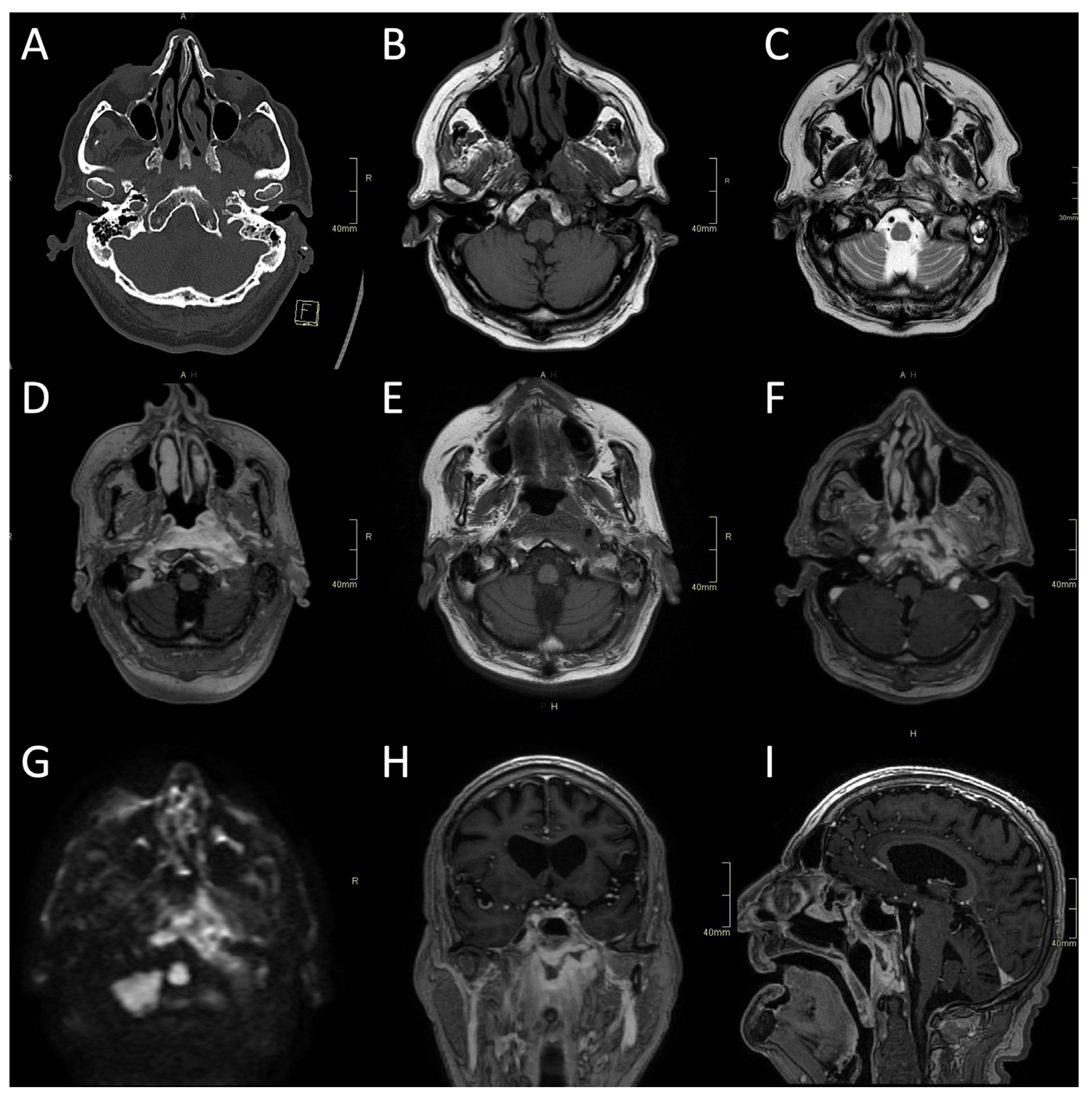
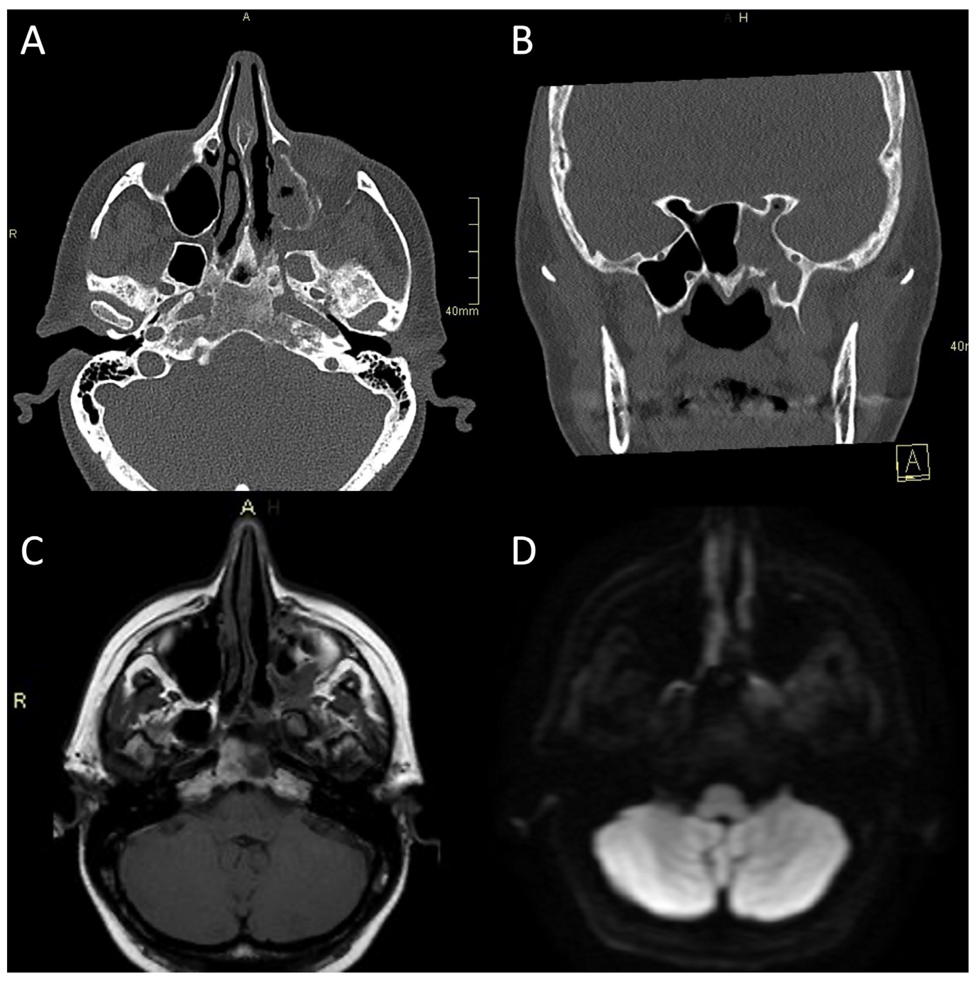
4.1. Imaging Studies
4.1.1. CT Imaging
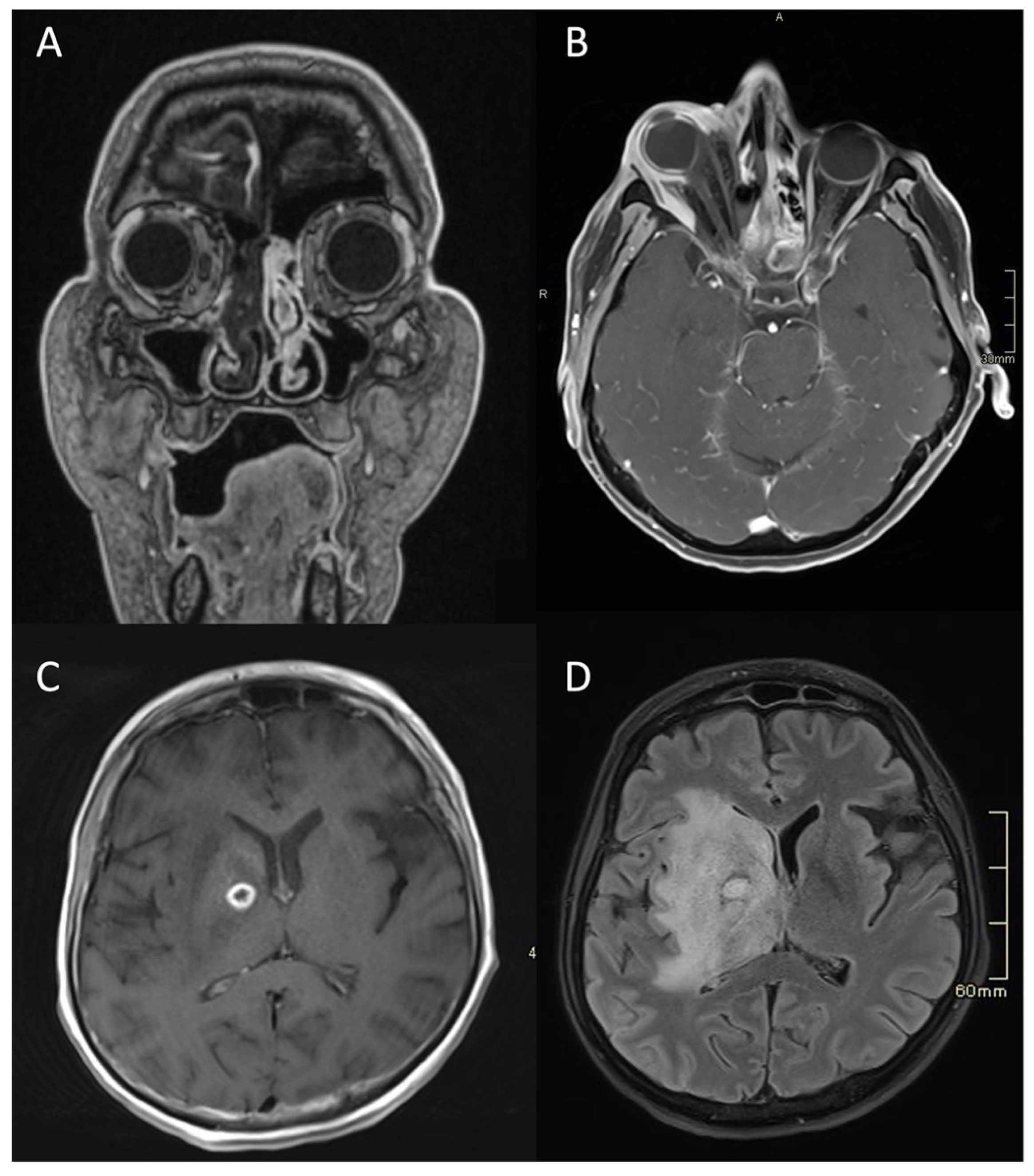
4.1.2. MR Imaging
4.1.3. Nuclear Medicine Studies
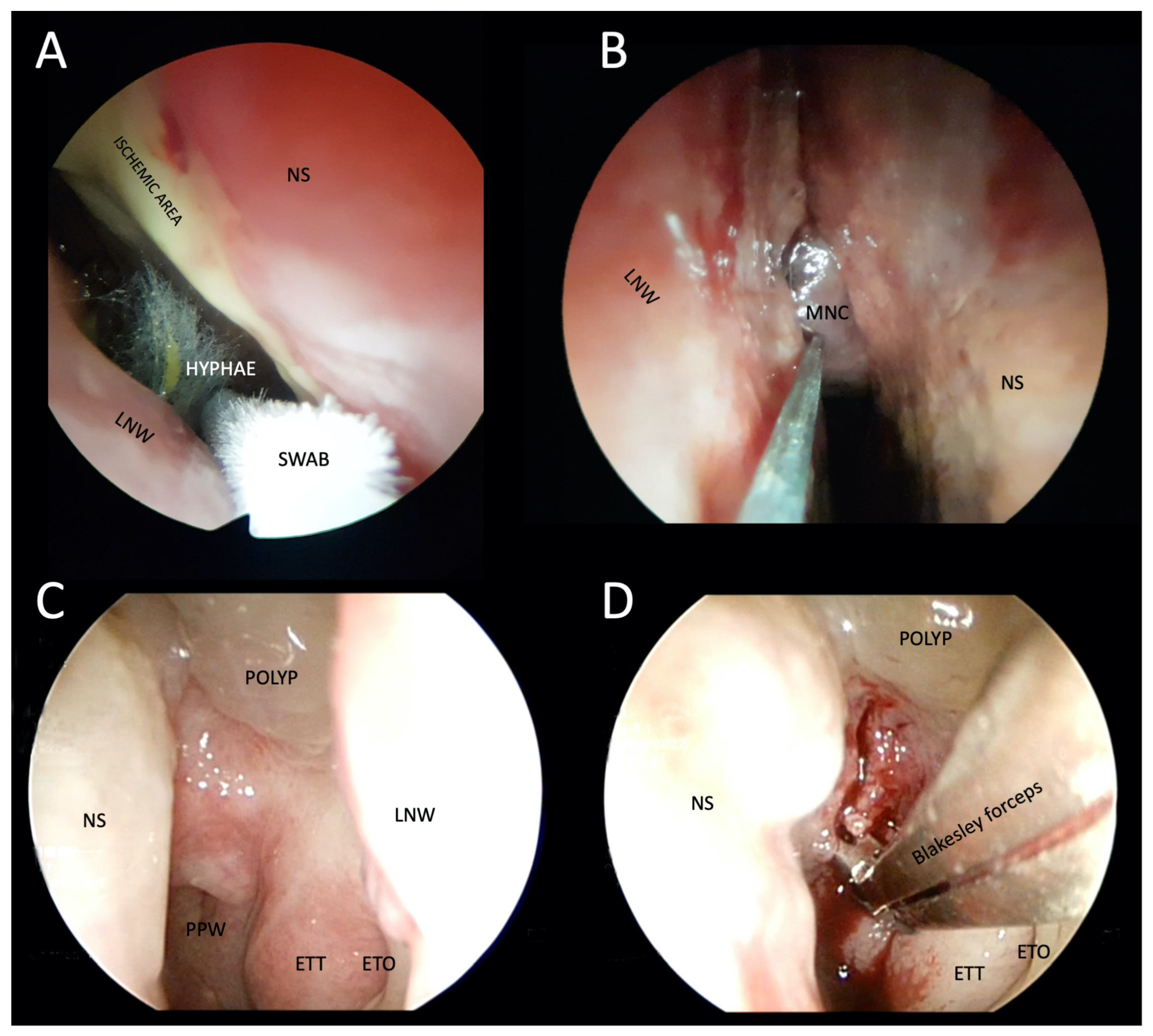
4.2. Otorhinolaryngological Examination and Tissue Specimen Sampling
4.3. Histopathology
4.4. Microbiology Studies
4.5. Blood Tests
5. Differential Diagnosis of Atypical Skull-Base Osteomyelitis
5.1. Typical (i.e., Otogenic) Skull-Base Osteomyelitis and Necrotising External Otitis
5.2. Rheumatological Diseases
5.3. Tumours
6. Treatment of Atypical Skull-Base Osteomyelitis
6.1. Surgery
6.2. Antimicrobial Treatment
6.3. Hyperbaric Oxygen Therapy
7. Prognosis of Atypical Skull-Base Osteomyelitis
8. Conclusions
Funding
Institutional Review Board Statement
Informed Consent Statement
Data Availability Statement
Conflicts of Interest
References
- Pott, P. Observations on the Nature and Consequences of Those Injuries to Which the Head Is Liable from External Violence: To Which Are Added, Some Few General Remarks on Fractures and Dislocations; Printed for L. Hawes, W. Clarke, R.; Collins: London, UK, 1768. [Google Scholar]
- Meltzer, P.E.; Kelemen, G. Pyocyaneous Osteomyelitis of the Temporal Bone, Mandible and Zygoma. Laryngoscope 1959, 69, 1300–1316. [Google Scholar] [CrossRef]
- Sie, K.C.Y.; Glenn, M.G.; Hillel, A.H. Osteomyelitis of the Skull Base, Etiology Unknown. Otolaryngol. Neck Surg. 1991, 104, 252–256. [Google Scholar] [CrossRef]
- Chapman, P.R.; Choudhary, G.; Singhal, A. Skull Base Osteomyelitis: A Comprehensive Imaging Review. Am. J. Neuroradiol. 2021, 42, 404–413. [Google Scholar] [CrossRef]
- Mortazavi, M.M.; Khan, M.A.; Quadri, S.A.; Suriya, S.S.; Fahimdanesh, K.M.; Fard, S.A.; Hassanzadeh, T.; Taqi, M.A.; Grossman, H.; Tubbs, R.S. Cranial Osteomyelitis: A Comprehensive Review of Modern Therapies. World Neurosurg. 2018, 111, 142–153. [Google Scholar] [CrossRef]
- Clark, M.P.A.; Pretorius, P.M.; Byren, I.; Milford, C.A. Central or Atypical Skull Base Osteomyelitis: Diagnosis and Treatment. Skull Base Off. J. North Am. Skull Base Soc. Al. 2009, 19, 247–254. [Google Scholar] [CrossRef]
- Oka, K.; Nakano, Y.; Sazumi, Y.; Michitani, T.; Horiguchi, S.; Ocho, K.; Iwamuro, M.; Otsuka, F. Clival Osteomyelitis with Cavernous Sinus Thrombosis Due to Fusobacterium nucleatum and Campylobacter rectus Induced by Tooth Extraction. Intern. Med. 2018, 57, 3325–3328. [Google Scholar] [CrossRef]
- Trück, J.; Thompson, A.; Dwivedi, R.; Segal, S.; Anand, G.; Kelly, D.F. Nonotogenic Skull Base Osteomyelitis in Children: Two Cases and a Review of the Literature. Pediatr. Infect. Dis. J. 2015, 34, 1025–1027. [Google Scholar] [CrossRef]
- See, A.; Tan, T.Y.; Gan, E.C. Atypical Culture-Negative Skull Base Osteomyelitis Masquerading as Advanced Nasopharyngeal Carcinoma. Am. J. Otolaryngol. 2016, 37, 236–239. [Google Scholar] [CrossRef]
- Shellman, Z.; Coates, M.; Kara, N. Polymicrobial Skull Base Osteomyelitis Related to Chronic Sphenoiditis and Endoscopic Sinus Surgery. Laryngoscope 2021, 131, E1086–E1087. [Google Scholar] [CrossRef]
- Kayode-Ajala, F.; Williams, N.; Ejikeme, C.; Walji, A.; Farrer, W. A Case of Adult Clival Osteomyelitis. J. Investig. Med. High Impact Case Rep. 2022, 10, 23247096221101856. [Google Scholar] [CrossRef]
- Moreddu, E.; Treut, C.L.; Triglia, J.-M.; Nicollas, R. Abscess with Osteomyelitis of the Clivus after Adenoidectomy: An Uncommon Complication of a Common Procedure. Eur. Ann. Otorhinolaryngol. Head Neck Dis. 2018, 135, 81–82. [Google Scholar] [CrossRef]
- Raskin, J.; Borrelli, M.; Wu, A.W. Skull Base Osteomyelitis After Nasopharyngeal Cyst Excision. Ear. Nose. Throat J. 2021, 100, 867S–869S. [Google Scholar] [CrossRef]
- Benadjaoud, Y.; Klopp-Dutote, N.; Choquet, M.; Brunel, E.; Guiheneuf, R.; Page, C. A Case of Acute Clival Osteomyelitis in a 7-Year-Old Boy Secondary to Infection of a Thornwaldt Cyst. Int. J. Pediatr. Otorhinolaryngol. 2017, 95, 87–90. [Google Scholar] [CrossRef]
- Czech, M.M.; Hwang, P.H.; Colevas, A.D.; Fischbein, N.; Ho, D.Y. Skull Base Osteomyelitis in Patients with Head and Neck Cancer: Diagnosis, Management, and Outcomes in a Case Series of 23 Patients. Laryngoscope Investig. Otolaryngology 2022, 7, 47–59. [Google Scholar] [CrossRef]
- Di Lullo, A.M.; Russo, C.; Grimaldi, G.; Capriglione, P.; Cantone, E.; del Vecchio, W.; Motta, G.; Iengo, M.; Elefante, A.; Cavaliere, M. Skull Base Fungal Osteomyelitis: A Case Report and Review of the Literature. Ear. Nose. Throat J. 2021, 100, 1089S–1094S. [Google Scholar] [CrossRef]
- Radhakrishnan, S.; Mujeeb, H.; Radhakrishnan, C. Central Skull Base Osteomyelitis Secondary to Invasive Aspergillus Sphenoid Sinusitis Presenting with Isolated 12th Nerve Palsy. IDCases 2020, 22, e00930. [Google Scholar] [CrossRef]
- Johnson, A.K.; Batra, P.S. Central Skull Base Osteomyelitis: An Emerging Clinical Entity. Laryngoscope 2014, 124, 1083–1087. [Google Scholar] [CrossRef]
- Jáñez, F.Á.; Barriga, L.Q.; Iñigo, T.R.; Lora, F.R. Diagnosis of Skull Base Osteomyelitis. Radiographics 2021, 41, 156–174. [Google Scholar] [CrossRef]
- Gómez, D.F.; Vargas-Osorio, M.P.; Hakim, F.; Tejada-Angarita, K.S. Skull Base Osteomyelitis Mimicking Skull Base Tumor in Immunocompetent Pediatric Patient: Case Report. Childs Nerv. Systems 2022, 38, 1833–1835. [Google Scholar] [CrossRef]
- Blyth, C.C.; Gomes, L.; Sorrell, T.C.; da Cruz, M.; Sud, A.; Chen, S.C.-A. Skull-Base Osteomyelitis: Fungal vs. Bacterial Infection. Clin. Microbiol. Infect. 2011, 17, 306–311. [Google Scholar] [CrossRef]
- Abou-Al-Shaar, H.; Mulvaney, G.G.; Alzhrani, G.; Gozal, Y.M.; Oakley, G.M.; Couldwell, W.T. Nocardial Clival Osteomyelitis Secondary to Sphenoid Sinusitis: An Atypical Skull Base Infection. Acta Neurochir. 2019, 161, 529–534. [Google Scholar] [CrossRef] [PubMed]
- Schreiber, A.; Ravanelli, M.; Rampinelli, V.; Ferrari, M.; Vural, A.; Mattavelli, D.; Mataj, E.; Mazza, V.; Zorza, I.; Bonù, M.L.; et al. Skull Base Osteomyelitis: Clinical and Radiologic Analysis of a Rare and Multifaceted Pathological Entity. Neurosurg. Rev. 2021, 44, 555–569. [Google Scholar] [CrossRef]
- Weitzman, R.E.; Parikh, A.S.; Gadkaree, S.K.; Corrales, C.E. Skull Base Osteomyelitis Complicated by Petrous Internal Carotid Artery Blowout. Ear. Nose. Throat J. 2021, 100, 790S–794S. [Google Scholar] [CrossRef] [PubMed]
- Chen, J.-C.; Yeh, C.-F.; Shiao, A.-S.; Tu, T.-Y. Temporal Bone Osteomyelitis: The Relationship with Malignant Otitis Externa, the Diagnostic Dilemma, and Changing Trends. Sci. World J. 2014, 2014, 591714. [Google Scholar] [CrossRef]
- Rubin Grandis, J.; Branstetter, B.F.; Yu, V.L. The Changing Face of Malignant (Necrotising) External Otitis: Clinical, Radiological, and Anatomic Correlations. Lancet Infect. Dis. 2004, 4, 34–39. [Google Scholar] [CrossRef] [PubMed]
- Khan, M.; Quadri, S.; Kazmi, A.; Kwatra, V.; Ramachandran, A.; Gustin, A.; Farooqui, M.; Suriya, S.; Zafar, A. A Comprehensive Review of Skull Base Osteomyelitis: Diagnostic and Therapeutic Challenges among Various Presentations. Asian J. Neurosurg. 2018, 13, 959–970. [Google Scholar] [CrossRef]
- Carr, M.M.; Cusimano, M.D.; Muller, P.J.; Bilbao, J. Aspergillosis of the Cranial Base. Skull Base Surg. 1994, 4, 159–163. [Google Scholar] [CrossRef]
- Carfrae, M.J.; Kesser, B.W. Malignant Otitis Externa. Otolaryngol. Clin. N. Am. 2008, 41, 537–549. [Google Scholar] [CrossRef]
- Adams, A.; Offiah, C. Central Skull Base Osteomyelitis as a Complication of Necrotizing Otitis Externa: Imaging Findings, Complications, and Challenges of Diagnosis. Clin. Radiol. 2012, 67, e7–e16. [Google Scholar] [CrossRef]
- Moss, W.J.; Finegersh, A.; Narayanan, A.; Chan, J.Y.K. Meta-Analysis Does Not Support Routine Traditional Nuclear Medicine Studies for Malignant Otitis. Laryngoscope 2020, 130, 1812–1816. [Google Scholar] [CrossRef]
- Rozenblum-Beddok, L.; Verillaud, B.; Paycha, F.; Vironneau, P.; Abulizi, M.; Benada, A.; Cross, T.; El-Deeb, G.; Vodovar, N.; Peretti, I.; et al. 99mTc-HMPAO-Leukocyte Scintigraphy for Diagnosis and Therapy Monitoring of Skull Base Osteomyelitis. Laryngoscope Investig. Otolaryngol. 2018, 3, 218–224. [Google Scholar] [CrossRef]
- Chakraborty, D.; Bhattacharya, A.; Gupta, A.; Panda, N.; Das, A.; Mittal, B. Skull Base Osteomyelitis in Otitis Externa: The Utility of Triphasic and Single Photon Emission Computed Tomography/Computed Tomography Bone Scintigraphy. Indian J. Nucl. Med. 2013, 28, 65–69. [Google Scholar] [CrossRef]
- Balakrishnan, R.; Dalakoti, P.; Nayak, D.R.; Pujary, K.; Singh, R.; Kumar, R. Efficacy of HRCT Imaging vs SPECT/CT Scans in the Staging of Malignant External Otitis. Otolaryngol. Head Neck Surg. Off. J. Am. Acad. Otolaryngol. Head Neck Surg. 2019, 161, 336–342. [Google Scholar] [CrossRef]
- Strashun, A.M.; Nejatheim, M.; Goldsmith, S.J. Malignant External Otitis: Early Scintigraphic Detection. Radiology 1984, 150, 541–545. [Google Scholar] [CrossRef] [PubMed]
- Stokkel, M.P.; Boot, C.N.; van Eck-Smit, B.L. SPECT Gallium Scintigraphy in Malignant External Otitis: Initial Staging and Follow-up. Case Reports. Laryngoscope 1996, 106, 338–340. [Google Scholar] [CrossRef] [PubMed]
- Seabold, J.E.; Simonson, T.M.; Weber, P.C.; Thompson, B.H.; Harris, K.G.; Rezai, K.; Madsen, M.T.; Hoffman, H.T. Cranial Osteomyelitis: Diagnosis and Follow-up with In-111 White Blood Cell and Tc-99m Methylene Diphosphonate Bone SPECT, CT, and MR Imaging. Radiology 1995, 196, 779–788. [Google Scholar] [CrossRef] [PubMed]
- Chang, P.C.; Fischbein, N.J.; Holliday, R.A. Central Skull Base Osteomyelitis in Patients without Otitis Externa: Imaging Findings. AJNR Am. J. Neuroradiol. 2003, 24, 1310–1316. [Google Scholar] [PubMed]
- Chandler, J.R.; Grobman, L.; Quencer, R.; Serafini, A. Osteomyelitis of the Base of the Skull. Laryngoscope 1986, 96, 245–251. [Google Scholar] [CrossRef] [PubMed]
- Ducic, Y. Skull Base Osteomyelitis. South. Med. J. 2006, 99, 1051. [Google Scholar] [CrossRef]
- Singh, A.; Khabori, M. Al Skull Base Osteomyelitis: Diagnostic and Therapeutic Challenges in Atypical Presentation. Otolaryngol. Neck Surg. 2005, 133, 121–125. [Google Scholar] [CrossRef]
- Ridder, G.J.; Breunig, C.; Kaminsky, J.; Pfeiffer, J. Central Skull Base Osteomyelitis: New Insights and Implications for Diagnosis and Treatment. Eur. Arch. Otorhinolaryngol. 2015, 272, 1269–1276. [Google Scholar] [CrossRef]
- Borges, A. Imaging of the Central Skull Base. Neuroimaging Clin. N. Am. 2009, 19, 669–696. [Google Scholar] [CrossRef]
- Bag, A.K.; Chapman, P.R. Neuroimaging: Intrinsic Lesions of the Central Skull Base Region. Semin. Ultrasound CT MRI 2013, 34, 412–435. [Google Scholar] [CrossRef]
- Chan, L.L.; Singh, S.; Jones, D.; Diaz, E.M.J.; Ginsberg, L.E. Imaging of Mucormycosis Skull Base Osteomyelitis. AJNR Am. J. Neuroradiol. 2000, 21, 828–831. [Google Scholar] [PubMed]
- Haider, A.S.; Esianor, B.I.; Shail, M.S.; Engelhardt, M.I.; Kafai Golahmadi, A.; Khan, R.; Khan, U.; Vayalumkal, S.; Thakur, R.; Layton, K.F. Mycotic Pseudoaneurysm Associated with Skull Base Osteomyelitis Treated with Endovascular Embolization. Cureus 2017, 9, e1622. [Google Scholar] [CrossRef] [PubMed]
- Honda, H.; Warren, D.K. Central Nervous System Infections: Meningitis and Brain Abscess. Infect. Dis. Clin. N. Am. 2009, 23, 609–623. [Google Scholar] [CrossRef]
- Ginat, D.T.; Mangla, R.; Yeaney, G.; Ekholm, S. Diffusion-Weighted Imaging of Skull Lesions. J. Neurol. Surg. Part B Skull Base 2014, 75, 204–213. [Google Scholar] [CrossRef]
- Ozgen, B.; Oguz, K.K.; Cila, A. Diffusion MR Imaging Features of Skull Base Osteomyelitis Compared with Skull Base Malignancy. AJNR Am. J. Neuroradiol. 2011, 32, 179–184. [Google Scholar] [CrossRef]
- Goh, J.P.N.; Karandikar, A.; Loke, S.C.; Tan, T.Y. Skull Base Osteomyelitis Secondary to Malignant Otitis Externa Mimicking Advanced Nasopharyngeal Cancer: MR Imaging Features at Initial Presentation. Am. J. Otolaryngol. 2017, 38, 466–471. [Google Scholar] [CrossRef]
- Lesser, F.D.; Derbyshire, S.G.; Lewis-Jones, H. Can Computed Tomography and Magnetic Resonance Imaging Differentiate between Malignant Pathology and Osteomyelitis in the Central Skull Base? J. Laryngol. Otol. 2015, 129, 852–859. [Google Scholar] [CrossRef]
- Jain, N.; Jasper, A.; Vanjare, H.A.; Mannam, P.; Mani, S.E. The Role of Imaging in Skull Base Osteomyelitis—Reviewed. Clin. Imaging 2020, 67, 62–67. [Google Scholar] [CrossRef]
- Van Kroonenburgh, A.M.J.L.; van der Meer, W.L.; Bothof, R.J.P.; van Tilburg, M.; van Tongeren, J.; Postma, A.A. Advanced Imaging Techniques in Skull Base Osteomyelitis Due to Malignant Otitis Externa. Curr. Radiol. Rep. 2018, 6, 3. [Google Scholar] [CrossRef]
- Kulkarni, S.C.; Padma, S.; Shanmuga Sundaram, P. In the Evaluation of Patients with Skull Base Osteomyelitis, Does 18F-FDG PET CT Have a Role? Nucl. Med. Commun. 2020, 41, 1187. [Google Scholar] [CrossRef]
- Redleaf, M.I.; Angeli, S.I.; McCabe, B.F. Indium 111-Labeled White Blood Cell Scintigraphy as an Unreliable Indicator of Malignant External Otitis Resolution. Ann. Otol. Rhinol. Laryngol. 1994, 103, 444–448. [Google Scholar] [CrossRef]
- Galldiks, N.; Lohmann, P.; Fink, G.R.; Langen, K.-J. Amino Acid PET in Neurooncology. J. Nucl. Med. 2023, 264859. [Google Scholar] [CrossRef] [PubMed]
- Guarner, J.; Brandt, M.E. Histopathologic Diagnosis of Fungal Infections in the 21st Century. Clin. Microbiol. Rev. 2011, 24, 247–280. [Google Scholar] [CrossRef]
- Montone, K.T.; Guarner, J. In Situ Hybridization for RRNA Sequences in Anatomic Pathology Specimens, Applications for Fungal Pathogen Detection: A Review. Adv. Anat. Pathol. 2013, 20, 168–174. [Google Scholar] [CrossRef]
- Roozeboom, M.H.; Westers-Attema, A.; Winnepenninckx, V.; Hurkens, K.P.G.M.; Kelleners-Smeets, N.W.J. The Different Faces of Actinomycosis. Clin. Exp. Dermatol. Print 2012, 37, 322–324. [Google Scholar] [CrossRef]
- Wong, V.K.; Turmezei, T.D.; Weston, V.C. Actinomycosis. BMJ 2011, 343, d6099. [Google Scholar] [CrossRef] [PubMed]
- Ponnaiyan, D.; Anitha, C.M.; Prakash, P.S.G.; Subramanian, S.; Rughwani, R.R.; Kumar, G.; Nandipati, S. Reddy Mucormycosis Diagnosis Revisited: Current and Emerging Diagnostic Methodologies for the Invasive Fungal Infection (Review). Exp. Ther. Med. 2023, 25, 47. [Google Scholar] [CrossRef] [PubMed]
- Lockhart, S.R.; Bialek, R.; Kibbler, C.C.; Cuenca-Estrella, M.; Jensen, H.E.; Kontoyiannis, D.P. Molecular Techniques for Genus and Species Determination of Fungi from Fresh and Paraffin-Embedded Formalin-Fixed Tissue in the Revised EORTC/MSGERC Definitions of Invasive Fungal Infection. Clin. Infect. Dis. 2021, 72, S109–S113. [Google Scholar] [CrossRef] [PubMed]
- Jillwin, J.; Rudramurthy, S.M.; Singh, S.; Bal, A.; Das, A.; Radotra, B.; Prakash, H.; Dhaliwal, M.; Kaur, H.; Ghosh, A.K.; et al. Molecular Identification of Pathogenic Fungi in Formalin-Fixed and Paraffin-Embedded Tissues. J. Med. Microbiol. 2021, 70, 1282. [Google Scholar] [CrossRef] [PubMed]
- Cornely, O.A.; Arikan-Akdagli, S.; Dannaoui, E.; Groll, A.H.; Lagrou, K.; Chakrabarti, A.; Lanternier, F.; Pagano, L.; Skiada, A.; Akova, M.; et al. ESCMID† and ECMM‡ Joint Clinical Guidelines for the Diagnosis and Management of Mucormycosis 2013. Clin. Microbiol. Infect. 2014, 20, 5–26. [Google Scholar] [CrossRef]
- Ullmann, A.J.; Aguado, J.M.; Arikan-Akdagli, S.; Denning, D.W.; Groll, A.H.; Lagrou, K.; Lass-Flörl, C.; Lewis, R.E.; Munoz, P.; Verweij, P.E.; et al. Diagnosis and Management of Aspergillus Diseases: Executive Summary of the 2017 ESCMID-ECMM-ERS Guideline. Clin. Microbiol. Infect. 2018, 24, e1–e38. [Google Scholar] [CrossRef]
- Griffith, D.E.; Aksamit, T.; Brown-Elliott, B.A.; Catanzaro, A.; Daley, C.; Gordin, F.; Holland, S.M.; Horsburgh, R.; Huitt, G.; Iademarco, M.F.; et al. An Official ATS/IDSA Statement: Diagnosis, Treatment, and Prevention of Nontuberculous Mycobacterial Diseases. Am. J. Respir. Crit. Care Med. 2007, 175, 367–416. [Google Scholar] [CrossRef]
- Kim, H.Y.; Kook, Y.; Yun, Y.J.; Park, C.G.; Lee, N.Y.; Shim, T.S.; Kim, B.J.; Kook, Y.H. Proportions of Mycobacterium massiliense and Mycobacterium molletii Strains among Korean Mycobacterium chelonae-Mycobacterium abscessus Group Isolates. J. Clin. Microbiol. 2008, 46, 3384–3390. [Google Scholar] [CrossRef]
- Nie, W.; Duan, H.; Huang, H.; Lu, Y.; Bi, D.; Chu, N. Species Identification of Mycobacterium abscessus Subsp. Abscessus and Mycobacterium abscessus Subsp. Bolletii Using RpoB and Hsp65, and Susceptibility Testing to Eight Antibiotics. Int. J. Infect. Dis. 2014, 25, 170–174. [Google Scholar] [CrossRef] [PubMed]
- Donnelly, J.P.; Chen, S.C.; Kauffman, C.A.; Steinbach, W.J.; Baddley, J.W.; Verweij, P.E.; Clancy, C.J.; Wingard, J.R.; Lockhart, S.R.; Groll, A.H.; et al. Revision and Update of the Consensus Definitions of Invasive Fungal Disease from the European Organization for Research and Treatment of Cancer and the Mycoses Study Group Education and Research Consortium. Clin. Infect. Dis. 2019, 71, 1367–1376. [Google Scholar] [CrossRef]
- Chandler, J.R. Malignant External Otitis. Laryngoscope 1968, 78, 1257–1294. [Google Scholar] [CrossRef]
- Slattery, W.H.; Brackmann, D.E. Skull Base Osteomyelitis. Malignant External Otitis. Otolaryngol. Clin. N. Am. 1996, 29, 795–806. [Google Scholar] [CrossRef]
- Gardiner, L.J. Complicated Frontal Sinusitis: Evaluation and Management. Otolaryngol. Neck Surg. Off. J. Am. Acad. Otolaryngol. Head Neck Surg. 1986, 95, 333–343. [Google Scholar] [CrossRef]
- von Itzstein, M.S.; Abeykoon, J.P.; Summerfield, D.D.; Whitaker, J.A. Severe Destructive Nasopharyngeal Granulomatosis with Polyangiitis with Superimposed Skull Base Pseudomonas Aeruginosa Osteomyelitis. Case Rep. 2017, bcr-2017-220135. [Google Scholar] [CrossRef]
- Cler, S.J.; Sharifai, N.; Baker, B.; Dowling, J.L.; Pipkorn, P.; Yaeger, L.; Clifford, D.B.; Dahiya, S.; Chicoine, M.R. IgG4-Related Disease of the Skull and Skull Base—A Systematic Review and Report of Two Cases. World Neurosurg. 2021, 150, 179–196.e1. [Google Scholar] [CrossRef] [PubMed]
- Burns, T.C.; Mindea, S.A.; Pendharkar, A.V.; Lapustea, N.B.; Irime, I.; Nayak, J.V. Endoscopic Transnasal Approach for Urgent Decompression of the Craniocervical Junction in Acute Skull Base Osteomyelitis. J. Neurol. Surg. Rep. 2015, 76, e37–e42. [Google Scholar] [CrossRef]
- Mejzlik, J.; Cerny, M.; Zeinerova, L.; Dedkova, J.; Kopriva, J.; Zadrobilek, K.; Adamkov, J.; Chrobok, V.; Pellantova, V. The Routes of Infection Spread in Central Skull-Base Osteomyelitis and the Diagnostic Role of CT and MRI Scans. BMC Med. Imaging 2019, 19, 60. [Google Scholar] [CrossRef] [PubMed]


| Atypical Skull-Base Osteomyelitis | Typical Skull-Base Osteomyelitis | |
|---|---|---|
| Usual cause | Advanced paranasal sinus infection | Necrotising external otitis |
| Usual causative organism | Staphylococcus aureus | Pseudomonas aeruginosa |
| Other causative organisms | More common fungi and other bacteria | Pseudomonas aeruginosa in 90–98% |
| Usual microbiological tests results | More often negative | More often positive |
| Occurrence | Very rare | Rare |
| The usual initial clinical picture | Nonspecific symptoms: headache and facial pain, the clinical picture of rhinosinusitis, facial cellulitis, furuncle, or pharyngitis | Headache, otalgia, aural discharge, conductive hearing loss, external auditory canal swelling, and granulation tissue |
| Usually affected cranial nerves after | VIth, IXth, Xth | VIIth |
Disclaimer/Publisher’s Note: The statements, opinions and data contained in all publications are solely those of the individual author(s) and contributor(s) and not of MDPI and/or the editor(s). MDPI and/or the editor(s) disclaim responsibility for any injury to people or property resulting from any ideas, methods, instructions or products referred to in the content. |
© 2023 by the authors. Licensee MDPI, Basel, Switzerland. This article is an open access article distributed under the terms and conditions of the Creative Commons Attribution (CC BY) license (https://creativecommons.org/licenses/by/4.0/).
Share and Cite
Urbančič, J.; Vozel, D.; Battelino, S.; Bošnjak, R.; Kokošar Ulčar, B.; Matos, T.; Munda, M.; Papst, L.; Steiner, N.; Vouk, M.; et al. Atypical Skull-Base Osteomyelitis: Comprehensive Review and Multidisciplinary Management Viewpoints. Trop. Med. Infect. Dis. 2023, 8, 254. https://doi.org/10.3390/tropicalmed8050254
Urbančič J, Vozel D, Battelino S, Bošnjak R, Kokošar Ulčar B, Matos T, Munda M, Papst L, Steiner N, Vouk M, et al. Atypical Skull-Base Osteomyelitis: Comprehensive Review and Multidisciplinary Management Viewpoints. Tropical Medicine and Infectious Disease. 2023; 8(5):254. https://doi.org/10.3390/tropicalmed8050254
Chicago/Turabian StyleUrbančič, Jure, Domen Vozel, Saba Battelino, Roman Bošnjak, Barbara Kokošar Ulčar, Tadeja Matos, Matic Munda, Lea Papst, Nejc Steiner, Matej Vouk, and et al. 2023. "Atypical Skull-Base Osteomyelitis: Comprehensive Review and Multidisciplinary Management Viewpoints" Tropical Medicine and Infectious Disease 8, no. 5: 254. https://doi.org/10.3390/tropicalmed8050254