
Hepatocellular Carcinoma: The Role of MicroRNAs


1
Division of Gastroenterology and Hepatology, Department of Medicine, University of Missouri, Columbia, MO 65212, USA
2
Harry S. Truman Veterans Hospital, Columbia, MO 65201, USA
3
Department of Medical Pharmacology and Physiology, University of Missouri, Columbia, MO 65212, USA
*
Author to whom correspondence should be addressed.
†
These authors contributed equally to this work.
Biomolecules 2022, 12(5), 645; https://doi.org/10.3390/biom12050645
Submission received: 6 February 2022
/
Revised: 21 April 2022
/
Accepted: 25 April 2022
/
Published: 27 April 2022
(This article belongs to the Special Issue Biomolecules and Cancer Prevention 2.0)
Abstract
:Hepatocellular carcinoma (HCC) is the second leading cause of cancer-related deaths worldwide. HCC is diagnosed in its advanced stage when limited treatment options are available. Substantial morphologic, genetic and epigenetic heterogeneity has been reported in HCC, which poses a challenge for the development of a targeted therapy. In this review, we discuss the role and involvement of several microRNAs (miRs) in the heterogeneity and metastasis of hepatocellular carcinoma with a special emphasis on their possible role as a diagnostic and prognostic tool in the risk prediction, early detection, and treatment of hepatocellular carcinoma.
1. Introduction
Hepatocellular carcinoma (HCC) is the most common and deadly liver cancer and the second leading cause of cancer-related deaths worldwide [1,2]. HCC, aggressive in nature, accounts for 90% of primary liver cancers [3]. HCC normally develops in the setting of cirrhosis and the process of tumorigenesis is further promoted by chronic viral hepatitis related to the hepatitis B (HBV) and hepatitis C viruses (HCV), alcohol-induced injury, non-alcoholic fatty liver disease, exposure to aflatoxins or genetic disposition [4]. Diet-induced HCC is an emerging problem in developed as well as in developing countries [5,6]. Over the past 15 years, reported cases of HCC have more than doubled due to late diagnosis [7] with limited treatment options and marginal clinical benefits to patients.
Among the limited potential curative strategies are liver resection and liver transplantation as well as loco-regional therapies, such as radiofrequency ablation and transarterial chemoembolization [8]. Because of shortage of healthy livers, the transplants are provided crucially to patients that have the best chance of long-term survival. Cytotoxic systemic therapy is limited by tumor chemoresistance and patient intolerance [9].
Multiple molecular regulations set a precedence for the high rate of recurrence and may lead to tumor heterogeneity in HCC. In this regard, substantial heterogeneity has been reported between the multiple tumor foci in a single patient. Therefore, more reliable biomarkers are needed for the diagnosis, treatment and surveillance of HCC [8,10].
2. Heterogeneity in HCC
Cancer heterogeneity has been recognized as an important clinical determinant of patient outcomes, such as response or resistance to anti-cancer therapies [11,12]. Heterogeneity is prevalent in cancer, both between and within individuals. Multiple morphologic (differentiation status and cytologic features), genetic (mutational background), epigenetic (DNA methylation) and microenvironment (hypoxia gradients and local oxidative stress) variations create heterogeneity amongst different tumors [10,13]. HCC is a highly heterogenous cancer with significant intratumor heterogeneity. The pathologic classification of HCC is based on the degree of cellular differentiation. In a single tumor, the cancerous tissue of two different histological grades may be present. In addition, tissue obtained from HCC may exhibit different immunohistochemical characteristics in the same tumor. Furthermore, genetic heterogeneity has been described in HCC. Thus, HCC is less likely to be caused by a single driver mutation. The intratumor heterogeneity of HCC plays an important role in the prognosis of the disease. Hence, the detection of HCC intratumor heterogeneity is important for the development of effective targeted therapies. Though liver transplantation, surgical resection and radiofrequency ablation (RFA) offer a curative treatment for HCC, they are not an option for patients with intermediate/advanced stage HCC. Sorafenib, a multikinase inhibitor of several tyrosine protein kinases, is implicated in the treatment of patients with intermediate/advanced HCC. It has shown a modest increase in median survival in clinical trials [14]. However, the adequate concentration of this drug is not achieved because of vascular heterogeneity within the tumor, resulting in a reduced response to therapy. Thus, intratumor heterogeneity also plays a role in drug resistance. Therefore, the better understanding of the intratumor heterogeneity of HCC should provide critical knowledge about the prognosis of the disease and response to potential future therapy.
2.1. Morphologic Heterogeneity in HCC
HCC can be classified pathologically on the basis of the degree of cellular differentiation, which include well differentiated to moderately and poorly differentiated and undifferentiated tumors. The two histological grades can be present in one tumor, thereby exhibiting different immunohistochemical characteristics in the same tumor. Apart from differentiation, intratumor heterogeneity also decides the size of the tumor and lymphovascular spread. Some of the histochemical markers used for HCC diagnosis include pCEA, CD10, alpha fetoprotein (AFP), hepatocyte paraffin1 (hepPar1), cytoplasmic thyroid transcription factor-1 (TTF1), glutamine synthetase, GPC3, CK8 and CK18, but unfortunately none of them is specific of early stage HCC. AFP is most insensitive of all because it is not expressed by all HCC cells [15]. Zhang Q et al. recently proposed an immunophenotypic classification of HCC using two markers (CD45 and Foxp3) that will facilitate prognostic prediction and decision making for the choice of therapies [16].
HCC is well known for morphologic intratumor heterogeneity, but very few systematic analyses of this phenomenon have been performed. Intratumor heterogeneity is detectable in the majority of HCC cases (87%), with 26% of cases at the level of morphology. Further, 39% of cases are classified at the combined morphologic and immunohistochemical level and 22% at the combined morphologic and immunohistochemical treatment target level with known mutational status, for example, TP53 and β-catenin [4].
Intratumor heterogeneity poses a challenge for the development of a robust HCC classification as well as a targeted therapy and may contribute to treatment failure and drug resistance in many cases of HCC. HCC is known to frequently display heterogeneous growth patterns and/or cytologic features within the same tumor. This kind of plasticity of phenotypes or intratumor heterogeneity has already been described in several solid tumors, from skin, breast and kidney diseases [17,18,19,20,21]. In small HCC, measuring 3 to 5 cm in diameter, up to 64% of cases display intratumor heterogeneity at the level of histologic differentiation grade and proliferative activity, whereas in HCC smaller than 2 cm, intratumor heterogeneity is 25–47% [22,23]. In larger HCC, on the other hand, the true extent of intratumor heterogeneity with respect to morphologic, immunohistochemical and molecular features has not been systematically assessed [4].
2.2. Genetic Heterogeneity in HCC
Nault and Villanueva identified TERT promoter mutations with an overall frequency of 60%, as the most frequent somatic genetic defect in HCC [24]. It is also recurrently mutated in precancerous nodules as the earliest genetic alteration involved in the malignant transformation of HCC and possibly considered as a tumor “gatekeeper”. They speculated that TERT promoter mutations are present in the common ancestor cell and transmitted to its progeny and, therefore, are present in most tumor cells. Further studies are still needed to decipher HCC intratumor genetic heterogeneity using unbiased approaches, such as whole-exome or whole-genome sequencing, preferable by ultra-deep sequencing [13,25].
Large and independent studies have validated several molecular prognostic signatures derived from the tumor, despite tumor heterogeneity [26,27]. All of the biomarker-based molecular therapies approved in solid malignancies also did not account for genetic heterogeneity and are mainly based on traditional genetic analysis, for example, vemurafenib for BRAF V600E-mutated melanoma, cetuximab for wild-type RAS colorectal cancer and orcrizotinib for ALK-translocated non-small cell lung cancer [28].
Within-patient heterogeneity in HCC is well studied because it often presents with multiple tumor foci. In patients with multifocal HCC, the individual lesions usually arise from either the local dissemination of the primary tumor or from the oncogenic predisposition of the diseased liver. In the latter case, a patient with multifocal HCC may have multiple tumors presumably because of distinct genomic profiles and clonal unrelation, a situation that poses a significant challenge to genomic analyses. A microRNA biomarker of HCC recurrence, following liver transplantation, accounting for within-patient heterogeneity has been described [8].
3. MicroRNAs and Heterogeneity in HCC
Altered genomic and transcriptional landscapes are associated with carcinogenesis and include protein-coding genes as well as several classes of structurally and functionally different noncoding RNAs. At least 90% of the human genome is transcribed into noncoding RNAs that do not translate into proteins. Amongst the noncoding RNAs, miRs are a highly conserved class of small noncoding RNAs of ~18–24 nucleotides in length, which are involved in the regulation of gene expression at the transcription level by binding to their target gene promoters [29,30] and post-transcriptional level by degrading their target mRNAs and/or inhibiting their translation by binding to the 3′-untranslated region (3′UTR) of target mRNAs [31,32]. A single miR may regulate hundreds of target mRNAs with the same short recognition region; simultaneously, the 3′-UTR of most mRNAs has more than one binding site for different miRs. Since the discovery of the first miR lin-4 in 1993, more than 2000 miRs, to date, have been discovered in humans [33] that regulate one third of the genes in genome [34,35].
miRs are differentially expressed in all cancers as oncogenic miRs are generally enriched, while tumor-suppressor miRs are downregulated [36]. MiRs in HCC progression and metastasis regulate proliferation, apoptosis, invasion, the epithelial–mesesnchymal transition (EMT), angiogenesis, drug resistance and autophagy [37,38,39,40]. Amongst the tumor-suppressor miRs, some are involved in tumor initiation and progression [41,42,43], while others are involved in proliferation, invasion, metastasis and recurrence [44,45,46,47,48,49,50,51]. Similarly, some oncogenic miRs are associated with tumor development and progression [52,53,54,55,56,57,58,59,60], while others are associated with recurrence and metastasis [61,62,63,64,65,66] (Table 1).
MiR-122, a tumor-suppressing liver-specific miR, accounts for 70% of the total miR in hepatic tissues. It modulates hepatic lipid metabolism and inhibits viral replication in HBV-related HCC by targeting NDRG3, a member of N-myc downstream-regulated gene family. Both miR-122 and NDRG3 are considered possible therapeutic targets for HBV-related HCC [92]. MiR-122 is downregulated in HCC with a cirrhotic background as well as with other tumor-suppressor miR let-7 family members, miR-221, and miR-145 [93]. Studies have shown that miR associated with cell-cycle inhibition (miR-34a, miR-101, miR-199-a-5p and miR-223) [68,77,94,95] were downregulated, whereas the ones associated with cell proliferation and the inhibition of apoptosis (miR-17-92 polycistron, miR-21, miR-96, miR-221 and miR-2240) are upregulated in HCC [96,97,98,99]. Studies have also reported the association of miR-221 with the multifocality of tumors [100]. Jovel et al. described heterogeneity in terms of the upregulation of let-7i-5p, miR941, miR-301b, miR-210-3p and miR-21-3p, and the downregulation of miR-483-3p, miR-429, miR200a-3p, miR200b-3p, miR-125a-5p, miR-203a and miR-335-3p in liver-transplant recipients and the upregulation of miR1269a, miR-10b-5p, miR-217 and miR-452-5p and the downregulation of miR-483-3p, mir-483-5p, miR-4485-3p and miR-214-3p in liver resection patients [10].
The differential expression level of miRs in HCC regulates apoptosis and the mTOR, Wnt, JAK/STAT and MAPK pathways [101]. Many studies have shown that the downregulation of tumor-suppressor or the upregulation of oncogenic miRs results in the activation of the mTOR pathway in HCC. The tumor-suppressor miRs involved in the suppression of the mTOR pathway are miR758-3p, miR-142, miR-199b-5p, miR-187, miR-497, miR-99a, miR-592, miR-296-5p, miR-139-5p, miR-15b-5p, miR-345 and miR-223 [102,103,104,105,106,107,108,109,110,111,112,113]. Conversely, the oncogenic miRs, involved in the activation of the mTOR pathway, include miR-33a, miR-302d, miR-23b, miR-181a, miR-155-5p and miR-25 [114,115,116,117,118,119].
On the basis of a miRNome study in HCC tissues, several important deregulated miRs have been suggested, such as miR-361 [78], miR-122 [74] and miR-199 [75]. However, as more than one miR is deregulated in cancer cells and a single miR can target multiple mRNAs, it is an ongoing process to identify the deregulated miRs and uncover their roles in HCC development. MiR-500a, which is upregulated in HCC tissues, targets the BH3-interacting death agonist (BID) protein and can also serve as a possible prognostic predictor and therapeutic target in HCC patients [91]. Wu H. et al. demonstrated that miR-206 is a robust tumor suppressor and strongly prevented the development of HCC in AKT/Ras and cMyc HCC mouse models [76]. Studies by Sun J.J. et al. demonstrated that miR-361-5p inhibits cancer cell growth by targeting CXCR6 in HCC. The knockdown of CXCR6 and the forced expression of miR-361-5p inhibited tumor growth both in vitro and in vivo. It, therefore, serves as a tumor suppressor and might serve as a novel therapeutic target for the treatment of HCC patients [78].
4. MicroRNAs in Metastasis of HCC
Increasing data suggest a role of miRs in liver development, regeneration and metabolism, in various liver diseases, including liver hepatitis, steatosis, cirrhosis and HCC [120]. The role of several miRs in human cancer onset and progression, including invasion and metastasis, has been demonstrated [71]. For the initiation and maintenance of a mobile phenotype, the responsible cellular mechanisms include the cessation of cell polarity, cytoskeletal reorganization, re-connection with the microenvironment and the activation of pro-migratory intracellular molecules. However, the upstream regulation, leading to a highly invasive cellular phenotype, is not completely understood. Recently, Chuang at al illustrated that the dysregulation of a single miR, miR-494, supports HCC invasiveness through the epigenetic regulation of a miR network [121]. Interestingly, the role of miR-494 is both tumor suppressing and oncogenic across different tumor types and HCC, which reflects its pleiotropic character targeting distinct mRNA sets according to the genomic background and microenvironment. Its target in HCC involves the tumor-suppressor gene PTEN and MCC [1].
In recent decades, several miRs have been associated with HCC progression and metastasis, for example, miR-148a [122], miR-124 and miR-203 [69], miR-138 [123], miR-122 [124] and miR-30a-5p [125]. However, there are many more miRs that play a role in the progression and metastasis of HCC. For example, miR-141-3p inhibits the progression and metastasis of HCC by inhibiting EMT through the targeting of the Golgi protein 73 (GP73). It induces the expression of E-cadherin (epithelial cell marker), occludin (a marker of tight junctions) and cytokeratin 18 (CK 18) (a noninvasive cell marker), but reduces the expression of two mesenchymal markers N-cadherin and vimentin [72]. GP73 restores the inhibitory effects of miR-141-3p on the invasion and metastasis of HCC cells. MiR-487a, on the other hand, promotes the proliferation and metastasis of HCC by binding to phosphoinositide-3-Kinase regulatory subunit 1 (PIK3R1) and Sprouty-related EVH1 domain containing 2 (SPRED2) [90]. MiR-874 negatively regulates δ opioid receptor (DOR), which can suppress the proliferation and metastasis in HCC tumor by targeting the DOR/EGFR/ERK pathway [80], whereas miR-501-3p controls the metastatic process of HCCs by targeting Lin-7 homolog A (LIN7A) [79]. Additionally, miR-219-5p promotes HCC cell proliferation, invasion and metastasis in nude mice models bearing human HCC tumors by targeting the cadherin 1 (CDH1) gene [87]. MiR-197, which is dysregulated in several cancers, including lung, breast, ovarian, colorectal, thyroid, prostate, head and neck carcinoma, HCC as well as in non-alcoholic fatty liver disease, plays an important role in EMT. It promotes the invasion and metastasis of HCC cells by activating Wnt/β-catenin signaling by targeting Axin-2, Naked cuticle 1 (NKD1) and Dickkopf-related protein 2 (DKK2) [86]. However, miR-197-3p, which is downregulated in HCC tissues, inhibits the metastasis of HCC cells both in vitro and in vivo. Its novel target in HCC cells is the zinc finger protein interacting with K protein 1 (ZIK1) [73]. MiR-424-5p, which is involved in the progression, invasion and intrahepatic metastasis of HCC regulates Tripartite motif-containing 29 (TRIM 29), a member of the TRIM protein family that participates in the formation of nucleic-acid-bound homodimers or heterodimers, acting as transcriptional regulators of carcinogenesis and differentiation. MiR-221 and 222 also promote metastasis in HCC by targeting Plant homeodomain finger 2 (PHF2), AKT pathway, PTEN, CDK inhibitor p27 and DDIT4 [88,89]. Figure 1 depicts the reported miRs associated with HCC heterogeneity as well as metastasis. Interestingly, four reported miRs (MiR 221, miR 21, miR 203 and miR 214) are common to both heterogeneity and metastasis in HCC (Figure 1). All these miRs can serve as possible diagnostic and prognostic markers for HCC.
5. Diagnostic and Prognostic MicroRNAs in HCC
The survival rate of HCC is at most 5 years, which is still very low, partly because of the unsatisfactory results of conventional biomarkers (e.g., DPC, AFP and AFP-L3) that are often unable to distinguish between cancer and inflammatory diseases, such as chronic hepatitis or liver cirrhosis [126]. On the other hand, miRs have a high specificity in cancer detection and classification. They are highly stable and can be accurately detected under extreme conditions in a wide variety of body fluids [127,128]. The dysregulation of miRs is considered an early event in tumorigenesis, so miRs are promising biomarkers for the early diagnosis of cancer [129,130,131]. However, variations in the isolation protocols, cohort specifications, detection platforms and tumor heterogeneity often result in poor consensus regarding circulating miR profiles in patients with HCC [132].
In cancer, miRs have shown promise as both diagnostic and prognostic biomarkers [133]. A recent study reported miR-718 from serum exosome samples serves as biomarker of HCC recurrence after liver transplantation [134]. Exosomes are a class of extracellular vesicles derived from most cell types and are present in biological fluids, such as serum, plasma, urine, saliva, ascites and cerebrospinal and amniotic fluids. Studies reported their role in mediating cell-to-cell communication. Several functions of exosomes have been characterized, including cellular proliferation, differentiation, apoptosis, angiogenesis and immune regulation. Exosomes exhibit these functions by interacting with the surface receptors of recipient cells, thus transmitting biomolecule miRNAs. Exosome miRs have the potential to be used as biomarkers for HCC diagnosis and prognosis. Sohn et al. used fluorescent quantitative PCR to detect the expression levels of serum exosomal miRs in patients with chronic hepatitis B, liver cirrhosis and HCC. They discovered that the serum levels of exo-miR-18a, exo-miR-221, exo-miR-222 and exo-miR-224 in patients with HCC were significantly higher than those in patients with chronic hepatitis B or liver, leading to the conclusion that serum exosomal miRs can be employed as novel biomarkers for HCC screening and diagnosis [135]. Patients with serum exo-miR-215-5p overexpression had a significantly lower disease-free survival than patients with low serum exo-miR-215-5p expression, according to a Kaplan–Meier analysis. Simultaneously, the expression level of exo-miR-215-5p rises with the progression of the tumor stage and can be employed as a predictive biomarker in HCC [136]. Another study indicated that, as compared to patients with liver cirrhosis, exo-miR-21 and exo-miR-96 expression levels in HCC patients’ exosomes and plasma were significantly higher, while exo-miR-122 expression was significantly lower. Exo-miR-122, exo-miR-21 and exo-miR-96 are substantially more accurate in the diagnosis of HCC in diverse populations than plasma microRNA and AFP levels, and are prospective biomarkers for the early identification of HCC [137]. Some exosomal miRs have recently been identified as recurrence-specific indicators, particularly in HCC patients. Exo-miR-92b was more expressed in patients with recurrence after surgery than in patients without recurrence, and it can be employed as a useful biomarker for predicting the probability of HCC recurrence [138,139]. Other studies have proposed miRs as biomarkers of HCC recurrence from solid tumor biopsies based on their miR expression profiles [67,70,140,141]. Yang et al. did a meta-analysis of miR expression in HCC and identified a meta-signature of five upregulated (miR-221, miR-222, miR-93, miR-21 and miR-224) and four downregulated (miR-130a, miR-195, miR-199a and miR-375) miRs. These nine miRs are associated with cell signaling and cancer pathogenesis and could serve as potential diagnostic and therapeutic targets of this malignancy [142]. Table 2 lists diagnostic and prognostic miRs in HCC.
In HCV-induced HCC, miR-1269, miR-224, miR-224-3p and miR-452 are upregulated, whereas miR-199a-5p, miR-199a-3p and miR-199b are downregulated as compared to healthy controls, HCV-induced cirrhosis and HBV-induced liver failure [143]. Furthermore, miR-122, miR-199a and miR-16 have been established as potential biomarkers of HCV-induced HCC in Egyptian patients [144]. Li et al. identified a 13-miR panel (miR-375, miR-92a, miR-10a, miR-223, miR-423, miR-23b, miR-23a, miR-342-3p, miR-99a, miR-122a, miR-125b, miR-150 and let-7c) as a novel noninvasive biomarker in HBV-mediated HCC and this panel has made possible the diagnosis and differentiation of HBV-induced HCC cases from healthy controls, HCV and subjects with HBV infection without HCC [146]. Recently, a panel of seven miRs (miR-29a, miR-29c, miR-133a, miR-143, miR-145, miR-192 and miR-505) is able to differentiate HCC patients from healthy volunteers, patients with cirrhosis and patients with chronic HBV infection [128,155]. Similarly, other studies represent the combination of AFP and a panel of three miRs (miR-92-3p, miR-107 and miR-3126-5p) as an effective diagnostic aid for early-stage and low-level AFP-HCC patients [147]. The overexpression of an eight-miR panel (miR-20a-5p, miR-25-3p, miR-30a-5p, miR-92a-3p, miR-132-3p, miR-185-5p, miR-320a and miR-324-3p) can be used to differentiate between HBV-positive cancer-free controls and HBV-positive HCC patients [148].
The use of the biomarkers for epigenetic changes involving miR for the early detection and risk prediction of HCC [156] and as prognostic or diagnostic markers in the clinical management of patients with HCC is a very promising area [157]. For example, miR-21 and miR-199a are potential biomarkers for HCC [149] and a panel of miRs (miR-192-5p, miR-21-5p and miR-375, alone or combined with AFP) may serve as a blood-based early detection biomarker for HCC screening. Circulating miR-21 is characterized as potential diagnostic biomarker for HCC because of some of its unique advantages over others. MiRs offer the advantages of being minimally invasive, the serum levels being stable and reproducible, and levels not being influenced by both cirrhosis and viral status with a significant overexpression even in early stage HCC patients. MiRs can serve as novel co-biomarkers to AFP to improve the diagnostic accuracy of early stage HCC [158]. Sorafenib administration has been reported to modulate the expression of miRs. Fourteen miRs are upregulated by Sorafenib treatment in HCC cell lines [159]. The overexpression of miR-122 in HCC cell lines makes them sensitive to Sorafenib treatment [160] and the overexpression of miR-122 in HCC cells makes them sensitive to doxorubicin treatment [161]. However, the decreased expression of miR-34a indicates the resistance of HCC cells to Sorafenib [162]. Recently, an artificial lncRNA was generated that overcomes the Sorafenib resistance of HCC cells by targeting multiple miRs [163].
MetastamiRs are miRs that promote or suppress the migration and metastasis of cancer cells, thereby exhibiting significant functional correlation with the prognosis of HCC. Unlike targeted therapy, metastamiRs have been shown to target multiple mRNAs and signaling pathways with the considerable suppression of cancer metastasis that might in future enable an anti-HCC miR drug development [164]. In addition to miRs, its upstream regulators and downstream target genes can also be used as alternative biomarkers and therapeutic targets for the diagnosis and therapy of HCC. Various miR target prediction software, such as MiRanda 3.0, TargetScan5.1 and miRecords, can be used to study miR targets and these targets can be analyzed further by gene ontology hierarchy (http://pantherdb.org, or https://david.ncifcrf.gov/ (accessed on 30 September 2021) [165]. RNA-seq and miR array analysis can add more miRs involved in HCC development and some of the specific regulated miRs can be used in targeted therapy. Instead of targeting specific miRs, signaling pathways involved in cancer development can also be used for precision treatment by miR and the possible method used for miR delivery include the sleeping beauty transposon via hydrodynamic tail vein injection [166]. The CRSIPR/CAS9 genome editor method and liver-specific gene knockout or knock-in mice are some of the approaches used for miRs and their target gene functions in HCC [167].
Although treatment options for patients with advanced HCC have improved in recent years, it is critical to develop prognostic markers that anticipate tumor growth and worsening liver function in order to move patients to more successful treatment lines [168,169]. The most important finding from Frundt et al. suggest that exosomal miR-192 levels in the plasma have a diagnostic and predictive value in an HCC patient cohort, and that exosomal miR-192 presence was linked to a lower overall survival rate (OS). Exosomal miR-192 levels were found to be higher in the blood of HCC patients by Xue et al. [150]. Furthermore, the high serum levels of exosome- and cell-free circulating miR-192 were linked to poor OS, according to Zhu et al. [170]. Because these patients were treated with surgical resection, microwave ablation (MWA) or transarterial chemoembolization (TACE), the enrichment of miR-192 in exosomes can offer predictive value, especially for patients at an early or intermediated tumor stage. Suheiro et al. recently found that changes in exosomal miR-122 expression are linked to survival in HCC patients treated with TACE, demonstrating miRNAs’ ability to act as biomarkers for therapeutic monitoring [171]. miR-16 is known to be downregulated in HCC cells, and overexpression suppresses HCC cell proliferation, invasion and metastasis [172], implying that miR-16 functions as a tumor suppressor. miR-221 is an oncogenic miRNA that regulates the PTEN/PI3K/AKT and JAK-STAT3 signaling pathways, which are important in the development of HCC [173,174]. Exosomal miR-221 levels were found to be greater in HCC patients than in liver cirrhosis patients by Sohn et al. [135]. These findings point to miR-221’s potential utility as a tumor marker for HCC screening in individuals with hepatic cirrhosis. Zhang et al. reported that miRs, such hsa-miR-139-3p, hsa-miR-760 and hsa-miR-7-5p, have independent prognostic relevance, and were found to be strongly linked with HCC patients’ overall survival [175]. The above studies show that miRNAs are also predictive markers in patients with liver cirrhosis, which could aid assessment in these individuals.
6. Strategies for MicroRNA Potential Use in HCC as Therapeutic Targets
The main challenge faced by miR-based therapy is to reach the required drug levels in the tumor. However, they can be achieved with the chemical modification of therapeutic miRs [176]. Table 3 summarizes some of the miRs used in HCC therapeutics. Two strategies are involved in use of miR for cancer therapeutics. The first strategy involves inhibiting oncogenic miRs (OncomiRs) to gain function using miR antagonists, such as locked nucleic acids (LNA), antagomiRs and antimiRs. The most commonly used miR inhibitors are LNAs and antisense oligonucleotides [177]. LNAs are RNA analogs with very high affinity and specificity for complimentary miR. LNAs can be used in low doses and are more resistant to digestion by nucleases [178]. For example, the use of LNAs specific for miR-122 in non-human primates chronically infected with HCV suppressed long-term viral growth, supporting its use as therapeutic agent in HCC [179]. Further, the antagonist of miR-122, miravirsen, is used in a multi-center phase IIA trial in HCC patients and exhibited sequestration of mature miRNA and reduction in viral load [180]. Additionally, the use of Morpholino-anti-miR 487a oligomers effectively silenced miR-487a in mouse models, resulting in the inhibition of HCC tumor progression with no toxicity to mice in terms of weight loss, other visible impairments and animal death [90,181,182].
The second strategy involved in use of miR is replacement by re-introducing tumor-suppressor miRs to restore the loss of function [183,184,185,186,187,188,189,190,191,192,193,194,195]. The replacement miRs are either short double-stranded oligonucleotides or miR mimics, which are double-stranded RNA molecules with inverted bases and alkyl groups [196]. For example, the use of oligonucleotides in a pre-clinical study targeting miR-221 in an orthotopic HCC mouse model resulted in the inhibition of cell transformation and improved survival. Similarly, the AAV-mediated delivery of miR-122, miR-26a and miR-199a and the systemic restoration of miR-124, miR-29 and miR-375 (2′O-methyl-modified and cholesterol-conjugated form) could inhibit tumorigenesis in HCC animal models [179,197]. Additionally, miR mimics have been used successfully as a strategy. For example, a miRNA mimic of miR34 (MRX34) has been used in HCC patients in a phase 1 clinical trial. Despite the fact that Mirna Therapeutics terminated the trial early due to substantial immune-mediated side effects that resulted in four patient deaths, the dose-dependent regulation of key target genes shows that a miRNA-based cancer therapy can be effective. MiRs are functional in diverse cellular events and because of these properties, several clinical trials in cancer research utilizing miRs are currently underway (Available online: http://ClinicalTrials.gov (accessed on 15 August 2021)). Many studies show that miRNA-based treatments in cancer provide a proof-of-concept; however, this class of medications still needs to be researched further to prevent immune-related toxicity in patients.
7. Conclusions
HCC is a complex disease with the involvement of a variety of risk factors and usually diagnosed when cancer is in advanced stage with poor survival, frequent recurrence and limited therapy. Tumor heterogeneity, both at the clinical and molecular level, is well known in HCC and poses a challenge for the development of a targeted therapy. The lack of specific diagnostic markers for HCC presents challenges for the early detection of the disease and cancer therapy. There is an urgent need of novel diagnostic biomarkers to achieve the risk stratification and earlier diagnosis of HCC. MiRs are endogenous transcriptional and post-transcriptional regulators of gene expression and have a critical role in pathogenesis of HCC. They are expressed differentially even at very early stages of cancer and are involved in cancer heterogeneity and metastasis. The emerging role of miRs as novel clinical biomarkers is definitely going to change the face of HCC clinical evaluation through risk prediction, early diagnosis and determining the appropriate therapeutic course of action.
Author Contributions
Conception: S.K., T.K. and J.A.I.; Review of the literature: S.K., T.K. and R.R.; Drafting of the article: S.K., T.K. and R.R.; Critical revision and final editing: J.A.I.; Final approval of the version to be published: S.K., T.K., R.R. and J.A.I. All authors have read and agreed to the published version of the manuscript.
Funding
Supported by the Raymond E. and Vaona H. Peck Chair in Cancer Research Endowment to J.A.I. and by institutional startup funds to S.K.
Conflicts of Interest
The authors declare no conflict of interest.
References
- Gonzalez-Vallinas, M.; Breuhahn, K. MicroRNAs are key regulators of hepatocellular carcinoma (HCC) cell dissemination-what we learned from microRNA-494. Hepatobiliary Surg. Nutr. 2016, 5, 372–376. [Google Scholar] [CrossRef] [PubMed] [Green Version]
- Parkin, D.M.; Bray, F.; Ferlay, J.; Pisani, P. Global cancer statistics, 2002. CA Cancer J. Clin. 2005, 55, 74–108. [Google Scholar] [CrossRef] [PubMed]
- Altekruse, S.F.; McGlynn, K.A.; Reichman, M.E. Hepatocellular Carcinoma Incidence, Mortality, and Survival Trends in the United States From 1975 to 2005. J. Clin. Oncol. 2009, 27, 1485–1491. [Google Scholar] [CrossRef] [PubMed] [Green Version]
- Friemel, J.; Rechsteiner, M.; Frick, L.; Bohm, F.; Struckmann, K.; Egger, M.; Moch, H.; Heikenwalder, M.; Weber, A. Intratumor heterogeneity in hepatocellular carcinoma. Clin. Cancer Res. 2015, 21, 1951–1961. [Google Scholar] [CrossRef] [Green Version]
- Ascha, M.S.; Hanouneh, I.A.; Lopez, R.; Tamimi, T.A.; Feldstein, A.F.; Zein, N.N. The incidence and risk factors of hepatocellular carcinoma in patients with nonalcoholic steatohepatitis. Hepatology 2010, 51, 1972–1978. [Google Scholar] [CrossRef] [PubMed]
- Michelotti, G.A.; Machado, M.V.; Diehl, A.M. NAFLD, NASH and liver cancer. Nat. Rev. Gastroenterol. Hepatol. 2013, 10, 656–665. [Google Scholar] [CrossRef]
- Bruix, J.; Sherman, M. Management of hepatocellular carcinoma. Hepatology 2005, 42, 1208–1236. [Google Scholar] [CrossRef]
- Xie, Q.Y.; Almudevar, A.; Whitney-Miller, C.L.; Barry, C.T.; McCall, M.N. A microRNA biomarker of hepatocellular carcinoma recurrence following liver transplantation accounting for within-patient heterogeneity. BMC Med. Genom. 2016, 9, 18. [Google Scholar] [CrossRef] [Green Version]
- Yang, J.D.; Roberts, L.R. Hepatocellular carcinoma: A global view. Nat. Rev. Gastroenterol. Hepatol. 2010, 7, 448–458. [Google Scholar] [CrossRef] [Green Version]
- Jovel, J.; Lin, Z.; O’Keefe, S.; Willows, S.; Wang, W.; Zhang, G.; Patterson, J.; Moctezuma-Velazquez, C.; Kelvin, D.J.; Ka-Shu Wong, G.; et al. A Survey of Molecular Heterogeneity in Hepatocellular Carcinoma. Hepatol. Commun. 2018, 2, 941–955. [Google Scholar] [CrossRef]
- Fedele, C.; Tothill, R.W.; McArthur, G.A. Navigating the challenge of tumor heterogeneity in cancer therapy. Cancer Discov. 2014, 4, 146–148. [Google Scholar] [CrossRef] [PubMed] [Green Version]
- McGranahan, N.; Swanton, C. Clonal Heterogeneity and Tumor Evolution: Past, Present, and the Future. Cell 2017, 168, 613–628. [Google Scholar] [CrossRef] [PubMed] [Green Version]
- Marusyk, A.; Almendro, V.; Polyak, K. Intra-tumour heterogeneity: A looking glass for cancer? Nat. Rev. Cancer 2012, 12, 323–334. [Google Scholar] [CrossRef]
- Llovet, J.M.; Ricci, S.; Mazzaferro, V.; Hilgard, P.; Gane, E.; Blanc, J.F.; de Oliveira, A.C.; Santoro, A.; Raoul, J.L.; Forner, A.; et al. Sorafenib in advanced hepatocellular carcinoma. N. Engl. J. Med. 2008, 359, 378–390. [Google Scholar] [CrossRef] [PubMed] [Green Version]
- Hammoud, G.M.; Ibdah, J.A. Are we getting closer to understanding intratumor heterogeneity in hepatocellular carcinoma? Hepatobiliary Surg. Nutr. 2016, 5, 188–190. [Google Scholar] [CrossRef]
- Zhang, Q.; Lou, Y.; Yang, J.; Wang, J.; Feng, J.; Zhao, Y.; Wang, L.; Huang, X.; Fu, Q.; Ye, M.; et al. Integrated multiomic analysis reveals comprehensive tumour heterogeneity and novel immunophenotypic classification in hepatocellular carcinomas. Gut 2019, 68, 2019–2031. [Google Scholar] [CrossRef]
- Almendro, V.; Marusyk, A.; Polyak, K. Cellular heterogeneity and molecular evolution in cancer. Annu. Rev. Pathol. 2013, 8, 277–302. [Google Scholar] [CrossRef] [Green Version]
- Berman, H.K.; Gauthier, M.L.; Tlsty, T.D. Premalignant breast neoplasia: A paradigm of interlesional and intralesional molecular heterogeneity and its biological and clinical ramifications. Cancer Prev. Res. 2010, 3, 579–587. [Google Scholar] [CrossRef] [Green Version]
- Gerlinger, M.; Rowan, A.J.; Horswell, S.; Math, M.; Larkin, J.; Endesfelder, D.; Gronroos, E.; Martinez, P.; Matthews, N.; Stewart, A.; et al. Intratumor heterogeneity and branched evolution revealed by multiregion sequencing. N. Engl. J. Med. 2012, 366, 883–892. [Google Scholar] [CrossRef] [Green Version]
- Holzel, M.; Bovier, A.; Tuting, T. Plasticity of tumour and immune cells: A source of heterogeneity and a cause for therapy resistance? Nat. Rev. Cancer 2013, 13, 365–376. [Google Scholar] [CrossRef]
- Raychaudhuri, M.; Schuster, T.; Buchner, T.; Malinowsky, K.; Bronger, H.; Schwarz-Boeger, U.; Hofler, H.; Avril, S. Intratumoral heterogeneity of microRNA expression in breast cancer. J. Mol. Diagn. 2012, 14, 376–384. [Google Scholar] [CrossRef] [PubMed]
- An, F.Q.; Matsuda, M.; Fujii, H.; Tang, R.F.; Amemiya, H.; Dai, Y.M.; Matsumoto, Y. Tumor heterogeneity in small hepatocellular carcinoma: Analysis of tumor cell proliferation, expression and mutation of p53 AND beta-catenin. Int. J. Cancer 2001, 93, 468–474. [Google Scholar] [CrossRef] [PubMed]
- Kenmochi, K.; Sugihara, S.; Kojiro, M. Relationship of histologic grade of hepatocellular carcinoma (HCC) to tumor size, and demonstration of tumor cells of multiple different grades in single small HCC. Liver 1987, 7, 18–26. [Google Scholar] [CrossRef] [PubMed]
- Nault, J.C.; Mallet, M.; Pilati, C.; Calderaro, J.; Bioulac-Sage, P.; Laurent, C.; Laurent, A.; Cherqui, D.; Balabaud, C.; Zucman-Rossi, J. High frequency of telomerase reverse-transcriptase promoter somatic mutations in hepatocellular carcinoma and preneoplastic lesions. Nat. Commun. 2013, 4, 2218. [Google Scholar] [CrossRef] [PubMed] [Green Version]
- Yates, L.R.; Campbell, P.J. Evolution of the cancer genome. Nat. Rev. Genet. 2012, 13, 795–806. [Google Scholar] [CrossRef] [PubMed] [Green Version]
- Nault, J.C.; De Reynies, A.; Villanueva, A.; Calderaro, J.; Rebouissou, S.; Couchy, G.; Decaens, T.; Franco, D.; Imbeaud, S.; Rousseau, F.; et al. A hepatocellular carcinoma 5-gene score associated with survival of patients after liver resection. Gastroenterology 2013, 145, 176–187. [Google Scholar] [CrossRef]
- Villanueva, A.; Hoshida, Y.; Battiston, C.; Tovar, V.; Sia, D.; Alsinet, C.; Cornella, H.; Liberzon, A.; Kobayashi, M.; Kumada, H.; et al. Combining clinical, pathology, and gene expression data to predict recurrence of hepatocellular carcinoma. Gastroenterology 2011, 140, 1501–1512.e1502. [Google Scholar] [CrossRef] [Green Version]
- Nault, J.C.; Villanueva, A. Intratumor molecular and phenotypic diversity in hepatocellular carcinoma. Clin. Cancer Res. 2015, 21, 1786–1788. [Google Scholar] [CrossRef] [Green Version]
- Janowski, B.A.; Younger, S.T.; Hardy, D.B.; Ram, R.; Huffman, K.E.; Corey, D.R. Activating gene expression in mammalian cells with promoter-targeted duplex RNAs. Nat. Chem. Biol. 2007, 3, 166–173. [Google Scholar] [CrossRef]
- Khraiwesh, B.; Arif, M.A.; Seumel, G.I.; Ossowski, S.; Weigel, D.; Reski, R.; Frank, W. Transcriptional control of gene expression by microRNAs. Cell 2010, 140, 111–122. [Google Scholar] [CrossRef]
- Hutvagner, G.; Zamore, P.D. A microRNA in a multiple-turnover RNAi enzyme complex. Science 2002, 297, 2056–2060. [Google Scholar] [CrossRef] [PubMed] [Green Version]
- Llave, C.; Xie, Z.; Kasschau, K.D.; Carrington, J.C. Cleavage of Scarecrow-like mRNA targets directed by a class of Arabidopsis miRNA. Science 2002, 297, 2053–2056. [Google Scholar] [CrossRef] [PubMed] [Green Version]
- Lee, R.C.; Feinbaum, R.L.; Ambros, V. The C. elegans heterochronic gene lin-4 encodes small RNAs with antisense complementarity to lin-14. Cell 1993, 75, 843–854. [Google Scholar] [CrossRef]
- Hammond, S.M. An overview of microRNAs. Adv. Drug Deliv. Rev. 2015, 87, 3–14. [Google Scholar] [CrossRef] [PubMed] [Green Version]
- Sidhu, K.; Kapoor, N.R.; Pandey, V.; Kumar, V. The "Macro" World of microRNAs in Hepatocellular Carcinoma. Front. Oncol. 2015, 5, 68. [Google Scholar] [CrossRef] [Green Version]
- Deng, S.; Calin, G.A.; Croce, C.M.; Coukos, G.; Zhang, L. Mechanisms of microRNA deregulation in human cancer. Cell Cycle 2008, 7, 2643–2646. [Google Scholar] [CrossRef]
- Huang, S.; He, X. The role of microRNAs in liver cancer progression. Br. J. Cancer 2011, 104, 235–240. [Google Scholar] [CrossRef] [Green Version]
- Khare, S.; Zhang, Q.; Ibdah, J.A. Epigenetics of hepatocellular carcinoma: Role of microRNA. World J. Gastroenterol. 2013, 19, 5439–5445. [Google Scholar] [CrossRef]
- Law, P.T.; Wong, N. Emerging roles of microRNA in the intracellular signaling networks of hepatocellular carcinoma. J. Gastroenterol. Hepatol. 2011, 26, 437–449. [Google Scholar] [CrossRef]
- Xu, X.; Tao, Y.; Shan, L.; Chen, R.; Jiang, H.; Qian, Z.; Cai, F.; Ma, L.; Yu, Y. The Role of MicroRNAs in Hepatocellular Carcinoma. J. Cancer 2018, 9, 3557–3569. [Google Scholar] [CrossRef]
- Kota, J.; Chivukula, R.R.; O’Donnell, K.A.; Wentzel, E.A.; Montgomery, C.L.; Hwang, H.W.; Chang, T.C.; Vivekanandan, P.; Torbenson, M.; Clark, K.R.; et al. Therapeutic microRNA delivery suppresses tumorigenesis in a murine liver cancer model. Cell 2009, 137, 1005–1017. [Google Scholar] [CrossRef] [PubMed] [Green Version]
- Wang, L.; Yao, J.; Zhang, X.; Guo, B.; Le, X.; Cubberly, M.; Li, Z.; Nan, K.; Song, T.; Huang, C. miRNA-302b suppresses human hepatocellular carcinoma by targeting AKT2. Mol. Cancer Res. 2014, 12, 190–202. [Google Scholar] [CrossRef] [PubMed] [Green Version]
- Zhang, L.; Liao, Y.; Tang, L. MicroRNA-34 family: A potential tumor suppressor and therapeutic candidate in cancer. J. Exp. Clin. Cancer Res. 2019, 38, 53. [Google Scholar] [CrossRef] [PubMed] [Green Version]
- Ahsani, Z.; Mohammadi-Yeganeh, S.; Kia, V.; Karimkhanloo, H.; Zarghami, N.; Paryan, M. WNT1 Gene from WNT Signaling Pathway Is a Direct Target of miR-122 in Hepatocellular Carcinoma. Appl. Biochem. Biotechnol. 2017, 181, 884–897. [Google Scholar] [CrossRef]
- Ding, J.; Huang, S.; Wang, Y.; Tian, Q.; Zha, R.; Shi, H.; Wang, Q.; Ge, C.; Chen, T.; Zhao, Y.; et al. Genome-wide screening reveals that miR-195 targets the TNF-alpha/NF-kappaB pathway by down-regulating IkappaB kinase alpha and TAB3 in hepatocellular carcinoma. Hepatology 2013, 58, 654–666. [Google Scholar] [CrossRef]
- Du, H.; Xu, Q.; Xiao, S.; Wu, Z.; Gong, J.; Liu, C.; Ren, G.; Wu, H. MicroRNA-424-5p acts as a potential biomarker and inhibits proliferation and invasion in hepatocellular carcinoma by targeting TRIM29. Life Sci. 2019, 224, 1–11. [Google Scholar] [CrossRef]
- He, X.X.; Chang, Y.; Meng, F.Y.; Wang, M.Y.; Xie, Q.H.; Tang, F.; Li, P.Y.; Song, Y.H.; Lin, J.S. MicroRNA-375 targets AEG-1 in hepatocellular carcinoma and suppresses liver cancer cell growth in vitro and in vivo. Oncogene 2012, 31, 3357–3369. [Google Scholar] [CrossRef] [Green Version]
- Sun, G.; Hou, Y.B.; Jia, H.Y.; Bi, X.H.; Yu, L.; Chen, D.J. MiR-370 promotes cell death of liver cancer cells by Akt/FoxO3a signalling pathway. Eur. Rev. Med. Pharmacol. Sci. 2016, 20, 2011–2019. [Google Scholar]
- Wang, Y.; Hu, C.; Cheng, J.; Chen, B.; Ke, Q.; Lv, Z.; Wu, J.; Zhou, Y. MicroRNA-145 suppresses hepatocellular carcinoma by targeting IRS1 and its downstream Akt signaling. Biochem. Biophys. Res. Commun. 2014, 446, 1255–1260. [Google Scholar] [CrossRef]
- Wong, C.M.; Wei, L.; Law, C.T.; Ho, D.W.; Tsang, F.H.; Au, S.L.; Sze, K.M.; Lee, J.M.; Wong, C.C.; Ng, I.O. Up-regulation of histone methyltransferase SETDB1 by multiple mechanisms in hepatocellular carcinoma promotes cancer metastasis. Hepatology 2016, 63, 474–487. [Google Scholar] [CrossRef] [Green Version]
- Xia, H.; Ooi, L.L.; Hui, K.M. MiR-214 targets beta-catenin pathway to suppress invasion, stem-like traits and recurrence of human hepatocellular carcinoma. PLoS ONE 2012, 7, e44206. [Google Scholar] [CrossRef]
- Li, S.; Fu, H.; Wang, Y.; Tie, Y.; Xing, R.; Zhu, J.; Sun, Z.; Wei, L.; Zheng, X. MicroRNA-101 regulates expression of the v-fos FBJ murine osteosarcoma viral oncogene homolog (FOS) oncogene in human hepatocellular carcinoma. Hepatology 2009, 49, 1194–1202. [Google Scholar] [CrossRef] [PubMed]
- Liu, W.H.; Yeh, S.H.; Lu, C.C.; Yu, S.L.; Chen, H.Y.; Lin, C.Y.; Chen, D.S.; Chen, P.J. MicroRNA-18a prevents estrogen receptor-alpha expression, promoting proliferation of hepatocellular carcinoma cells. Gastroenterology 2009, 136, 683–693. [Google Scholar] [CrossRef] [PubMed]
- Ma, S.; Tang, K.H.; Chan, Y.P.; Lee, T.K.; Kwan, P.S.; Castilho, A.; Ng, I.; Man, K.; Wong, N.; To, K.F.; et al. miR-130b Promotes CD133+ liver tumor-initiating cell growth and self-renewal via tumor protein 53-induced nuclear protein 1. Cell Stem Cell 2010, 7, 694–707. [Google Scholar] [CrossRef] [Green Version]
- Ohta, K.; Hoshino, H.; Wang, J.; Ono, S.; Iida, Y.; Hata, K.; Huang, S.K.; Colquhoun, S.; Hoon, D.S. MicroRNA-93 activates c-Met/PI3K/Akt pathway activity in hepatocellular carcinoma by directly inhibiting PTEN and CDKN1A. Oncotarget 2015, 6, 3211–3224. [Google Scholar] [CrossRef] [Green Version]
- Shan, S.W.; Fang, L.; Shatseva, T.; Rutnam, Z.J.; Yang, X.; Du, W.; Lu, W.Y.; Xuan, J.W.; Deng, Z.; Yang, B.B. Mature miR-17-5p and passenger miR-17-3p induce hepatocellular carcinoma by targeting PTEN, GalNT7 and vimentin in different signal pathways. J. Cell Sci. 2013, 126, 1517–1530. [Google Scholar] [CrossRef] [Green Version]
- Su, Z.X.; Zhao, J.; Rong, Z.H.; Geng, W.M.; Wu, Y.G.; Qin, C.K. Upregulation of microRNA-25 associates with prognosis in hepatocellular carcinoma. Diagn. Pathol. 2014, 9, 47. [Google Scholar] [CrossRef] [Green Version]
- Yang, X.W.; Shen, G.Z.; Cao, L.Q.; Jiang, X.F.; Peng, H.P.; Shen, G.; Chen, D.; Xue, P. MicroRNA-1269 promotes proliferation in human hepatocellular carcinoma via downregulation of FOXO1. BMC Cancer 2014, 14, 909. [Google Scholar] [CrossRef] [Green Version]
- Zheng, Z.; Liu, J.; Yang, Z.; Wu, L.; Xie, H.; Jiang, C.; Lin, B.; Chen, T.; Xing, C.; Liu, Z.; et al. MicroRNA-452 promotes stem-like cells of hepatocellular carcinoma by inhibiting Sox7 involving Wnt/beta-catenin signaling pathway. Oncotarget 2016, 7, 28000–28012. [Google Scholar] [CrossRef]
- Zhuang, L.K.; Yang, Y.T.; Ma, X.; Han, B.; Wang, Z.S.; Zhao, Q.Y.; Wu, L.Q.; Qu, Z.Q. MicroRNA-92b promotes hepatocellular carcinoma progression by targeting Smad7 and is mediated by long non-coding RNA XIST. Cell Death Dis. 2016, 7, e2203. [Google Scholar] [CrossRef] [Green Version]
- Dundar, H.Z.; Aksoy, F.; Aksoy, S.A.; Tasar, P.; Ugras, N.; Tunca, B.; Egeli, U.; Cecener, G.; Yerci, O.; Kaya, E. Overexpression of miR-21 Is Associated With Recurrence in Patients With Hepatitis B Virus-Mediated Hepatocellular Carcinoma Undergoing Liver Transplantation. Transpl. Proc. 2019, 51, 1157–1161. [Google Scholar] [CrossRef] [PubMed]
- Higgs, G.; Slack, F. The multiple roles of microRNA-155 in oncogenesis. J. Clin. Bioinform. 2013, 3, 17. [Google Scholar] [CrossRef] [PubMed] [Green Version]
- Hung, J.H.; Li, C.H.; Yeh, C.H.; Huang, P.C.; Fang, C.C.; Chen, Y.F.; Lee, K.J.; Chou, C.H.; Cheng, H.Y.; Huang, H.D.; et al. MicroRNA-224 down-regulates Glycine N-methyltransferase gene expression in Hepatocellular Carcinoma. Sci. Rep. 2018, 8, 12284. [Google Scholar] [CrossRef] [PubMed]
- Ma, D.; Tao, X.; Gao, F.; Fan, C.; Wu, D. miR-224 functions as an onco-miRNA in hepatocellular carcinoma cells by activating AKT signaling. Oncol. Lett. 2012, 4, 483–488. [Google Scholar] [CrossRef] [Green Version]
- Yan, S.Y.; Chen, M.M.; Li, G.M.; Wang, Y.Q.; Fan, J.G. MiR-32 induces cell proliferation, migration, and invasion in hepatocellular carcinoma by targeting PTEN. Tumour Biol. 2015, 36, 4747–4755. [Google Scholar] [CrossRef]
- Yau, W.L.; Lam, C.S.; Ng, L.; Chow, A.K.; Chan, S.T.; Chan, J.Y.; Wo, J.Y.; Ng, K.T.; Man, K.; Poon, R.T.; et al. Over-expression of miR-106b promotes cell migration and metastasis in hepatocellular carcinoma by activating epithelial-mesenchymal transition process. PLoS ONE 2013, 8, e57882. [Google Scholar] [CrossRef] [Green Version]
- Han, Z.B.; Zhong, L.; Teng, M.J.; Fan, J.W.; Tang, H.M.; Wu, J.Y.; Chen, H.Y.; Wang, Z.W.; Qiu, G.Q.; Peng, Z.H. Identification of recurrence-related microRNAs in hepatocellular carcinoma following liver transplantation. Mol. Oncol. 2012, 6, 445–457. [Google Scholar] [CrossRef] [Green Version]
- Su, H.; Yang, J.R.; Xu, T.; Huang, J.; Xu, L.; Yuan, Y.; Zhuang, S.M. MicroRNA-101, down-regulated in hepatocellular carcinoma, promotes apoptosis and suppresses tumorigenicity. Cancer Res. 2009, 69, 1135–1142. [Google Scholar] [CrossRef] [Green Version]
- Furuta, M.; Kozaki, K.I.; Tanaka, S.; Arii, S.; Imoto, I.; Inazawa, J. miR-124 and miR-203 are epigenetically silenced tumor-suppressive microRNAs in hepatocellular carcinoma. Carcinogenesis 2010, 31, 766–776. [Google Scholar] [CrossRef] [Green Version]
- Gong, J.; He, X.X.; Tian, A. Emerging role of microRNA in hepatocellular carcinoma (Review). Oncol. Lett. 2015, 9, 1027–1033. [Google Scholar] [CrossRef] [Green Version]
- Lou, W.; Chen, J.; Ding, B.; Chen, D.; Zheng, H.; Jiang, D.; Xu, L.; Bao, C.; Cao, G.; Fan, W. Identification of invasion-metastasis-associated microRNAs in hepatocellular carcinoma based on bioinformatic analysis and experimental validation. J. Transl. Med. 2018, 16, 266. [Google Scholar] [CrossRef] [PubMed]
- Hou, X.; Yang, L.; Jiang, X.; Liu, Z.; Li, X.; Xie, S.; Li, G.; Liu, J. Role of microRNA-141-3p in the progression and metastasis of hepatocellular carcinoma cell. Int. J. Biol. Macromol. 2019, 128, 331–339. [Google Scholar] [CrossRef] [PubMed]
- Ni, J.S.; Zheng, H.; Huang, Z.P.; Hong, Y.G.; Ou, Y.L.; Tao, Y.P.; Wang, M.C.; Wang, Z.G.; Yang, Y.; Zhou, W.P. MicroRNA-197-3p acts as a prognostic marker and inhibits cell invasion in hepatocellular carcinoma. Oncol. Lett. 2019, 17, 2317–2327. [Google Scholar] [CrossRef] [PubMed] [Green Version]
- Hou, J.; Lin, L.; Zhou, W.; Wang, Z.; Ding, G.; Dong, Q.; Qin, L.; Wu, X.; Zheng, Y.; Yang, Y.; et al. Identification of miRNomes in human liver and hepatocellular carcinoma reveals miR-199a/b-3p as therapeutic target for hepatocellular carcinoma. Cancer Cell 2011, 19, 232–243. [Google Scholar] [CrossRef] [Green Version]
- Zhan, Y.; Zheng, N.; Teng, F.; Bao, L.; Liu, F.; Zhang, M.; Guo, M.; Guo, W.; Ding, G.; Wang, Q. MiR-199a/b-5p inhibits hepatocellular carcinoma progression by post-transcriptionally suppressing ROCK1. Oncotarget 2017, 8, 67169–67180. [Google Scholar] [CrossRef]
- Wu, H.; Tao, J.; Li, X.; Zhang, T.; Zhao, L.; Wang, Y.; Zhang, L.; Xiong, J.; Zeng, Z.; Zhan, N.; et al. MicroRNA-206 prevents the pathogenesis of hepatocellular carcinoma by modulating expression of met proto-oncogene and cyclin-dependent kinase 6 in mice. Hepatology 2017, 66, 1952–1967. [Google Scholar] [CrossRef]
- Wong, Q.W.; Lung, R.W.; Law, P.T.; Lai, P.B.; Chan, K.Y.; To, K.F.; Wong, N. MicroRNA-223 is commonly repressed in hepatocellular carcinoma and potentiates expression of Stathmin1. Gastroenterology 2008, 135, 257–269. [Google Scholar] [CrossRef]
- Sun, J.J.; Chen, G.Y.; Xie, Z.T. MicroRNA-361-5p Inhibits Cancer Cell Growth by Targeting CXCR6 in Hepatocellular Carcinoma. Cell Physiol. Biochem. 2016, 38, 777–785. [Google Scholar] [CrossRef]
- Luo, C.; Yin, D.; Zhan, H.; Borjigin, U.; Li, C.; Zhou, Z.; Hu, Z.; Wang, P.; Sun, Q.; Fan, J.; et al. microRNA-501-3p suppresses metastasis and progression of hepatocellular carcinoma through targeting LIN7A. Cell Death Dis. 2018, 9, 535. [Google Scholar] [CrossRef]
- Zhang, Y.; Wei, Y.; Li, X.; Liang, X.; Wang, L.; Song, J.; Zhang, X.; Zhang, C.; Niu, J.; Zhang, P.; et al. microRNA-874 suppresses tumor proliferation and metastasis in hepatocellular carcinoma by targeting the DOR/EGFR/ERK pathway. Cell Death Dis. 2018, 9, 130. [Google Scholar] [CrossRef]
- Lu, Z.; Yu, Y.; Ding, X.; Jin, D.; Wang, G.; Zhou, Y.; Zhu, Y.; Na, L.; He, Y.; Wang, Q. LncRNA FLJ33360 accelerates the metastasis in hepatocellular carcinoma by targeting miRNA-140/MMP9 axis. Am. J. Transl. Res. 2020, 12, 583–591. [Google Scholar] [PubMed]
- Ye, Y.; Guo, J.; Xiao, P.; Ning, J.; Zhang, R.; Liu, P.; Yu, W.; Xu, L.; Zhao, Y.; Yu, J. Macrophages-induced long noncoding RNA H19 up-regulation triggers and activates the miR-193b/MAPK1 axis and promotes cell aggressiveness in hepatocellular carcinoma. Cancer Lett. 2020, 469, 310–322. [Google Scholar] [CrossRef] [PubMed]
- Liu, C.; Wang, H.; Tang, L.; Huang, H.; Xu, M.; Lin, Y.; Zhou, L.; Ho, L.; Lu, J.; Ai, X. LncRNA BACE1-AS enhances the invasive and metastatic capacity of hepatocellular carcinoma cells through mediating miR-377-3p/CELF1 axis. Life Sci. 2021, 275, 119288. [Google Scholar] [CrossRef] [PubMed]
- Hu, Y.L.; Feng, Y.; Chen, Y.Y.; Liu, J.Z.; Su, Y.; Li, P.; Huang, H.; Mao, Q.S.; Xue, W.J. SNHG16/miR-605-3p/TRAF6/NF-κB feedback loop regulates hepatocellular carcinoma metastasis. J. Cell Mol. Med. 2020, 24, 7637–7651. [Google Scholar] [CrossRef] [PubMed]
- Wu, J.; Pang, R.; Li, M.; Chen, B.; Huang, J.; Zhu, Y. m6A-Induced LncRNA MEG3 Suppresses the Proliferation, Migration and Invasion of Hepatocellular Carcinoma Cell Through miR-544b/BTG2 Signaling. Onco Targets Ther. 2021, 14, 3745–3755. [Google Scholar] [CrossRef] [PubMed]
- Hu, Z.; Wang, P.; Lin, J.; Zheng, X.; Yang, F.; Zhang, G.; Chen, D.; Xie, J.; Gao, Z.; Peng, L.; et al. MicroRNA-197 Promotes Metastasis of Hepatocellular Carcinoma by Activating Wnt/beta-Catenin Signaling. Cell Physiol. Biochem. 2018, 51, 470–486. [Google Scholar] [CrossRef]
- Yang, J.; Sheng, Y.Y.; Wei, J.W.; Gao, X.M.; Zhu, Y.; Jia, H.L.; Dong, Q.Z.; Qin, L.X. MicroRNA-219-5p Promotes Tumor Growth and Metastasis of Hepatocellular Carcinoma by Regulating Cadherin 1. Biomed. Res. Int. 2018, 2018, 4793971. [Google Scholar] [CrossRef]
- Pineau, P.; Volinia, S.; McJunkin, K.; Marchio, A.; Battiston, C.; Terris, B.; Mazzaferro, V.; Lowe, S.W.; Croce, C.M.; Dejean, A. miR-221 overexpression contributes to liver tumorigenesis. Proc. Natl. Acad. Sci. USA 2010, 107, 264–269. [Google Scholar] [CrossRef] [Green Version]
- Fu, Y.; Liu, M.; Li, F.; Qian, L.; Zhang, P.; Lv, F.; Cheng, W.; Hou, R. MiR-221 Promotes Hepatocellular Carcinoma Cells Migration via Targeting PHF2. Biomed. Res. Int. 2019, 2019, 4371405. [Google Scholar] [CrossRef]
- Chang, R.M.; Xiao, S.; Lei, X.; Yang, H.; Fang, F.; Yang, L.Y. miRNA-487a Promotes Proliferation and Metastasis in Hepatocellular Carcinoma. Clin. Cancer Res. 2017, 23, 2593–2604. [Google Scholar] [CrossRef] [Green Version]
- Bao, L.; Zhang, M.; Han, S.; Zhan, Y.; Guo, W.; Teng, F.; Liu, F.; Guo, M.; Zhang, L.; Ding, G.; et al. MicroRNA-500a Promotes the Progression of Hepatocellular Carcinoma by Post-Transcriptionally Targeting BID. Cell Physiol. Biochem. 2018, 47, 2046–2055. [Google Scholar] [CrossRef] [PubMed]
- Fan, C.G.; Wang, C.M.; Tian, C.; Wang, Y.; Li, L.; Sun, W.S.; Li, R.F.; Liu, Y.G. miR-122 inhibits viral replication and cell proliferation in hepatitis B virus-related hepatocellular carcinoma and targets NDRG3. Oncol. Rep. 2011, 26, 1281–1286. [Google Scholar] [CrossRef] [PubMed]
- Gramantieri, L.; Ferracin, M.; Fornari, F.; Veronese, A.; Sabbioni, S.; Liu, C.G.; Calin, G.A.; Giovannini, C.; Ferrazzi, E.; Grazi, G.L.; et al. Cyclin G1 is a target of miR-122a, a microRNA frequently down-regulated in human hepatocellular carcinoma. Cancer Res. 2007, 67, 6092–6099. [Google Scholar] [CrossRef] [Green Version]
- Datta, J.; Kutay, H.; Nasser, M.W.; Nuovo, G.J.; Wang, B.; Majumder, S.; Liu, C.G.; Volinia, S.; Croce, C.M.; Schmittgen, T.D.; et al. Methylation mediated silencing of MicroRNA-1 gene and its role in hepatocellular carcinogenesis. Cancer Res. 2008, 68, 5049–5058. [Google Scholar] [CrossRef] [PubMed] [Green Version]
- Li, N.; Fu, H.; Tie, Y.; Hu, Z.; Kong, W.; Wu, Y.; Zheng, X. miR-34a inhibits migration and invasion by down-regulation of c-Met expression in human hepatocellular carcinoma cells. Cancer Lett. 2009, 275, 44–53. [Google Scholar] [CrossRef] [PubMed]
- Connolly, E.; Melegari, M.; Landgraf, P.; Tchaikovskaya, T.; Tennant, B.C.; Slagle, B.L.; Rogler, L.E.; Zavolan, M.; Tuschl, T.; Rogler, C.E. Elevated expression of the miR-17-92 polycistron and miR-21 in hepadnavirus-associated hepatocellular carcinoma contributes to the malignant phenotype. Am. J. Pathol. 2008, 173, 856–864. [Google Scholar] [CrossRef] [Green Version]
- Fornari, F.; Gramantieri, L.; Ferracin, M.; Veronese, A.; Sabbioni, S.; Calin, G.A.; Grazi, G.L.; Giovannini, C.; Croce, C.M.; Bolondi, L.; et al. MiR-221 controls CDKN1C/p57 and CDKN1B/p27 expression in human hepatocellular carcinoma. Oncogene 2008, 27, 5651–5661. [Google Scholar] [CrossRef] [Green Version]
- Meng, F.; Henson, R.; Wehbe-Janek, H.; Ghoshal, K.; Jacob, S.T.; Patel, T. MicroRNA-21 regulates expression of the PTEN tumor suppressor gene in human hepatocellular cancer. Gastroenterology 2007, 133, 647–658. [Google Scholar] [CrossRef] [Green Version]
- Wang, Y.; Lee, A.T.; Ma, J.Z.; Wang, J.; Ren, J.; Yang, Y.; Tantoso, E.; Li, K.B.; Ooi, L.L.; Tan, P.; et al. Profiling microRNA expression in hepatocellular carcinoma reveals microRNA-224 up-regulation and apoptosis inhibitor-5 as a microRNA-224-specific target. J. Biol. Chem. 2008, 283, 13205–13215. [Google Scholar] [CrossRef] [Green Version]
- Gramantieri, L.; Fornari, F.; Ferracin, M.; Veronese, A.; Sabbioni, S.; Calin, G.A.; Grazi, G.L.; Croce, C.M.; Bolondi, L.; Negrini, M. MicroRNA-221 targets Bmf in hepatocellular carcinoma and correlates with tumor multifocality. Clin. Cancer Res. 2009, 15, 5073–5081. [Google Scholar] [CrossRef] [Green Version]
- Vasuri, F.; Visani, M.; Acquaviva, G.; Brand, T.; Fiorentino, M.; Pession, A.; Tallini, G.; D’Errico, A.; de Biase, D. Role of microRNAs in the main molecular pathways of hepatocellular carcinoma. World J. Gastroenterol. 2018, 24, 2647–2660. [Google Scholar] [CrossRef] [PubMed]
- Cheng, H.; Xue, J.; Yang, S.; Chen, Y.; Wang, Y.; Zhu, Y.; Wang, X.; Kuang, D.; Ruan, Q.; Duan, Y.; et al. Co-targeting of IGF1R/mTOR pathway by miR-497 and miR-99a impairs hepatocellular carcinoma development. Oncotarget 2017, 8, 47984–47997. [Google Scholar] [CrossRef] [PubMed]
- Dong, Z.; Qi, R.; Guo, X.; Zhao, X.; Li, Y.; Zeng, Z.; Bai, W.; Chang, X.; Hao, L.; Chen, Y.; et al. MiR-223 modulates hepatocellular carcinoma cell proliferation through promoting apoptosis via the Rab1-mediated mTOR activation. Biochem. Biophys. Res. Commun. 2017, 483, 630–637. [Google Scholar] [CrossRef] [PubMed] [Green Version]
- Han, X.; Wang, X.; Zhao, B.; Chen, G.; Sheng, Y.; Wang, W.; Teng, M. MicroRNA-187 inhibits tumor growth and metastasis via targeting of IGF-1R in hepatocellular carcinoma. Mol. Med. Rep. 2017, 16, 2241–2246. [Google Scholar] [CrossRef] [Green Version]
- Jiang, D.; Cho, W.; Li, Z.; Xu, X.; Qu, Y.; Jiang, Z.; Guo, L.; Xu, G. MiR-758-3p suppresses proliferation, migration and invasion of hepatocellular carcinoma cells via targeting MDM2 and mTOR. Biomed. Pharmacother. 2017, 96, 535–544. [Google Scholar] [CrossRef]
- Li, H.; Zhang, J.; Lee, M.J.; Yu, G.R.; Han, X.; Kim, D.G. OIP5, a target of miR-15b-5p, regulates hepatocellular carcinoma growth and metastasis through the AKT/mTORC1 and beta-catenin signaling pathways. Oncotarget 2017, 8, 18129–18144. [Google Scholar] [CrossRef] [Green Version]
- Ma, X.; Zhuang, B.; Li, W. MicroRNA2965p downregulated AKT2 to inhibit hepatocellular carcinoma cell proliferation, migration and invasion. Mol. Med. Rep. 2017, 16, 1565–1572. [Google Scholar] [CrossRef]
- Mo, Y.; Lu, Y.; Wang, P.; Huang, S.; He, L.; Li, D.; Li, F.; Huang, J.; Lin, X.; Li, X.; et al. Long non-coding RNA XIST promotes cell growth by regulating miR-139-5p/PDK1/AKT axis in hepatocellular carcinoma. Tumour Biol. 2017, 39, 1010428317690999. [Google Scholar] [CrossRef] [Green Version]
- Tovar, V.; Alsinet, C.; Villanueva, A.; Hoshida, Y.; Chiang, D.Y.; Sole, M.; Thung, S.; Moyano, S.; Toffanin, S.; Minguez, B.; et al. IGF activation in a molecular subclass of hepatocellular carcinoma and pre-clinical efficacy of IGF-1R blockage. J. Hepatol. 2010, 52, 550–559. [Google Scholar] [CrossRef] [Green Version]
- Wang, W.; Zhang, H.; Tang, M.; Liu, L.; Zhou, Z.; Zhang, S.; Wang, L. MicroRNA-592 targets IGF-1R to suppress cellular proliferation, migration and invasion in hepatocellular carcinoma. Oncol. Lett. 2017, 13, 3522–3528. [Google Scholar] [CrossRef] [Green Version]
- Yu, M.; Xue, H.; Wang, Y.; Shen, Q.; Jiang, Q.; Zhang, X.; Li, K.; Jia, M.; Jia, J.; Xu, J.; et al. miR-345 inhibits tumor metastasis and EMT by targeting IRF1-mediated mTOR/STAT3/AKT pathway in hepatocellular carcinoma. Int. J. Oncol. 2017, 50, 975–983. [Google Scholar] [CrossRef] [PubMed] [Green Version]
- Yu, Q.; Xiang, L.; Yin, L.; Liu, X.; Yang, D.; Zhou, J. Loss-of-function of miR-142 by hypermethylation promotes TGF-beta-mediated tumour growth and metastasis in hepatocellular carcinoma. Cell Prolif. 2017, 50, e12384. [Google Scholar] [CrossRef] [PubMed] [Green Version]
- Zhou, S.J.; Liu, F.Y.; Zhang, A.H.; Liang, H.F.; Wang, Y.; Ma, R.; Jiang, Y.H.; Sun, N.F. MicroRNA-199b-5p attenuates TGF-beta1-induced epithelial-mesenchymal transition in hepatocellular carcinoma. Br. J. Cancer 2017, 117, 233–244. [Google Scholar] [CrossRef] [PubMed]
- Chang, S.; Chen, B.; Wang, X.; Wu, K.; Sun, Y. Long non-coding RNA XIST regulates PTEN expression by sponging miR-181a and promotes hepatocellular carcinoma progression. BMC Cancer 2017, 17, 248. [Google Scholar] [CrossRef] [PubMed]
- Chang, W.; Zhang, L.; Xian, Y.; Yu, Z. MicroRNA-33a promotes cell proliferation and inhibits apoptosis by targeting PPARalpha in human hepatocellular carcinoma. Exp. Ther. Med. 2017, 13, 2507–2514. [Google Scholar] [CrossRef] [PubMed] [Green Version]
- Chen, Y.L.; Xu, Q.P.; Guo, F.; Guan, W.H. MicroRNA-302d downregulates TGFBR2 expression and promotes hepatocellular carcinoma growth and invasion. Exp. Ther. Med. 2017, 13, 681–687. [Google Scholar] [CrossRef] [Green Version]
- Feng, X.; Jiang, J.; Shi, S.; Xie, H.; Zhou, L.; Zheng, S. Knockdown of miR-25 increases the sensitivity of liver cancer stem cells to TRAIL-induced apoptosis via PTEN/PI3K/Akt/Bad signaling pathway. Int. J. Oncol. 2016, 49, 2600–2610. [Google Scholar] [CrossRef] [Green Version]
- Fu, X.; Wen, H.; Jing, L.; Yang, Y.; Wang, W.; Liang, X.; Nan, K.; Yao, Y.; Tian, T. MicroRNA-155-5p promotes hepatocellular carcinoma progression by suppressing PTEN through the PI3K/Akt pathway. Cancer Sci. 2017, 108, 620–631. [Google Scholar] [CrossRef]
- Zhuang, L.; Wang, X.; Wang, Z.; Ma, X.; Han, B.; Zou, H.; Wu, Z.; Dong, S.; Qu, Z.; Zang, Y.; et al. MicroRNA-23b functions as an oncogene and activates AKT/GSK3beta/beta-catenin signaling by targeting ST7L in hepatocellular carcinoma. Cell Death Dis. 2017, 8, e2804. [Google Scholar] [CrossRef]
- Szabo, G.; Bala, S. MicroRNAs in liver disease. Nat. Rev. Gastroenterol. Hepatol. 2013, 10, 542–552. [Google Scholar] [CrossRef] [Green Version]
- Chuang, K.H.; Whitney-Miller, C.L.; Chu, C.Y.; Zhou, Z.; Dokus, M.K.; Schmit, S.; Barry, C.T. MicroRNA-494 is a master epigenetic regulator of multiple invasion-suppressor microRNAs by targeting ten eleven translocation 1 in invasive human hepatocellular carcinoma tumors. Hepatology 2015, 62, 466–480. [Google Scholar] [CrossRef] [PubMed]
- Pan, L.; Huang, S.; He, R.; Rong, M.; Dang, Y.; Chen, G. Decreased expression and clinical significance of miR-148a in hepatocellular carcinoma tissues. Eur. J. Med. Res. 2014, 19, 68. [Google Scholar] [CrossRef] [PubMed] [Green Version]
- Wang, W.; Zhao, L.J.; Tan, Y.X.; Ren, H.; Qi, Z.T. MiR-138 induces cell cycle arrest by targeting cyclin D3 in hepatocellular carcinoma. Carcinogenesis 2012, 33, 1113–1120. [Google Scholar] [CrossRef] [PubMed]
- Coulouarn, C.; Factor, V.M.; Andersen, J.B.; Durkin, M.E.; Thorgeirsson, S.S. Loss of miR-122 expression in liver cancer correlates with suppression of the hepatic phenotype and gain of metastatic properties. Oncogene 2009, 28, 3526–3536. [Google Scholar] [CrossRef] [Green Version]
- Zhang, S.; Liu, Q.; Zhang, Q.; Liu, L. MicroRNA-30a-5p suppresses proliferation, invasion and tumor growth of hepatocellular cancer cells via targeting FOXA1. Oncol. Lett. 2017, 14, 5018–5026. [Google Scholar] [CrossRef] [Green Version]
- Wang, Y.; Tian, Y. miRNA for diagnosis and clinical implications of human hepatocellular carcinoma. Hepatol. Res. 2016, 46, 89–99. [Google Scholar] [CrossRef]
- Di Leva, G.; Garofalo, M.; Croce, C.M. MicroRNAs in cancer. Annu. Rev. Pathol. 2014, 9, 287–314. [Google Scholar] [CrossRef] [Green Version]
- Wang, L.; Yue, Y.; Wang, X.; Jin, H. Function and clinical potential of microRNAs in hepatocellular carcinoma. Oncol. Lett. 2015, 10, 3345–3353. [Google Scholar] [CrossRef] [Green Version]
- Acunzo, M.; Romano, G.; Wernicke, D.; Croce, C.M. MicroRNA and cancer—A brief overview. Adv. Biol. Regul. 2015, 57, 1–9. [Google Scholar] [CrossRef]
- Chen, G.; Wang, J.; Cui, Q. Could circulating miRNAs contribute to cancer therapy? Trends Mol. Med. 2013, 19, 71–73. [Google Scholar] [CrossRef]
- Cortez, M.A.; Bueso-Ramos, C.; Ferdin, J.; Lopez-Berestein, G.; Sood, A.K.; Calin, G.A. MicroRNAs in body fluids—The mix of hormones and biomarkers. Nat. Rev. Clin. Oncol. 2011, 8, 467–477. [Google Scholar] [CrossRef] [PubMed] [Green Version]
- Fornari, F.; Ferracin, M.; Trere, D.; Milazzo, M.; Marinelli, S.; Galassi, M.; Venerandi, L.; Pollutri, D.; Patrizi, C.; Borghi, A.; et al. Circulating microRNAs, miR-939, miR-595, miR-519d and miR-494, Identify Cirrhotic Patients with HCC. PLoS ONE 2015, 10, e0141448. [Google Scholar] [CrossRef] [PubMed] [Green Version]
- Pichler, M.; Calin, G.A. MicroRNAs in cancer: From developmental genes in worms to their clinical application in patients. Br. J. Cancer 2015, 113, 569–573. [Google Scholar] [CrossRef] [PubMed] [Green Version]
- Sugimachi, K.; Matsumura, T.; Hirata, H.; Uchi, R.; Ueda, M.; Ueo, H.; Shinden, Y.; Iguchi, T.; Eguchi, H.; Shirabe, K.; et al. Identification of a bona fide microRNA biomarker in serum exosomes that predicts hepatocellular carcinoma recurrence after liver transplantation. Br. J. Cancer 2015, 112, 532–538. [Google Scholar] [CrossRef] [PubMed]
- Sohn, W.; Kim, J.; Kang, S.H.; Yang, S.R.; Cho, J.Y.; Cho, H.C.; Shim, S.G.; Paik, Y.H. Serum exosomal microRNAs as novel biomarkers for hepatocellular carcinoma. Exp. Mol. Med. 2015, 47, e184. [Google Scholar] [CrossRef]
- Cho, H.J.; Eun, J.W.; Baek, G.O.; Seo, C.W.; Ahn, H.R.; Kim, S.S.; Cho, S.W.; Cheong, J.Y. Serum Exosomal MicroRNA, miR-10b-5p, as a Potential Diagnostic Biomarker for Early-Stage Hepatocellular Carcinoma. J. Clin. Med. 2020, 9, 281. [Google Scholar] [CrossRef] [Green Version]
- Wang, S.; Yang, Y.; Sun, L.; Qiao, G.; Song, Y.; Liu, B. Exosomal MicroRNAs as Liquid Biopsy Biomarkers in Hepatocellular Carcinoma. OncoTargets Ther. 2020, 13, 2021–2030. [Google Scholar] [CrossRef] [Green Version]
- Nakano, T.; Chen, I.H.; Wang, C.C.; Chen, P.J.; Tseng, H.P.; Huang, K.T.; Hu, T.H.; Li, L.C.; Goto, S.; Cheng, Y.F.; et al. Circulating exosomal miR-92b: Its role for cancer immunoediting and clinical value for prediction of posttransplant hepatocellular carcinoma recurrence. Am. J. Transplant. 2019, 19, 3250–3262. [Google Scholar] [CrossRef]
- Liu, C.; Wu, H.; Mao, Y.; Chen, W.; Chen, S. Exosomal microRNAs in hepatocellular carcinoma. Cancer Cell Int. 2021, 21, 254. [Google Scholar] [CrossRef]
- Huang, Y.H.; Lin, K.H.; Chen, H.C.; Chang, M.L.; Hsu, C.W.; Lai, M.W.; Chen, T.C.; Lee, W.C.; Tseng, Y.H.; Yeh, C.T. Identification of postoperative prognostic microRNA predictors in hepatocellular carcinoma. PLoS ONE 2012, 7, e37188. [Google Scholar] [CrossRef] [Green Version]
- Xia, H.; Ooi, L.L.; Hui, K.M. MicroRNA-216a/217-induced epithelial-mesenchymal transition targets PTEN and SMAD7 to promote drug resistance and recurrence of liver cancer. Hepatology 2013, 58, 629–641. [Google Scholar] [CrossRef] [PubMed]
- Yang, J.; Han, S.; Huang, W.; Chen, T.; Liu, Y.; Pan, S.; Li, S. A meta-analysis of microRNA expression in liver cancer. PLoS ONE 2014, 9, e114533. [Google Scholar] [CrossRef] [PubMed] [Green Version]
- Hou, W.; Bonkovsky, H.L. Non-coding RNAs in hepatitis C-induced hepatocellular carcinoma: Dysregulation and implications for early detection, diagnosis and therapy. World J. Gastroenterol. 2013, 19, 7836–7845. [Google Scholar] [CrossRef] [PubMed]
- El-Abd, N.E.; Fawzy, N.A.; El-Sheikh, S.M.; Soliman, M.E. Circulating miRNA-122, miRNA-199a, and miRNA-16 as Biomarkers for Early Detection of Hepatocellular Carcinoma in Egyptian Patients with Chronic Hepatitis C Virus Infection. Mol. Diagn. Ther. 2015, 19, 213–220. [Google Scholar] [CrossRef]
- Zhao, X.F.; Li, N.; Lin, D.D.; Sun, L.B. Circulating MicroRNA-122 for the Diagnosis of Hepatocellular Carcinoma: A Meta-Analysis. Biomed. Res. Int. 2020, 2020, 5353695. [Google Scholar] [CrossRef] [Green Version]
- Li, L.M.; Hu, Z.B.; Zhou, Z.X.; Chen, X.; Liu, F.Y.; Zhang, J.F.; Shen, H.B.; Zhang, C.Y.; Zen, K. Serum microRNA profiles serve as novel biomarkers for HBV infection and diagnosis of HBV-positive hepatocarcinoma. Cancer Res. 2010, 70, 9798–9807. [Google Scholar] [CrossRef] [Green Version]
- Zhang, Y.; Li, T.; Qiu, Y.; Zhang, T.; Guo, P.; Ma, X.; Wei, Q.; Han, L. Serum microRNA panel for early diagnosis of the onset of hepatocellular carcinoma. Medicine 2017, 96, e5642. [Google Scholar] [CrossRef]
- Wen, Y.; Han, J.; Chen, J.; Dong, J.; Xia, Y.; Liu, J.; Jiang, Y.; Dai, J.; Lu, J.; Jin, G.; et al. Plasma miRNAs as early biomarkers for detecting hepatocellular carcinoma. Int. J. Cancer 2015, 137, 1679–1690. [Google Scholar] [CrossRef] [Green Version]
- Amr, K.S.; Ezzat, W.M.; Elhosary, Y.A.; Hegazy, A.E.; Fahim, H.H.; Kamel, R.R. The potential role of miRNAs 21 and 199-a in early diagnosis of hepatocellular carcinoma. Gene 2016, 575, 66–70. [Google Scholar] [CrossRef]
- Xue, X.; Zhao, Y.; Wang, X.; Qin, L.; Hu, R. Development and validation of serum exosomal microRNAs as diagnostic and prognostic biomarkers for hepatocellular carcinoma. J. Cell Biochem. 2019, 120, 135–142. [Google Scholar] [CrossRef] [Green Version]
- Mohamed, A.A.; Omar, A.A.A.; El-Awady, R.R.; Hassan, S.M.A.; Eitah, W.M.S.; Ahmed, R.; Khater, A.; Tantawi, O.M.S. MiR-155 and MiR-665 Role as Potential Non-invasive Biomarkers for Hepatocellular Carcinoma in Egyptian Patients with Chronic Hepatitis C Virus Infection. J. Transl. Int. Med. 2020, 8, 32–40. [Google Scholar] [CrossRef] [PubMed]
- Han, J.; Li, J.; Qian, Y.; Liu, W.; Liang, J.; Huang, Z.; Wang, S.; Zhao, C. Identification of plasma miR-148a as a noninvasive biomarker for hepatocellular carcinoma. Clin. Res. Hepatol. Gastroenterol. 2019, 43, 585–593. [Google Scholar] [CrossRef] [PubMed]
- Ghosh, A.; Datta, S.; Dasgupta, D.; Das, S.; Ray, S.; Gupta, S.; Chowdhury, A.; Chatterjee, R.; Mohapatra, S.K.; Banerjee, S. Hepatic miR-126 is a potential plasma biomarker for detection of hepatitis B virus infected hepatocellular carcinoma. Int. J. Cancer 2016, 138, 2732–2744. [Google Scholar] [CrossRef] [PubMed] [Green Version]
- Yang, C.; Du, P.; Lu, W. Acts as a Novel Biomarker in the Diagnosis of Patients with Hepatocellular Carcinoma. Cancer Biother. Radiopharm. 2021. [Google Scholar] [CrossRef]
- Lin, X.J.; Chong, Y.; Guo, Z.W.; Xie, C.; Yang, X.J.; Zhang, Q.; Li, S.P.; Xiong, Y.; Yuan, Y.; Min, J.; et al. A serum microRNA classifier for early detection of hepatocellular carcinoma: A multicentre, retrospective, longitudinal biomarker identification study with a nested case-control study. Lancet Oncol. 2015, 16, 804–815. [Google Scholar] [CrossRef]
- Zhang, Y. Detection of epigenetic aberrations in the development of hepatocellular carcinoma. Methods Mol. Biol. 2015, 1238, 709–731. [Google Scholar] [CrossRef]
- Mizuguchi, Y.; Takizawa, T.; Yoshida, H.; Uchida, E. Dysregulated miRNA in progression of hepatocellular carcinoma: A systematic review. Hepatol. Res. 2016, 46, 391–406. [Google Scholar] [CrossRef]
- Liao, Q.; Han, P.; Huang, Y.; Wu, Z.; Chen, Q.; Li, S.; Ye, J.; Wu, X. Potential Role of Circulating microRNA-21 for Hepatocellular Carcinoma Diagnosis: A Meta-Analysis. PLoS ONE 2015, 10, e0130677. [Google Scholar] [CrossRef] [Green Version]
- Zhou, C.; Liu, J.; Li, Y.; Liu, L.; Zhang, X.; Ma, C.Y.; Hua, S.C.; Yang, M.; Yuan, Q. microRNA-1274a, a modulator of sorafenib induced a disintegrin and metalloproteinase 9 (ADAM9) down-regulation in hepatocellular carcinoma. FEBS Lett. 2011, 585, 1828–1834. [Google Scholar] [CrossRef] [Green Version]
- Bai, S.; Nasser, M.W.; Wang, B.; Hsu, S.H.; Datta, J.; Kutay, H.; Yadav, A.; Nuovo, G.; Kumar, P.; Ghoshal, K. MicroRNA-122 inhibits tumorigenic properties of hepatocellular carcinoma cells and sensitizes these cells to sorafenib. J. Biol. Chem. 2009, 284, 32015–32027. [Google Scholar] [CrossRef] [Green Version]
- Fornari, F.; Gramantieri, L.; Giovannini, C.; Veronese, A.; Ferracin, M.; Sabbioni, S.; Calin, G.A.; Grazi, G.L.; Croce, C.M.; Tavolari, S.; et al. MiR-122/cyclin G1 interaction modulates p53 activity and affects doxorubicin sensitivity of human hepatocarcinoma cells. Cancer Res. 2009, 69, 5761–5767. [Google Scholar] [CrossRef] [PubMed] [Green Version]
- Yang, F.; Li, Q.J.; Gong, Z.B.; Zhou, L.; You, N.; Wang, S.; Li, X.L.; Li, J.J.; An, J.Z.; Wang, D.S.; et al. MicroRNA-34a targets Bcl-2 and sensitizes human hepatocellular carcinoma cells to sorafenib treatment. Technol. Cancer Res. Treat. 2014, 13, 77–86. [Google Scholar] [CrossRef] [PubMed]
- Tang, S.; Tan, G.; Jiang, X.; Han, P.; Zhai, B.; Dong, X.; Qiao, H.; Jiang, H.; Sun, X. An artificial lncRNA targeting multiple miRNAs overcomes sorafenib resistance in hepatocellular carcinoma cells. Oncotarget 2016, 7, 73257–73269. [Google Scholar] [CrossRef] [PubMed] [Green Version]
- Wu, L.; Bai, X.; Xie, Y.; Yang, Z.; Yang, X.; Lin, J.; Zhu, C.; Wang, A.; Zhang, H.; Miao, R.; et al. MetastamiRs: A promising choice for antihepatocellular carcinoma nucleic acid drug development. Hepatol. Res. 2017, 47, 80–94. [Google Scholar] [CrossRef] [Green Version]
- Alisi, A.; Da Sacco, L.; Bruscalupi, G.; Piemonte, F.; Panera, N.; De Vito, R.; Leoni, S.; Bottazzo, G.F.; Masotti, A.; Nobili, V. Mirnome analysis reveals novel molecular determinants in the pathogenesis of diet-induced nonalcoholic fatty liver disease. Lab. Investig. 2011, 91, 283–293. [Google Scholar] [CrossRef]
- Tao, J.; Ji, J.; Li, X.; Ding, N.; Wu, H.; Liu, Y.; Wang, X.W.; Calvisi, D.F.; Song, G.; Chen, X. Distinct anti-oncogenic effect of various microRNAs in different mouse models of liver cancer. Oncotarget 2015, 6, 6977–6988. [Google Scholar] [CrossRef] [Green Version]
- Senis, E.; Fatouros, C.; Grosse, S.; Wiedtke, E.; Niopek, D.; Mueller, A.K.; Borner, K.; Grimm, D. CRISPR/Cas9-mediated genome engineering: An adeno-associated viral (AAV) vector toolbox. Biotechnol. J. 2014, 9, 1402–1412. [Google Scholar] [CrossRef]
- Kudo, M.; Finn, R.S.; Qin, S.; Han, K.H.; Ikeda, K.; Piscaglia, F.; Baron, A.; Park, J.W.; Han, G.; Jassem, J.; et al. Lenvatinib versus sorafenib in first-line treatment of patients with unresectable hepatocellular carcinoma: A randomised phase 3 non-inferiority trial. Lancet 2018, 391, 1163–1173. [Google Scholar] [CrossRef] [Green Version]
- Zhu, A.X.; Kang, Y.K.; Yen, C.J.; Finn, R.S.; Galle, P.R.; Llovet, J.M.; Assenat, E.; Brandi, G.; Pracht, M.; Lim, H.Y.; et al. Ramucirumab after sorafenib in patients with advanced hepatocellular carcinoma and increased α-fetoprotein concentrations (REACH-2): A randomised, double-blind, placebo-controlled, phase 3 trial. Lancet Oncol. 2019, 20, 282–296. [Google Scholar] [CrossRef]
- Zhu, H.T.; Hasan, A.M.; Liu, R.B.; Zhang, Z.C.; Zhang, X.; Wang, J.; Wang, H.Y.; Wang, F.; Shao, J.Y. Serum microRNA profiles as prognostic biomarkers for HBV-positive hepatocellular carcinoma. Oncotarget 2016, 7, 45637–45648. [Google Scholar] [CrossRef] [Green Version]
- Suehiro, T.; Miyaaki, H.; Kanda, Y.; Shibata, H.; Honda, T.; Ozawa, E.; Miuma, S.; Taura, N.; Nakao, K. Serum exosomal microRNA-122 and microRNA-21 as predictive biomarkers in transarterial chemoembolization-treated hepatocellular carcinoma patients. Oncol. Lett. 2018, 16, 3267–3273. [Google Scholar] [CrossRef] [PubMed]
- Wu, W.L.; Wang, W.Y.; Yao, W.Q.; Li, G.D. Suppressive effects of microRNA-16 on the proliferation, invasion and metastasis of hepatocellular carcinoma cells. Int. J. Mol. Med. 2015, 36, 1713–1719. [Google Scholar] [CrossRef] [PubMed] [Green Version]
- Kannan, M.; Jayamohan, S.; Moorthy, R.K.; Chabattula, S.C.; Ganeshan, M.; Arockiam, A.J.V. AEG-1/miR-221 Axis Cooperatively Regulates the Progression of Hepatocellular Carcinoma by Targeting PTEN/PI3K/AKT Signaling Pathway. Int. J. Mol. Sci. 2019, 20, 5526. [Google Scholar] [CrossRef] [PubMed] [Green Version]
- Kim, J.; Jiang, J.; Badawi, M.; Schmittgen, T.D. miR-221 regulates CD44 in hepatocellular carcinoma through the PI3K-AKT-mTOR pathway. Biochem. Biophys. Res. Commun. 2017, 487, 709–715. [Google Scholar] [CrossRef] [PubMed]
- Zhang, X.; Ma, L.; Zhai, L.; Chen, D.; Li, Y.; Shang, Z.; Zhang, Z.; Gao, Y.; Yang, W.; Pan, Y. Construction and validation of a three-microRNA signature as prognostic biomarker in patients with hepatocellular carcinoma. Int. J. Med. Sci. 2021, 18, 984–999. [Google Scholar] [CrossRef]
- Lim, L.; Balakrishnan, A.; Huskey, N.; Jones, K.D.; Jodari, M.; Ng, R.; Song, G.; Riordan, J.; Anderton, B.; Cheung, S.T.; et al. MicroRNA-494 within an oncogenic microRNA megacluster regulates G1/S transition in liver tumorigenesis through suppression of mutated in colorectal cancer. Hepatology 2014, 59, 202–215. [Google Scholar] [CrossRef] [Green Version]
- Stenvang, J.; Silahtaroglu, A.N.; Lindow, M.; Elmen, J.; Kauppinen, S. The utility of LNA in microRNA-based cancer diagnostics and therapeutics. Semin. Cancer Biol. 2008, 18, 89–102. [Google Scholar] [CrossRef]
- Obad, S.; dos Santos, C.O.; Petri, A.; Heidenblad, M.; Broom, O.; Ruse, C.; Fu, C.; Lindow, M.; Stenvang, J.; Straarup, E.M.; et al. Silencing of microRNA families by seed-targeting tiny LNAs. Nat. Genet. 2011, 43, 371–378. [Google Scholar] [CrossRef]
- Callegari, E.; Gramantieri, L.; Domenicali, M.; D’Abundo, L.; Sabbioni, S.; Negrini, M. MicroRNAs in liver cancer: A model for investigating pathogenesis and novel therapeutic approaches. Cell Death Differ. 2015, 22, 46–57. [Google Scholar] [CrossRef] [Green Version]
- Janssen, H.L.; Reesink, H.W.; Lawitz, E.J.; Zeuzem, S.; Rodriguez-Torres, M.; Patel, K.; van der Meer, A.J.; Patick, A.K.; Chen, A.; Zhou, Y.; et al. Treatment of HCV infection by targeting microRNA. N. Engl. J. Med. 2013, 368, 1685–1694. [Google Scholar] [CrossRef] [Green Version]
- Callegari, E.; Elamin, B.K.; Giannone, F.; Milazzo, M.; Altavilla, G.; Fornari, F.; Giacomelli, L.; D’Abundo, L.; Ferracin, M.; Bassi, C.; et al. Liver tumorigenicity promoted by microRNA-221 in a mouse transgenic model. Hepatology 2012, 56, 1025–1033. [Google Scholar] [CrossRef] [PubMed]
- Tomimaru, Y.; Eguchi, H.; Nagano, H.; Wada, H.; Tomokuni, A.; Kobayashi, S.; Marubashi, S.; Takeda, Y.; Tanemura, M.; Umeshita, K.; et al. MicroRNA-21 induces resistance to the anti-tumour effect of interferon-alpha/5-fluorouracil in hepatocellular carcinoma cells. Br. J. Cancer 2010, 103, 1617–1626. [Google Scholar] [CrossRef] [PubMed] [Green Version]
- Xiong, Y.; Fang, J.H.; Yun, J.P.; Yang, J.; Zhang, Y.; Jia, W.H.; Zhuang, S.M. Effects of microRNA-29 on apoptosis, tumorigenicity, and prognosis of hepatocellular carcinoma. Hepatology 2010, 51, 836–845. [Google Scholar] [CrossRef] [PubMed]
- Jia, X.Q.; Cheng, H.Q.; Qian, X.; Bian, C.X.; Shi, Z.M.; Zhang, J.P.; Jiang, B.H.; Feng, Z.Q. Lentivirus-mediated overexpression of microRNA-199a inhibits cell proliferation of human hepatocellular carcinoma. Cell Biochem. Biophys. 2012, 62, 237–244. [Google Scholar] [CrossRef]
- Hsu, S.H.; Wang, B.; Kota, J.; Yu, J.; Costinean, S.; Kutay, H.; Yu, L.; Bai, S.; La Perle, K.; Chivukula, R.R.; et al. Essential metabolic, anti-inflammatory, and anti-tumorigenic functions of miR-122 in liver. J. Clin. Investig. 2012, 122, 2871–2883. [Google Scholar] [CrossRef] [Green Version]
- Zhang, J.; Yang, Y.; Yang, T.; Yuan, S.; Wang, R.; Pan, Z.; Yang, Y.; Huang, G.; Gu, F.; Jiang, B.; et al. Double-negative feedback loop between microRNA-422a and forkhead box (FOX)G1/Q1/E1 regulates hepatocellular carcinoma tumor growth and metastasis. Hepatology 2015, 61, 561–573. [Google Scholar] [CrossRef]
- Devulapally, R.; Foygel, K.; Sekar, T.V.; Willmann, J.K.; Paulmurugan, R. Gemcitabine and Antisense-microRNA Co-encapsulated PLGA-PEG Polymer Nanoparticles for Hepatocellular Carcinoma Therapy. ACS Appl. Mater. Interfaces 2016, 8, 33412–33422. [Google Scholar] [CrossRef] [Green Version]
- Xue, H.Y.; Liu, Y.; Liao, J.Z.; Lin, J.S.; Li, B.; Yuan, W.G.; Lee, R.J.; Li, L.; Xu, C.R.; He, X.X. Gold nanoparticles delivered miR-375 for treatment of hepatocellular carcinoma. Oncotarget 2016, 7, 86675–86686. [Google Scholar] [CrossRef]
- Hsu, S.H.; Yu, B.; Wang, X.; Lu, Y.; Schmidt, C.R.; Lee, R.J.; Lee, L.J.; Jacob, S.T.; Ghoshal, K. Cationic lipid nanoparticles for therapeutic delivery of siRNA and miRNA to murine liver tumor. Nanomedicine 2013, 9, 1169–1180. [Google Scholar] [CrossRef] [Green Version]
- Ling, H.; Fabbri, M.; Calin, G.A. MicroRNAs and other non-coding RNAs as targets for anticancer drug development. Nat. Rev. Drug Discov. 2013, 12, 847–865. [Google Scholar] [CrossRef] [Green Version]
- Hatziapostolou, M.; Polytarchou, C.; Aggelidou, E.; Drakaki, A.; Poultsides, G.A.; Jaeger, S.A.; Ogata, H.; Karin, M.; Struhl, K.; Hadzopoulou-Cladaras, M.; et al. An HNF4alpha-miRNA inflammatory feedback circuit regulates hepatocellular oncogenesis. Cell 2011, 147, 1233–1247. [Google Scholar] [CrossRef] [PubMed] [Green Version]
- Jin, H.; Lv, S.; Yang, J.; Wang, X.; Hu, H.; Su, C.; Zhou, C.; Li, J.; Huang, Y.; Li, L.; et al. Use of microRNA Let-7 to control the replication specificity of oncolytic adenovirus in hepatocellular carcinoma cells. PLoS ONE 2011, 6, e21307. [Google Scholar] [CrossRef] [PubMed] [Green Version]
- Cawood, R.; Wong, S.L.; Di, Y.; Baban, D.F.; Seymour, L.W. MicroRNA controlled adenovirus mediates anti-cancer efficacy without affecting endogenous microRNA activity. PLoS ONE 2011, 6, e16152. [Google Scholar] [CrossRef] [PubMed]
- Fu, X.; Rivera, A.; Tao, L.; De Geest, B.; Zhang, X. Construction of an oncolytic herpes simplex virus that precisely targets hepatocellular carcinoma cells. Mol. Ther. 2012, 20, 339–346. [Google Scholar] [CrossRef] [PubMed] [Green Version]
- Callegari, E.; Elamin, B.K.; D’Abundo, L.; Falzoni, S.; Donvito, G.; Moshiri, F.; Milazzo, M.; Altavilla, G.; Giacomelli, L.; Fornari, F.; et al. Anti-tumor activity of a miR-199-dependent oncolytic adenovirus. PLoS ONE 2013, 8, e73964. [Google Scholar] [CrossRef] [Green Version]
- Bader, A.G.; Brown, D.; Stoudemire, J.; Lammers, P. Developing therapeutic microRNAs for cancer. Gene Ther. 2011, 18, 1121–1126. [Google Scholar] [CrossRef] [Green Version]
- Yang, N.; Ekanem, N.R.; Sakyi, C.A.; Ray, S.D. Hepatocellular carcinoma and microRNA: New perspectives on therapeutics and diagnostics. Adv. Drug Deliv. Rev. 2015, 81, 62–74. [Google Scholar] [CrossRef]
Figure 1.
Venn diagram showing a list of microRNAs involved in HCC heterogeneity (19) and metastasis (33). Overlapping region shows microRNAs (4) that are common to both.
Figure 1.
Venn diagram showing a list of microRNAs involved in HCC heterogeneity (19) and metastasis (33). Overlapping region shows microRNAs (4) that are common to both.


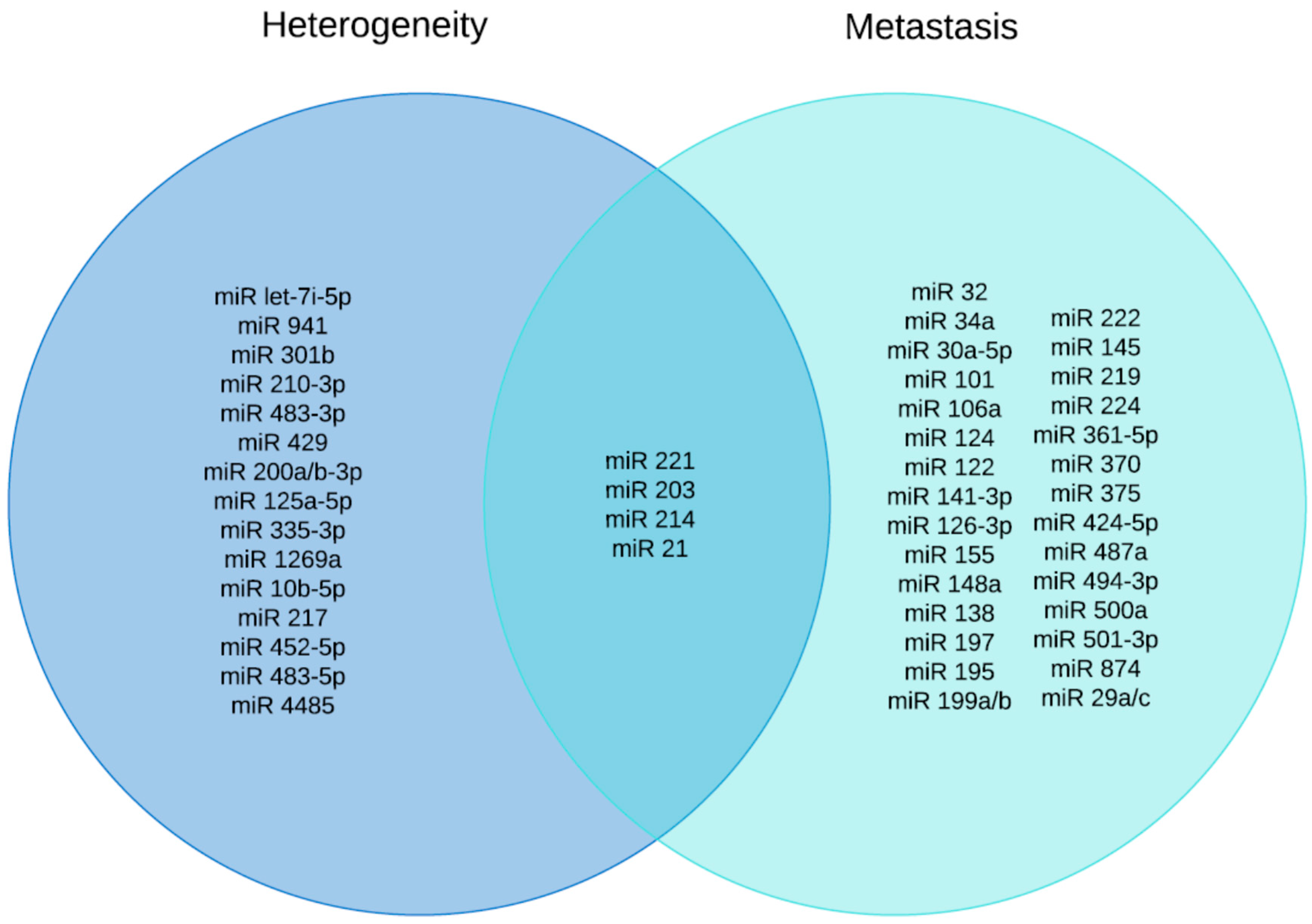
Table 1.
Differentially expressed miRs in HCC.
| MicroRNAs | Cellular Target/Mechanism | Related To | Reference | |
|---|---|---|---|---|
| Tumor-Suppressor miRNAs | miR-26a | FBXO11, Cyclin D2, Cyclin E2 | Tumor initiation/ progression | [41] |
| miR-17, miR19a, miR-20a, miR-24, miR-27a, miR-106a, miR-205, miR886-5p | E2F1, c-Myc, PTEN, SOX4,l c-Myc, E2F2, MMP14, FLT1 APC, CYP1B1, HNF3α, STAT3, MMP7 ZEB2, Bmi, ABL1, CAV1, K-RAS, Bax | Recurrence after liver Transplantation | [67] | |
| miR-29 a-c | SETDB1 | Metastasis | [50] | |
| miR-34a | p53, EMT-transcription factor, Notch/Wnt/TGF-β/SMAD signaling | Progression/EMT | [43] | |
| miR-101 | COX-2, Mcl-1 | Proliferation/Migration/ Recurrence after transplant | [68] | |
| miR-122 | WNT1, Bcl-W, p53 | Proliferation/Metastasis/ Recurrence after transplant | [44] [67] | |
| miR-124 | CDK6, VIM, SMYD3, IQGAP1 | Metastasis | [69] | |
| miR-125a/b | SIRT7, LIN28B, mcl-1, IL-6R, Bcl2 | Tumor differentiation | [70] | |
| miR-126-3p | Focal adhesion and MAPK signaling | Invasion/Metastasis/ Recurrence after transplant | [71] [67] | |
| miR-141-3p | GP73, E-cadherin, N-cadherin, occludin, vimentin and cytokeratin | Tumor growth/ Metastasis | [72] | |
| miR-145 | IRS1, AKT/FOXO1 | Proliferation/Migration/ Invasion | [49] | |
| miR-147 | HOXC6 | Recurrence after transplant | [67] | |
| miR-195 | Cyclin D1, CDK6, E2F3, IKKα, TGF-beta-activated kinase1, MAP3K7 binding protein 3 (TAb3), NF-κB and PCMT1 | Proliferation/ Tumorigenesis/Metastasis | [45] | |
| miR-197-3p | ZIK1 | Metastasis | [73] | |
| miR199a/b-3p | PAK4/Raf/MEK/ERK pathway | Progression | [74] | |
| miR199a/b-5p | ROCK1/MLC and PI3K/Akt signaling | Proliferation/Invasion/Migration | [75] | |
| miR-203 | CDK6,VIM, SMYD3, IQGAP1 and ABCE1 | Epigenetic control of HCC/ Recurrence after transplant | [69] | |
| miR-206 | cMET, CCND1 and CDK6 | Growth of HCC | [76] | |
| miR-214 | EZH2, β-catenin | Recurrence/Progression/ Metastasis | [51] | |
| miR-223 | STMN1 | Proliferation/HCC Development/Recurrence after transplant | [77] | |
| miR-302b | AKT2 | Tumor growth | [42] | |
| miR-339 | ZNF689 | Proliferation/Invasion | [78] | |
| miR-15a, miR16 and miR-107 | WNT3A | Proliferation/Cell cycle/Invasion | [48] | |
| miR-375 | YAP and AEG-1 | Proliferation/Migration/ Invasion | [47] | |
| miR-424-5p | TRIM 29 | Proliferation/Invasion | [46] | |
| miR-501-3p | LIN7A | Progression/Metastasis | [79] | |
| miR-874 | DOR, EGFR and ERK | Proliferation/Metastasis | [80] | |
| miR-140 | MMP9 | Invastion/Metastasis | [81] | |
| miR-193b | MAPK1 | Invasion/Migration | [82] | |
| miR-377-3p | CELF1 | Proliferation/Migration | [83] | |
| miR-605-3p | TRAF6 | Metastasis/EMT | [84] | |
| miR-544b | BTG2 | Proliferation/Migration/Invasion | [85] | |
| Oncogenic miRNAs | ||||
| miR-25 | Bim | Development/Progression/ Poor prognosis | [57] | |
| miR-17-5p | PTEN, GalNT7, Vimentin | HCV-induced HCC | [56] | |
| miR-18a | ESR1 | Proliferation | [53] | |
| miR-32 | PTEN | Proliferation/Migration/ Invasion | [65] | |
| miR-92b | SMAD7 | HCV-induced HCC | [60] | |
| miR-93 | PTEN, CDKN1A, c-Met/p13k/AKT pathway | Development/ Progression | [55] | |
| miR-101 | FOS oncogene | Viral HCC | [52] | |
| miR-106a | Tumor recurrence | [61] | ||
| miR-106b | EMT | Migration/Metastasis | [66] | |
| miR-130b | PPAR-γ, TP53INP1 | Tumorigenesis | [54] | |
| miR-155 | c-Myc, C/EBPβ, APC, β-catenin, cyclin D1, TP53INP1 | Invasion/Tumorigenesis/ Recurrence after transplant | [62] | |
| miR-197 | Wnt/β-catenin, Axin-2, NKD1 and DKK2 | Metastasis | [86] | |
| miR-219-5p | cadherin 1 | Tumor growth/Metatasis | [87] | |
| miR-221 and miR-222 | PHF2, AKT pathway, PTEN, CDK inhibitor p27 and DDIT4 | Occurrence/Development/ metastasis | [88] [89] | |
| miR-224 | CDC42, CDH1, PAK2, BCL-2, MAPK1, SMAD4 and HOXD10, PPP2R1B, AKT-signaling | Proliferation/Invasion/ Migration/HCV-induced HCC | [63] [64] | |
| miR-452 | Sox7, Wnt/β-catenin signaling | CSC in HCC | [59] | |
| miR-487a | PIK3R1-AKTsignaling SPRED2-protein kinase signaling | Proliferation/Metastasis | [90] | |
| miR-494-3p | Focal adhesion and MAPK signaling | Invasion/Metastasis | [71] | |
| miR-500a | BID, SFRP2 and GSK-3β, and Wnt/β-catenin signaling | Progression/Migration/ Invasion | [91] | |
| miR10b, miR147, miR338, miR-597 | HOXD10, TPM1, TRAIL3, NOL3, ZAP-70, SMAD2, LODH1, IRF2 and KLF3 | Recurrence after liver transplant | [67] | |
| miR-1269 | FOXO1, p21, cyclinD1 | Proliferation | [58] |
Table 2.
Diagnostic and prognostic miRs in HCC.
| miRNA | Expression | Observed Location | Type of HCC | References |
|---|---|---|---|---|
| miR-718 | Upregulated | Serum Exosome | Biomarker of HCC | [134] |
| exo-miR-18a, exo-miR-221, exo-miR-222 and exo-miR-224 | Upregulated | Serum Exosome | Biomarker of HCC | [135] |
| exo-miR-215-5p | Upregulated | Serum Exosome | Predictive biomarker of HCC | [136] |
| exo-miR-122a, exo-miR-21 and exo-miR-96 | Upregulated | Serum Exosome and plasma | Biomarker for the early identification of HCC | [137] |
| exo-miR-92b | Upregulated | Serum Exosome | Predicting the probability of HCC recurrance | [139] |
| (a) miR-221, miR-222, miR-93, miR-21 and miR-224 (b) miR-130a, miR-195, miR-199a and miR-375 | Upregulated Downregulated | ------ | Diagnosis and therapeutics targest to malignancy | [142] |
| (a) miR-1269, miR-224, miR-224-3p and miR-452 (b) miR-199a-5p, miR-199a-3p and miR-199b | Upregulated Downregulated | ------ | HCV-induced HCC | [143] |
| miR-122, miR-199a and miR-16 | Upregulated | Serum | Biomarkers of HCV-induced HCC | [144,145] |
| miR-375, miR-92a, miR-10a, miR-223, miR-423, miR-23b, miR-23a, miR-342-3p, miR-99a, miR-122a, miR-125b and miR-150 | Upregulated | Serum | noninvasive biomarker in HBV-mediated HCC | [146] |
| miR-92-3p, miR-107 and miR-3126-5p | Upregulated Downregulated | Serum | Early stage and low-level AFP-HCC | [147] |
| miR-20a-5p, miR-25-3p, miR-30a-5p, miR-92a-3p, miR-132-3p, miR-185-5p, miR-320a and miR-324-3p | Upregulated | Serum and tissue | Diagnosis of HBV-positive HCC | [148] |
| miR-192-5p, miR-21-5p and miR-375 | Upregulated | Serum and tissue | Biomarker for HCC screening | [149] |
| miR-192 | Upregulated | Serum Exosome | Biomarker of HCC | [150] |
| miR-665 | Upregulated | Serum | Diagnosis of HCC | [151] |
| miR-148a | Downregulated | Serum | Prognosis of HCC | [152] |
| miR-126 | Upregulated | Plasma | HBV HCC | [153] |
| miR-424 | Downregulated | Serum | Prognosis of HCC | [154] |
Table 3.
miRs in HCC therapeutics.
| Therapeutic Strategy | miRNA | Delivery System | Mouse Model | References |
|---|---|---|---|---|
| miRNA | miR-221 | Anti-miR-221-AMO | Orthotopic model | [181] |
| miR-487a | Morpholino-anti-miRNA miR-487a | Orthotopic model | [90] | |
| miR-500a-3p | antagomiR-500a-3p | Xenograft | [91] | |
| miR-122 | Miravirsen, anti-mir-122 | Phase III Clinical | [180] | |
| miR-21 | anti-miR-21 | Xenograft | [182] | |
| miR-494 | miR-494-anti-miR | tet-o-MYC; LAP-tTA | [176] | |
| miRNA replacement therapy | miR-26a | AAV vector | tet-o-MYC; LAP-tTA | [41] |
| miR-29b | 2’O-methyl modified miRNA | Xenograft | [183] | |
| miR-206 | Plenti_CMV-puromycin- miR206 | Xenograft | [76] | |
| miR-302b | Lipofectamine RNAi-MAX | Xenograft | [42] | |
| miR-361-5p | oligonucleoides miR-361-5p mimics in Lentivirus | Xenograft | [78] | |
| miR-101 | Lipofectamine RNAi-MAX | Xenograft | [68] | |
| miR-199a | oligonucleoides Lenti-miR-199a | Xenograft | [184] | |
| miR-375 | Cholesterol-conjugated 2’O-methyl modified Oligo mimics | Xenograft | [47] | |
| miR-122 | AAV | tet-o-MYC; LAP-tTA | [185] | |
| miR-422a | Oligo mimics; Lentivirus | Xenograft; DEN-induced mouse model | [186] | |
| miR-21 | Antisense MiR-21 co- encapsulated with gemci- tabine in PEGylated-PL GA nanoparticles | in vitro only | [187] | |
| miR-375 | AuNP-miR-375 | Xenograft | [188] | |
| miR-122 | Cationic lipid nanoparticles | Xenograft | [189] | |
| miR-34 | Liposome-based miR-34 mimic (MRX34) | Phase I clinical trial | [190] | |
| miR-124 | Liposomes | DEN-induced HCC mouse model | [191] | |
| Oncolytic Virus | let-7 | Adenovirus | Xenograft | [192] |
| miR-122 | Adenovirus | Xenograft | [193] | |
| miR-122; let-7 miR-124 | Herpes Simplex virus | Xenograft | [194] | |
| miR-199 | Adenovirus | Xenograft; TG-221 | [195] |
Publisher’s Note: MDPI stays neutral with regard to jurisdictional claims in published maps and institutional affiliations. |
© 2022 by the authors. Licensee MDPI, Basel, Switzerland. This article is an open access article distributed under the terms and conditions of the Creative Commons Attribution (CC BY) license (https://creativecommons.org/licenses/by/4.0/).
Share and Cite
MDPI and ACS Style
Khare, S.; Khare, T.; Ramanathan, R.; Ibdah, J.A. Hepatocellular Carcinoma: The Role of MicroRNAs. Biomolecules 2022, 12, 645. https://doi.org/10.3390/biom12050645
AMA Style
Khare S, Khare T, Ramanathan R, Ibdah JA. Hepatocellular Carcinoma: The Role of MicroRNAs. Biomolecules. 2022; 12(5):645. https://doi.org/10.3390/biom12050645
Chicago/Turabian StyleKhare, Sharad, Tripti Khare, Raghu Ramanathan, and Jamal A. Ibdah. 2022. "Hepatocellular Carcinoma: The Role of MicroRNAs" Biomolecules 12, no. 5: 645. https://doi.org/10.3390/biom12050645
Note that from the first issue of 2016, this journal uses article numbers instead of page numbers. See further details here.