Temperature-Independent Lifetime and Thermometer Operated in a Biological Window of Upconverting NaErF4 Nanocrystals


1
College of Physics and Optoelectronic Engineering, Harbin Engineering University, Harbin 150001, China
2
Key Lab of In-fiber Integrated Optics, Ministry Education of China, Harbin Engineering University, Harbin 150001, China
3
School of Economics and Management Engineering, Harbin Engineering University, Harbin 150001, China
*
Authors to whom correspondence should be addressed.
Nanomaterials 2020, 10(1), 24; https://doi.org/10.3390/nano10010024
Submission received: 23 November 2019
/
Revised: 12 December 2019
/
Accepted: 18 December 2019
/
Published: 20 December 2019
(This article belongs to the Section Nanophotonics Materials and Devices)
Abstract
:Lifetime of lanthanide luminescence basically decreases with increasing the ambient temperature. In this work, we developed NaErF4 core–shell nanocrystals with compensation of the lifetime variation with temperature. Upconversion lifetime of various emissions remains substantially unchanged as increasing the ambient temperature, upon 980/1530 nm excitation. The concentrated dopants, leading to extremely strong interactions between them, are responsible for the unique temperature-independent lifetime. Besides, upconversion mechanisms of NaErF4 core-only and core–shell nanocrystals under 980 and 1530 nm excitations were comparatively investigated. On the basis of luminescent ratiometric method, we demonstrated the optical thermometry using non-thermally coupled 4F9/2 and 4I9/2 emissions upon 1530 nm excitation, favoring the temperature monitoring in vivo due to both excitation and emissions fall in the biological window. The formed NaErF4 core–shell nanocrystals with ultra-small particle size, highly efficient upconversion luminescence, unique temperature-independent lifetimes, and thermometry operated in a biological window, are versatile in applications such as anti-counterfeiting, time-domain manipulation, and biological thermal probes.
1. Introduction
Lanthanide (Ln) heavily doped nanocrystals (NCs), with intense energy harvest and, thus, both potentials of efficient upconversion (UC) luminescence as well as light-to-heat conversion, are promising for a broad range of applications spanning background-free biolabeling and biosensing, light-triggered drug delivery, multimodal phototherapy, 3D display, solar cell, and light-emitting diodes [1,2,3,4,5,6,7]. However, Ln ions have abundant ladder-like energy levels, and cross-relaxation (CR) between ions and energy transfer (ET) to the surface quenchers become more sensitive with increasing the doping level; thus, the UC intensity of activating ions (such as Er3+, Tm3+, Ho3+, and Nd3+) heavily doped upconversion nanocrystals (UCNCs) has been strictly limited [8,9]. Over the past decade, researchers have developed various strategies to suppress the concentration quenching effect, mainly through coating an inert shell [10,11,12], increasing excitation power density [13], choosing host NCs with a large unit cell size [14], and engineering the distribution of dopants [15].
Among the above-mentioned strategies, the core–shell structure is one of the most effective ways to suppress the high-doping concentration quenching in UCNCs. Very recently, Johnson et al. [16] and Zuo et al. [17] have shown that the most intense luminescence is achieved when the host lattice is fully occupied by activators (100 mol.% doping), after inert layer passivation. Since then, Chen et al. [18] and Shang et al. [19] achieved enhanced UC luminescence in Er3+ heavily doped NCs via energy condensation through combined effects of mediated transient energy trapping and inert-shell coating. Although highly efficient UC luminescence of high-doping NCs has been reported in recent years, UC mechanisms have not been discussed in detail, and especially some unique optical properties aroused by high concentration doping remain undiscovered [20,21].
Conventional applications of UCNCs are generally based on the emission intensity and color. Recently, time-domain of UC is increasingly concerned, as it provides new dimension to exploit the UC emission, and thus multiplexing applications such as anti-counterfeiting, optical coding, and multichannel labeling are possible [22,23]. However, it is well known that the lifetime of Ln luminescence is sensitive to temperature, which decreases with heating, resulting in difficulty of manipulating lifetime under various ambient temperatures [24]. Thus, achieving temperature-independent lifetime of UCNCs is highly desired.
In addition, remote, sensitive, and accurate temperature monitoring is of vital importance for in vivo applications such as drug delivery and photothermal therapy [25]. The fluorescence intensity ratio (FIR), usually based on luminescent intensities from two thermally coupled levels of lanthanide ions, is the most popular method adopted for UC thermometer [26,27]. In principle, the populations of two thermally coupled levels follow the Boltzmann distribution, which requires the energy gap below 2000 cm−1, and thus sensitivity of the conventional FIR thermometer is restrained, due to it is proportional to the energy gap. Alternatively, using non-thermally coupled energy levels for FIR thermometer can greatly increase the sensitivity [28]. Further, choosing proper non-thermally coupled UC bands within the biological window not only improves the sensitivity, but also deepens the in vivo operation. Finally, high-doping core–shell UCNCs possessing highly efficient UC luminescent can improve sensing accuracy due to the high signal to noise ratio. Based on above discussions, Ln heavily doped core–shell UCNCs are promising thermal probes for in vivo applications.
In this paper, we investigated the UC luminescence behaviors of ultra-small NaErF4 core-only and core–shell NCs excited by 980 and 1530 nm laser. Effects of inert shell coating were discussed on the basis of structural and spectral measurements, thus enabling the analysis of the UC mechanisms. Anomalous variation of UC lifetimes in Er3+ heavy-doping NCs at various ambient temperatures were, for the first time, found and discussed. On the basis of the bright UC emissions, the FIR sensing behaviors of the thermal coupling levels (2H11/2 and 4S3/2) and non-thermally coupled levels (4S3/2/4F9/2, and 4I9/2) of Er3+ are comparatively explored.
2. Experiment Section
2.1. Synthesis of NaErF4 Core-Only NCs
The ultra-small NaErF4 core-only NCs were prepared via a slightly modified literature procedure [29], 1 M ErCl3, 0.5 M NaOH and NH4F methanol solutions were prepared firstly. Then, 1 mL of ErCl3 methanol solution, 8 mL of oleic acid (OA), and 15 mL of 1-octadecene (ODE) were added to a 50 mL three-necked flask. The mixture was heated at 180 °C with stir for 45 min before cooling down to 50 °C. Subsequently, 5 mL of NaOH and 8 mL of NH4F methanol solution were added into the mixture and stirred for another 45 min. After the reaction mixture was heated at 110 °C for 15 min to remove the residual methanol and water, the solution was quickly heated to 280 °C and kept for 1.25 h before cooling down to room temperature. The as-prepared NCs were precipitated by addition of ethanol, collected by centrifugation at 8000 rpm for 5 min, and washed with ethanol and methanol for several times. The final product was dispersed in 10 mL cyclohexane. Argon gas was adopted throughout the entire experiment to protect the reaction.
2.2. Synthesis of NaErF4@NaGdF4 Core–shell NCs
The as-prepared NaErF4 core-only NCs were used as seeds for shell modification. In a typical experiment, the shell solution was first prepared by mixing GdCl3 methanol solution (1 M, 0.5 mL), OA (5 mL), and ODE (7.5 mL) in a 50 mL three-necked flask. The resulting mixture was heated at 180 °C for 45 min before cooling down to 50 °C. 2.5 mL of NaOH and 4 mL of NH4F methanol solution and 5 mL of NaErF4 seed solution were then added and stirred for 45 min. After heating at 110 °C for 15 min to remove the residual methanol and water, the reaction mixture was quickly heated and kept at 280 °C for 1 h. The resulting core–shell NCs were washed and dispersed in cyclohexane following the above mentioned route. Argon gas was used throughout the entire experiment to protect the reaction.
2.3. Characterization and Spectral Measurements
The crystal structures of the sample was identified by powder X-ray diffraction (XRD, Smartlab, Rigaku Corp., Akishima, Japan) manufacturer, city, state abbreviation if US or Canada, country under various temperatures, with a resolution of 0.03°/step from 15 to 70°. The particle morphologies were recorded on a transmission electron microscopy (TEM, Tecnai G2, FEI Co., Ltd., Hillsboro, OH, USA). Room temperature UC luminescence measurements were performed by irradiating samples via a variable-power 980 or 1530 nm diode laser (LWIRL980-5W and LWIRL1530-1W, Laserwave Optoelectronics Technology Co., Ltd., Beijing, China). The emissions of core-only and core–shell β-NaErF4 NCs, dispersed into cyclohexane with identical concentration, were recorded by a spectrometer (FLMS03177, Ocean Optics Co., Ltd., Orlando, FL, USA). For time resolved luminescence measurements, the continuous wave lasers were modulated into square-wave output by a signal generator (VC2002, Victor Instruments Co., Ltd., Shenzhen, China), and the decay profiles were recorded by a photomultiplier tube (CR131, Zolix Instruments Co., Ltd., Beijing, China), connected to an oscilloscope (TBS1102, Tektronix Co., Ltd., Shanghai, China). To obtain the temperature dependent UC spectra and lifetimes, the powder form samples were pressed into small disks, which were further placed onto an electric heating plate (IKAC-MAG HP4, JKI Co., Ltd., Shanghai, China), with the temperature resolution of 0.1 K.
3. Results and Discussion
3.1. Characterizations
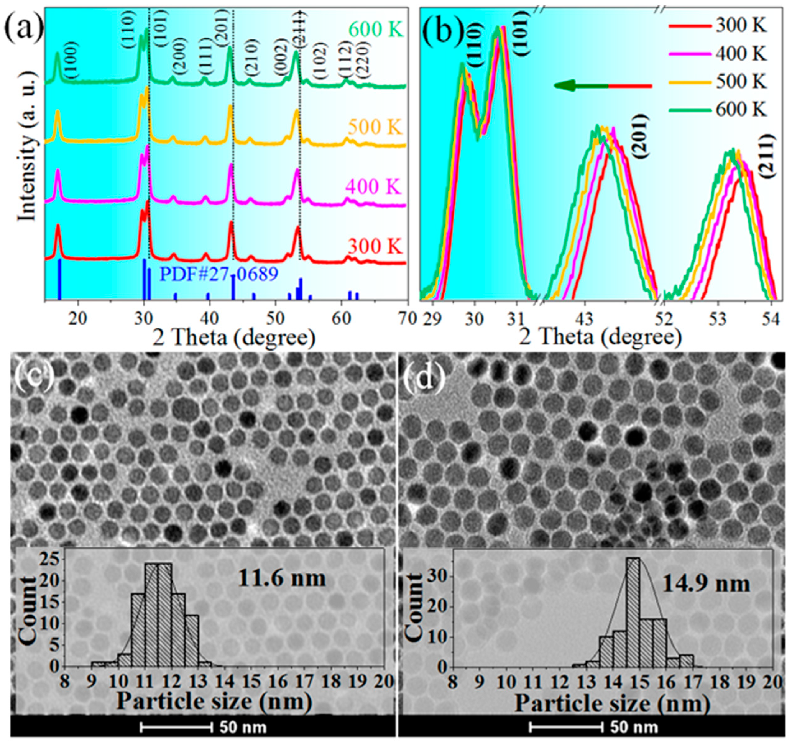
Figure 1a depicted the temperature-dependent XRD peaks of NaErF4, which are consistent with the standard data of hexagonal NaErF4 (PDF# 27-0689). No additional peaks appear, indicating the as-prepared sample is highly pure β-NaErF4. The crystal phase remains unchanged with increasing the ambient temperature up to 600 K, whereas the refraction peaks gradually blue-shift (Figure 1b), indicating the lattice expansion. The core-only NCs are well mono-disperse due to the good surface modification of OA. Spherical NaErF4 NCs are highly uniform in size, with an average diameter of 11.6 nm (Figure 1c). After epitaxial growth of pure NaGdF4 shell (~1.6 nm) on the core seeds, the mean diameter of core–shell NCs increases to ~14.9 nm, evidencing the successful coating of the inert shell (Figure 1d).
3.2. UC luminescence Properties
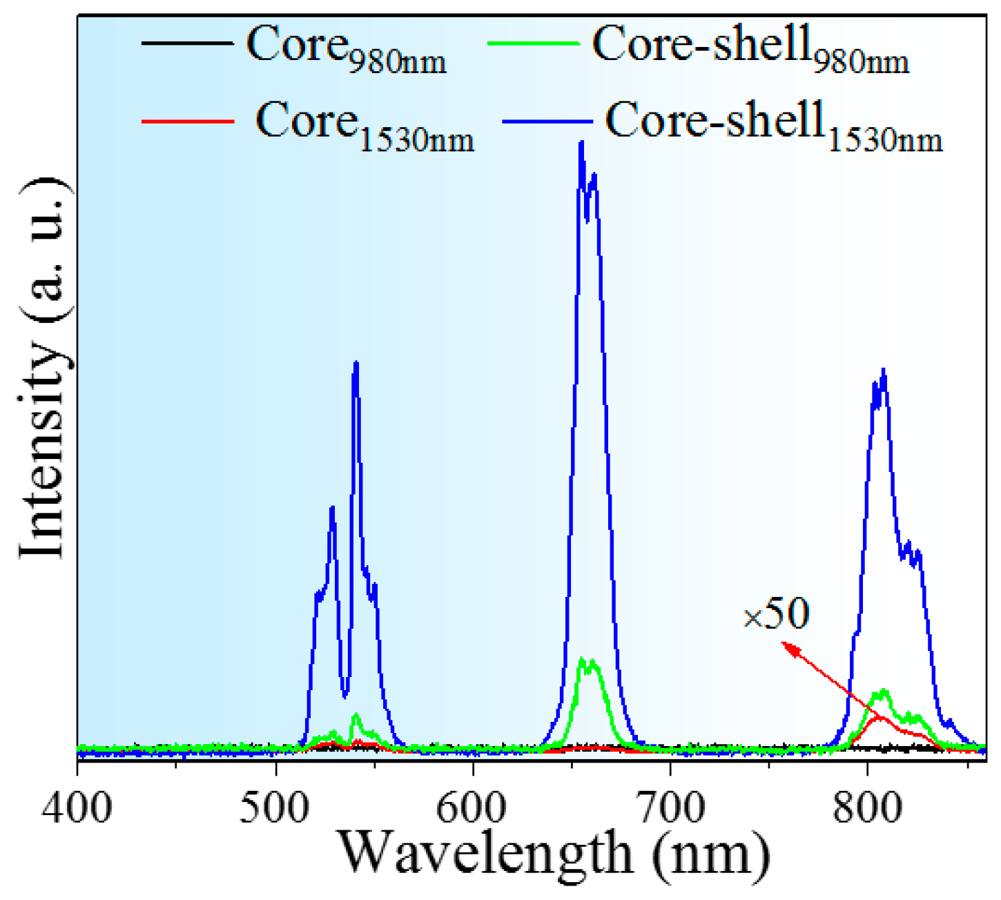
To illustrate the effect of inert coating, UC spectra of NaErF4 core-only and core–shell NCs were recorded (Figure 2). Upon NIR (980 or 1530 nm) laser excitation, the NCs exhibit green, red, and 800 nm UC emission bands, respectively corresponding to the transitions of 2H11/2/4S3/2→4I15/2, 4F9/2→4I15/2, and 4I9/2→4I15/2. The luminescence intensity of core-only NCs is extremely weak owing to strong concentration quenching, leading to no emission peak detected at 980 nm excitation. As more “dark ions” are activated through ~4-fold absorption intensity of Er3+ at 1.5 μm than that at 1 μm [21], slight emissions appear when pumped by 1530 nm laser.
In stark contrast, core–shell NCs yield highly efficient UC emissions (980 or 1530 nm excitation). The enhanced factor of overall intensity between core and core–shell (Icore–shell/Icore) NCs obtained by 1530 nm excitation goes up to ~1100, and it reaches ~8 for 1530 and 980 nm (I1530/I980) in core–shell NCs. The significant enhancement can be mainly attributed to the effective suppression of surface quenching, as core–shell structure extends the distance between luminescence centers and surface vibration modes. It is noteworthy that the relative intensity of NIR to visible emission in core NCs is stronger than that in their core–shell counterparts, due to visible luminescence originated from higher energy levels is more susceptible to the surface defects compared to the lower transition of 4I9/2→4I15/2.
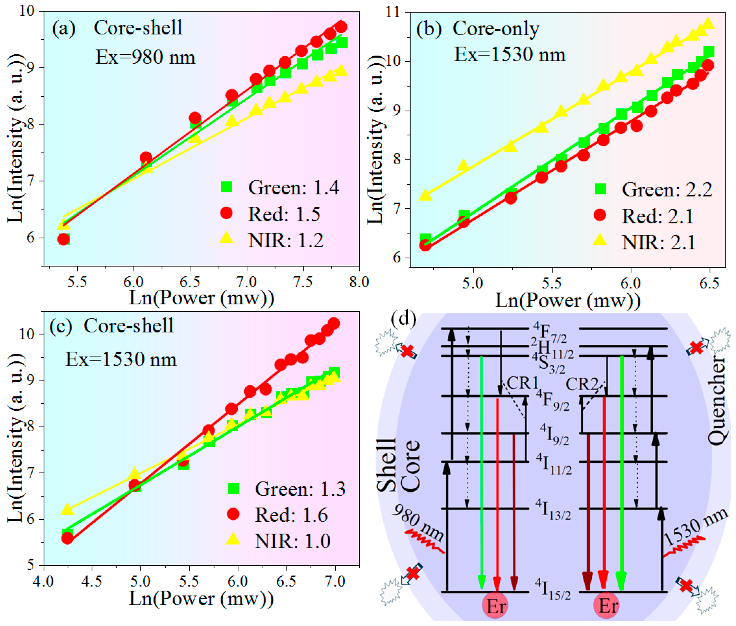
Further, pumping power dependent UC intensity of NaErF4 core-only and core–shell NCs under NIR excitation were investigated. The relationship between the UC intensity and the excitation power, IUP ∝ PnNIR, can be deduced by a simplified rate equation model [30], where IUP, PNIR, and n refer to the UC intensity, excitation power, and the number of NIR photons involved in the UC processes, respectively.
As plotted in Figure 3, the slopes obtained by linearly fitting are 1.2–1.5 (Figure 3a) and 2.1–2.2 (Figure 3b), indicating 2-photon UC process in core–shell NCs stimulated at 980 nm and 3-photon process in core-only NCs at 1530 nm, respectively [31,32]. However, slope values lower than 2, obtained in core–shell NCs upon 1530 nm excitation (Figure 3c), obviously differ from its core-only counterpart. These decreased slopes can be attributed to the effects of inert passivation. Specifically, owing to core–shell structure effectively suppresses surface quenching, upward transitions dominate downward relaxations in Er3+ intermediate levels of 4I13/2 and 4I9/2, and thus the slopes in the core–shell NCs decreases [33,34]. The detailed UC pathways of Er3+ excited by 980 or 1530 nm were illustrated in Figure 3d. Due to the inert passivation eliminating the high-energy surface vibration modes, red emitting level 4F9/2 should be less populated by nonradiative decay from green level 4S3/2. As a consequence, CR1 and CR2 are, respectively, responsible for the populations of red emission upon 980 and 1530 nm excitation [21].
3.3. Anomalous Variation of Lifetime Versus Temperature
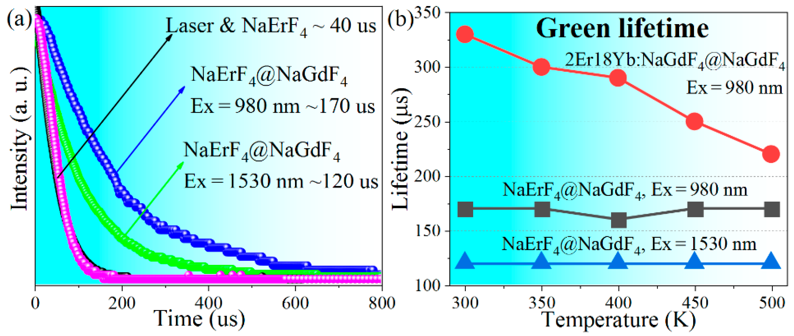
Time-resolved luminescent intensities were measured to shed more light on the UC behaviors. As shown in Figure 4a, the decay curves of laser and core-only sample are almost identical, indicating a rapid decay of core-only NCs (≤40 μs). After inert coating, the lifetime prolongs to 120 and 170 μs. The rapid decay of core-only NCs can be attributed to the strong surface quenching induced by ultra-high concentration (100 mol.%). When surface quenchers are isolated from Er3+ by an inert shell, the detrimental ETs to the surface quenchers are eliminated. Consequently, lifetime increases in the core–shell NCs, consistent with the highly enhanced luminescent intensity. It is noteworthy that the measured lifetime of core–shell NCs is smaller than that obtained in NaErF4@NaLuF4 (10 nm shell thickness) [16], which can stem from the thinner shell used in the present work (~1.5 nm).
Also, the measured lifetimes are obviously smaller than expected. On the basis of the J-O calculation of 80 mol.% Er3+ doped NaGdF4, which is similar to NaErF4, transition probabilities of Er3+ fall within 101~103 s–1 (4S3/2:240 s–1, 4F9/2:640 s–1, and 4I9/2:70 s–1) [20].
From another side, the non-radiative decay rates follows
where W0 = 1 × 10⁸ s−1 and α = 5 × 10⁻³ cm are constants, estimated by using LaF3 crystal [35], which is similar to NaErF4 (hexagonal structure and cut-off phonon energy of ~350 cm−1); ΔE is the energy gap given in cm−1, within 2000~3000 cm−1 for Er3+ UC emitting levels (4S3/2:3117 cm−1, 4F9/2:2885 cm−1, and 4I9/2:2228 cm−1) [36]. According to Equation (1), the decay rates of 4S3/2, 4F9/2, and 4I9/2 levels are 20, 50, and 1450 s−1, respectively. Combining the radiative transition and phonon-assistant decays and omitting the ETs and surface quenching, UC lifetimes of NaErF4 core–shell NCs should be within 0.7~4 ms. However, the measured UC lifetimes are all below 0.3 ms, indicating other processes dominating the lifetime. We attributed them to the strong interactions between Er3+, accelerating the depopulation of excited levels through CR processes, which were not labeled in Figure 3d as there are numerous possibilities.
Besides, it is found that the 980 nm laser excited green and NIR lifetimes are generally larger than that obtained by using 1530 nm laser, whereas lifetimes of red emission are similar (Figure 4a and Table 1). In principle, lifetime of an energy level depends on several processes including radiative transitions, non-radiative decays (from its upper level and to its lower level), and ETs involved. The spontaneous transition probabilities of lanthanide ions are the intrinsic natures of the luminescent centers and surrounding crystal field; multiphonon-assistant decay rates are related to the energy gap and lattice vibration modes; ET rates are decided by the spectral overlap of donor and acceptor as well as their spatial distance. Therefore, for a given luminescent materials with fixed composition, above three parameters should be independent to the incident wavelength. As a consequence, the lifetime variation aroused by incident wavelength can stem from the different UC pathways. As shown in Figure 3d, green emitting levels of 4S3/2 and NIR level of 4I9/2 are not directly populated by the incident 980 nm photons, whereas 1530 nm photons directly feed these levels. This results in the prolonged lifetimes excited by 980 nm laser, as electrons populating 4S3/2 and 4I9/2 levels undergo additional decays from their upper levels. As for the red emission, 4F9/2 level is fed by CR processes for both 980 and 1530 nm excitations, and extremely strong interactions of Er3+ lead to similar lifetime upon various excitations.
Most importantly, lifetime of NaErF4 core–shell NCs remains substantially unchanged with increasing the ambient temperature (Figure 4b and Table 1). It is well known that lanthanide luminescence is susceptible to ambient temperature. In general, lifetime decreases with increasing the temperature (see Figure 4b, 1/3 reduction in conventional 2Er/18Yb NCs), due to the stronger non-radiative decay processes associated with phonon vibrations [37].
This temperature-independent variation might be the consequence of the lattice expansion with heating, which can be proven by the blue-shift of the XRD peaks with increasing temperature [Figure 1b] and is in good agreement with the observations elsewhere [38,39,40]. As mentioned above, CR processes are dominant for Er3+ UC lifetimes. Similarly, there should be considerable energy migrations between excited Er3+ in high-doping situation. The energy migrations occur through dipolar-dipolar interactions, where the efficiency is inversely proportional to the 6th power of the average donor-acceptor distance r. Thus, for the energy migration process through the consecutive ETs of n pairs of Er3+, the efficiency is proportion to r−6n [41]. As a consequence, Er3+ luminescence process is strongly influenced by the energy migration distance. Increased temperature causes the lattice to expand, resulting in a larger transfer distance r and a longer transfer time, and finally increase the Er3+ lifetimes. Herein, the prolonged lifetime aroused by lattice expansion compensates the shortened lifetime caused by thermal quenching, leading to the temperature-independent lifetimes observed. This phenomenon relies on high-doping concentration, thus does not occur in traditional 2Er/18Yb sample (Figure 4b), and is also consistent with the observation that the prolonged lifetimes of Yb3+ (higher concentration) are more evident than that of Eu3+ (lower concentration) in NaGdF4 with increasing temperature [39].
3.4. Temperature Sensing
Typical UC spectra of NaErF4 core–shell NCs under 1530 nm excitation show the quenched intensities with raising temperature, and the relative intensity also varies with temperature (Figure 5). Therefore, based on the FIR technique, temperature sensing behavior of NaErF4 core–shell NCs under 1530 nm excitation was investigated. The FIR from thermally coupled levels (2H11/2 and 4S3/2) can be expressed as follows [42],
and FIR from non-thermally coupled levels (4S3/2, 4F9/2 and 4I9/2) can be fitted by a polynomial function [43]
where Ii is the integrated intensity of the emission band; k is the Boltzmann constant; T is the absolute temperature; A, Bi, and C are the constants need to be determined.
For thermometers, the sensitivities (absolute sensitivity Sa and relative sensitivity Sr) are critical for evaluating their sensing performance, which can be determined according to the expressions:
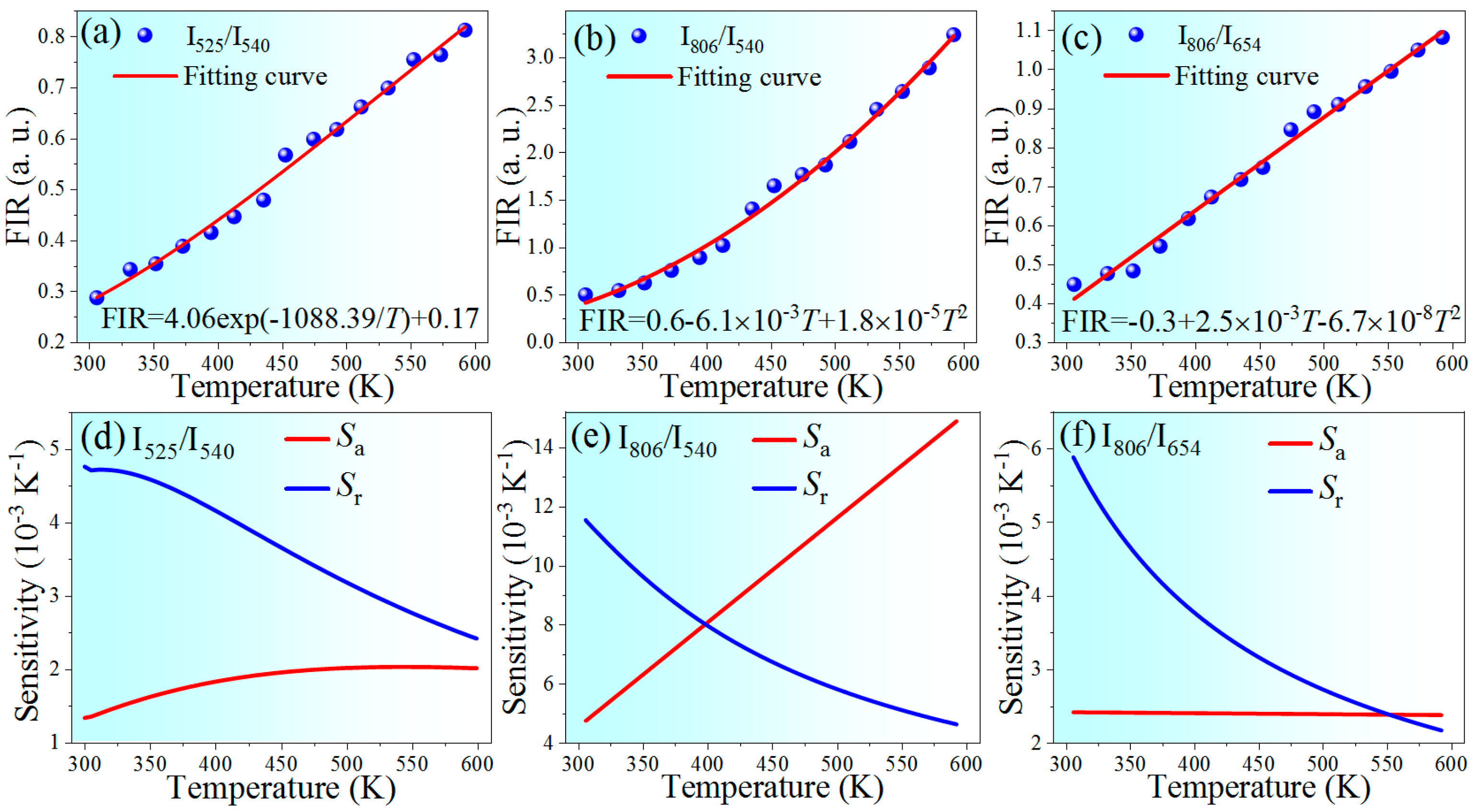
Figure 6a,b depict the FIR values as a function of temperature within the range of 303–593 K, by using thermally coupled and non-thermally coupled levels. The experimental data can be well fitted according to Equation (2) or (3), and the corresponding sensitivities are also shown in Figure 6d–e. The sensitivity obtained by using non-thermally coupled levels of 4S3/2 and 4I9/2, both for Sa or Sr, are higher than that obtained by using thermally coupled levels, the conventionally used thermally-coupled levels. The maximum Sa and Sr reach 0.0149 K−1 at 593 K and 1.15% K−1 at 303 K, respectively, within the operating temperature range, comparable to results reported recently, as shown in Table 2.
It is noteworthy that both the excitation wavelength and the Er3+ emissions of 4F9/2 and 4I9/2 level fall in the so-called biological windows (І-BW spinning 650–950 nm and Ш-BW within 1500–1750 nm [51]), the low-loss wavelength region in biological tissue. Combining this with other fascinating features of the newly developed UCNCs such as the ultra-small particle size (low biotoxicity) and the highly efficient UC emissions (high signal to noise ratio), using red and NIR UC emissions are promising for the in vivo temperature feedback. Well fitted data shown in Figure 6c evidences the feasibility of this biological thermometer. The maximum sensitivities of 0.0024 K−1 and 0.59% K−1, both obtained at 303 K which is close to the body temperature (Figure 6f), are slightly higher than that from the traditional 2H11/2/4S3/2 FIR.
4. Conclusions
In summary, the ultra-small hexagonal-NaErF4 core-only and core–shell NCs were synthesized by thermal decomposition method. Optical properties, including luminescence intensity, emitting lifetime, and UC mechanisms were investigated. The inert shell significantly enhances the luminescent intensity by a factor of ~1100, upon 1530 nm excitation. The unique temperature-independent lifetimes were obtained, mainly attributed to the balance between lattice expansion (prolong the lifetime) and thermal quenching (shorten the lifetime). Moreover, use of non-thermally coupled levels for FIR thermometry exhibited the maximum absolute and relative sensitivities of 0.0149 K−1 and 1.15% K−1, respectively, evidently higher than that obtained through traditional thermally coupled FIR. Finally, thermometer on the basis NIR/red FIR, suitable for in vivo temperature feedback, was demonstrated. These meaningful results will deepen the understanding of UC luminescence processes in Er3+ heavily doped systems, and also manifest the wide application prospects of NaErF4 core–shell UCNCs.
Author Contributions
All authors have read and agree to the published version of the manuscript. Conceptualization, L.L., and H.L.; investigation, K.L., L.X., and X.S.; writing—original draft preparation, K.L.; writing—review and editing, Y.Y., and L.L.; supervision, H.L. All authors have read and agreed to the published version of the manuscript.
Funding
This work was funded by the Fundamental Research Funds for the Central Universities (3072019CF2516); Natural Science Foundation of Heilongjiang Province (LC2018026); 111 project (B13015) to the Harbin Engineering University.
Conflicts of Interest
The authors declare no conflict of interest.
References
- Wen, S.H.; Zhou, J.J.; Zheng, K.Z.; Bednarkiewicz, A.; Liu, X.G.; Jin, D.Y. Advances in highly doped upconversion nanoparticles. Nat. Commun. 2018, 9, 1–12. [Google Scholar] [CrossRef]
- Drees, C.; Raj, A.N.; Kurre, R.; Busch, K.B.; Haase, M.; Piehler, J. Engineered upconversion nanoparticles for resolving protein interactions inside living cells. Angew. Chem. Int. Ed. Engl. 2016, 55, 11668–11672. [Google Scholar] [CrossRef] [PubMed]
- Zhang, Y.W.; Huang, L.; Li, Z.J.; Ma, G.L.; Zhou, Y.B.; Han, G. Illuminating cell signaling with near-infrared light-responsive nanomaterials. ACS Nano 2016, 10, 3881–3885. [Google Scholar] [CrossRef] [PubMed]
- Ai, X.; Wang, Z.; Cheong, H.; Wang, Y.; Zhang, R.; Lin, J.; Zheng, Y.; Gao, M.; Xing, B. Multispectral optoacoustic imaging of dynamic redox correlation and pathophysiological progression utilizing upconversion nanoprobes. Nat. Commun. 2019, 10, 1–11. [Google Scholar] [CrossRef] [PubMed] [Green Version]
- Wilhelm, S. Perspectives for upconverting nanoparticles. ACS Nano 2017, 11, 10644–10653. [Google Scholar] [CrossRef]
- Chan, E.M. Combinatorial approaches for developing upconverting nanomaterials: High-throughput screening, modeling, and applications. Chem. Soc. Rev. 2015, 44, 1653–1679. [Google Scholar] [CrossRef]
- Wang, F.; Han, Y.; Lim, C.S.; Lu, Y.H.; Wang, J.; Xu, J.; Chen, H.Y.; Zhang, C.; Hong, M.H.; Liu, X.G. Simultaneous phase and size control of upconversion nanocrystals through lanthanide doping. Nature 2010, 463, 1061–1065. [Google Scholar] [CrossRef]
- Zhou, B.; Shi, B.Y.; Jin, D.Y.; Liu, X.G. Controlling upconversion nanocrystals for emerging applications. Nat. Nanotechnol. 2015, 10, 924–936. [Google Scholar] [CrossRef]
- Boyer, J.C.; van Veggel, F.C.J.M. Absolute quantum yield measurements of colloidal NaYF4:Er3+, Yb3+upconverting nanoparticles. Nanoscale 2010, 2, 1417–1419. [Google Scholar] [CrossRef]
- Wang, F.; Wang, J.A.; Liu, X.G. Direct evidence of a surface quenching effect on size-dependent luminescence of upconversion nanoparticles. Angew. Chem. Int. Ed. Engl. 2010, 122, 7618–7622. [Google Scholar] [CrossRef]
- Viger, M.L.; Live, L.S.; Therrien, O.D.; Boudreau, D. Reduction of self-quenching in fluorescent silica-coated silver nanoparticles. Plasmonics 2008, 3, 33–40. [Google Scholar] [CrossRef]
- Chen, G.Y.; Agren, H.; Ohulchanskyy, T.Y.; Prasad, P.N. Light upconverting core-shell nanostructures: Nanophotonic control for emerging applications. Chem. Soc. Rev. 2015, 44, 1680–1713. [Google Scholar] [CrossRef] [PubMed]
- Gargas, D.J.; Chan, E.M.; Ostrowski, A.D.; Aloni, S.; Altoe, M.V.P.; Barnard, E.S.; Sanii, B.; Urban, J.J.; Milliron, D.J.; Cohen, B.E. Engineering bright sub-10 nm upconverting nanocrystals for single-molecule imaging. Nat. Nanotechnol. 2014, 9, 300–305. [Google Scholar] [CrossRef] [PubMed]
- Marciniak, L.; Strek, W.; Bednarkiewicz, A.; Hreniak, D. Bright upconversion emission of Nd3+ in LiLa1-xNdxP4O12 nanocrystalline powders. Opt. Mater. 2011, 33, 1492–1494. [Google Scholar] [CrossRef]
- Li, X.M.; Wang, R.; Zhang, F.; Zhao, D.Y. Engineering homogeneous doping in single nanoparticle to enhance upconversion efficiency. Nano Lett. 2014, 14, 3634–3639. [Google Scholar] [CrossRef]
- Johnson, N.J.J.; He, S.; Diao, S.; Chan, E.M.; Dai, H.; Almutairi, A. Direct evidence for coupled surface and concentration quenching dynamics in lanthanide-doped nanocrystals. J. Am. Chem. Soc. 2017, 139, 3275–3282. [Google Scholar] [CrossRef]
- Zuo, J.; Li, Q.Q.; Bai, X.; Li, C.X.; Chang, Y.L.; Zhang, Y.L.; Liu, X.M.; Tu, L.P.; Zhang, H.; Kong, X.G. Employing shells to eliminate concentration quenching in photonic upconversion nanostructure. Nanoscale 2017, 9, 7941–7946. [Google Scholar] [CrossRef]
- Chen, Q.S.; Xie, X.J.; Huang, B.L.; Liang, L.L.; Han, S.Y.; Yi, Z.G.; Wang, Y.; Li, Y.; Fan, D.Y.; Huang, L.; et al. Confining excitation energy in Er3+-sensitized upconversion nanocrystals through Tm3+-mediated transient energy trapping. Angew. Chem. Int. Ed. 2017, 56, 7605–7609. [Google Scholar] [CrossRef]
- Shang, Y.F.; Hao, S.W.; Lv, W.Q.; Chen, T.; Tian, L.; Lei, Z.T.; Yang, C.H. Confining excitation energy of Er3+-sensitized upconversion nanoparticles through introducing various energy trapping centers. J. Mater. Chem. C 2018, 6, 3869–3875. [Google Scholar] [CrossRef]
- Liu, L.; Lu, K.L.; Yan, D.; Zhao, E.; Li, H.; Shahzad, M.; Zhang, Y. Concentration dependent optical transition probabilities in ultra-small upconversion nanocrystals. Opt. Express 2018, 26, 23471–23479. [Google Scholar] [CrossRef]
- Liu, L.; Lu, K.L.; Xu, L.; Tang, D.; Liu, C.; Shahzad, M.; Yan, D.; Khan, F.; Zhao, E.; Li, H. Highly efficient upconversion luminescence of Er heavily doped nanocrystals through 1530, nm excitation. Opt. Lett. 2019, 44, 711–714. [Google Scholar] [CrossRef] [PubMed]
- Lu, Y.; Zhao, Y.J.; Zhang, R.; Liu, Y.; Liu, D.; Goldys, E.M.; Yang, X.; Xi, P.; Sunna, A.; Lu, J.; et al. Tunable lifetime multiplexing using luminescent nanocrystals. Nat. Photon. 2013, 8, 32–36. [Google Scholar] [CrossRef] [Green Version]
- Gu, Y.; Guo, Z.; Yuan, W.; Kong, M.; Liu, Y.; Gao, Y.; Feng, W.; Wang, F.; Zhou, J.; Jin, D.; et al. High-sensitivity imaging of time-domain near-infrared light transducer. Nat. Photon. 2019, 8, 32–36. [Google Scholar] [CrossRef]
- Yu, W.; Xu, W.; Song, H.; Zhang, S. Temperature-dependent upconversion luminescence and dynamics of NaYF4:Yb3+/Er3+ nanocrystals: Influence of particle size and crystalline phase. Dalton Trans. 2014, 43, 6139–6147. [Google Scholar] [CrossRef] [PubMed]
- Vetrone, F.; Naccache, R.; Zamarrón, A.; de la Fuente, A.J.; Sanz-Rodríguez, F.; Maestro, L.M.; Rodríguez, E.M.; Jaque, D.; García Solé, J.; Capobianco, J. Temperature sensing using fluorescent nanothermometers. ACS Nano 2010, 4, 3254–3258. [Google Scholar] [CrossRef]
- Xu, X.; Wang, Z.; Lei, P.; Yu, Y.; Yao, S.; Song, S.; Liu, X.; Su, Y.; Dong, L.; Feng, J.; et al. α-NaYb(Mn)F4:Er3+/Tm3+@NaYF4 UCNPs as “band-shape” luminescent nanothermometers over a wide temperature range. ACS Appl. Mater. Inter. 2015, 7, 20813–20819. [Google Scholar] [CrossRef]
- Tang, W.; Wang, S.; Li, Z.; Su, Y.; Zheng, L.; Zhang, R.; Yang, B.; Cao, W.; Yu, M. Ultrahigh-sensitive optical temperature sensing based on ferroelectric Pr3+-doped (K0.5Na0.5)NbO3. Appl. Phys. Lett. 2016, 108, 1–6. [Google Scholar] [CrossRef]
- Lu, H.; Hao, H.; Shi, G.; Gao, Y.; Wang, R.; Song, Y.; Wang, Y.; Zhang, X. Optical temperature sensing in β-NaLuF4:Yb3+/Er3+/Tm3+ based on thermal, quasi-thermal and non-thermal coupling levels. RSC Adv. 2016, 6, 55307–55311. [Google Scholar] [CrossRef]
- Wang, F.; Deng, R.R.; Liu, X.G. Preparation of core-shell NaGdF4 nanoparticles doped with luminescent lanthanide ions to be used as upconversion-based probes. Nat. Protoc. 2014, 9, 1634–1644. [Google Scholar] [CrossRef]
- Pandozzi, F.; Vetrone, F.; Boyer, J.C.; Naccache, R.; Capobianco, J.A.; Speghini, A.; Bettinelli, M. A spectroscopic analysis of blue and ultraviolet upconverted emissions from Gd3Ga5O12:Tm3+, Yb3+ nanocrystals. J. Phys. Chem. B 2005, 109, 17400–17405. [Google Scholar] [CrossRef]
- Li, L.; Xu, W.; Zheng, L.; Qin, F.; Zhou, Y.; Liang, Z.; Zhang, Z.; Cao, W. Valley-to-peak intensity ratio thermometry based on the red upconversion emission of Er3+. Opt. Express 2016, 24, 13244–13249. [Google Scholar] [CrossRef] [PubMed]
- Kumar, G.A.; Pokhrel, M.; Sardar, D.K. Intense visible and near infrared upconversion in M2O2S:Er (M = Y, Gd, La) phosphor under 1550, nm excitation. Mater. Lett. 2012, 68, 395–398. [Google Scholar] [CrossRef]
- Pollnau, M.; Gamelin, D.R.; Lüthi, S.R.; Güdel, H.U.; Hehlen, M.P. Power dependence of upconversion luminescence in lanthanide and transition-metal-ion systems. Phys. Rev. B 2000, 61, 3337–3346. [Google Scholar] [CrossRef]
- Jacinto, C.; Vermelho, M.V.D.; Gouveia, E.A.; de Araujo, M.T.; Udo, P.T.; Astrath, N.G.C.; Baesso, M.L. Pump-power-controlled luminescence switching in Yb3+/Tm3+ codoped water-free low silica calcium aluminosilicate glasses. Appl. Phys. Lett. 2007, 91, 1–4. [Google Scholar] [CrossRef]
- Miyakawa, T.; Dexter, D.L. Phonon sidebands, multiphonon relaxation of excited states, and photon-assisted energy transfer between ions in solids. Phys. Rev. B 1970, 1, 2961–2969. [Google Scholar] [CrossRef]
- Weber, M.J. Probabilities for radiative and nonradiative decay of Er3+ in LaF3. Phys. Rev. 1967, 157, 262–272. [Google Scholar] [CrossRef]
- Yao, L.; Li, Y.; Xu, D.; Lin, H.; Peng, Y.; Yang, S.; Zhang, Y. Upconversion luminescence enhancement and lifetime based thermometry of Na(Gd/Lu)F4 solid solutions. New J. Chem. 2019, 43, 3848–3855. [Google Scholar] [CrossRef]
- Janjua, R.A.; Gao, C.; Dai, R.; Sui, Z.; Raja, M.A.A.; Wang, Z.; Zhen, X.; Zhang, Z. Na+-driven nucleation of NaYF4:Yb, Er nanocrystals and effect of temperature on their structural transformations and luminescent properties. J. Phys. Chem. C 2018, 122, 23242–23250. [Google Scholar] [CrossRef]
- Cui, X.S.; Cheng, Y.; Lin, H.; Huang, F.; Wu, Q.P.; Wang, Y.S. Size-dependent abnormal thermo-enhanced luminescence of ytterbium-doped nanoparticles. Nanoscale 2017, 9, 13794–13799. [Google Scholar] [CrossRef]
- Janjua, R.A.; Farooq, U.; Dai, R.; Wang, Z.; Zhang, Z. Wide-range ratiometric upconversion luminescence thermometry based on non-thermally coupled levels of Er in high-temperature cubic phase NaYF4:Yb, Er. Opt. Lett. 2019, 44, 4678–4681. [Google Scholar] [CrossRef]
- Wang, F.; Deng, R.R.; Wang, J.; Wang, Q.X.; Han, Y.; Zhu, H.M.; Chen, X.Y.; Liu, X.G. Tuning upconversion through energy migration in core-shell nanoparticles. Nat. Mater. 2011, 10, 968–973. [Google Scholar] [CrossRef] [PubMed]
- Wade, S.A.; Collins, S.F.; Baxter, G.W. Fluorescence intensity ratio technique for optical fiber point temperature sensing. J. Appl. Phys. 2003, 94, 4743–4756. [Google Scholar] [CrossRef]
- Lu, H.; Hao, H.; Gao, Y.; Li, D.; Shi, G.; Song, Y.; Wang, Y.; Zhang, X. Optical sensing of temperature based on non-thermally coupled levels and upconverted white light emission of a Gd2 (WO4)3 phosphor co-doped with in Ho(III), Tm(III), and Yb (III). Microchim. Acta 2017, 184, 641–646. [Google Scholar] [CrossRef]
- Ma, Y.; Xiang, G.; Zhang, J.; Liu, Z.; Zhou, P.; Liu, W.; Tang, X.; Jiang, S.; Zhou, X.; Li, L.; et al. Upconversion properties and temperature sensing behaviors in visible and near-infrared region based on fluorescence intensity ratio in LuVO4:Yb3+/Er3+. J. Alloys Compd. 2018, 769, 325–331. [Google Scholar] [CrossRef]
- Wang, X.; Liu, Q.; Bu, Y.; Liu, C.; Liu, T.; Yan, X. Optical temperature sensing of rare-earth ion doped phosphors. RSC Adv. 2015, 5, 86219–86236. [Google Scholar] [CrossRef]
- Tang, J.; Zhang, Y.; Gou, J.; Ma, Z.; Li, G.; Man, Y.; Cheng, N. Sol-gel prepared Yb3+/Er3+ co-doped RE2O3 (RE=La, Gd, Lu) nanocrystals: Structural characterization and temperature-dependent upconversion behavior. J. Alloys Compd. 2018, 740, 229–236. [Google Scholar] [CrossRef]
- Geitenbeek, R.G.; Prins, P.T.; Albrecht, W. NaYF4:Er3+, Yb3+/SiO2 core/shell upconverting nanocrystals for luminescence thermometry up to 900 K. J. Phys. Chem. C 2017, 121, 3503–3510. [Google Scholar] [CrossRef] [Green Version]
- Zhang, D.; Yue, Z.; Liu, Z. Dual-mode luminescent core-shell nanoarchitectures for highly sensitive optical nanothermometry. J. Alloys Compd. 2019, 787, 585–593. [Google Scholar] [CrossRef]
- Zheng, K.Z.; Song, W.Y.; He, G.H.; Yuan, Z.; Qin, W.P. Five-photon UV upconversion emissions of Er3+ for temperature sensing. Opt. Express 2015, 23, 7653–7658. [Google Scholar] [CrossRef]
- Du, P.; Deng, A.M.; Luo, L.; Yu, J.S. Simultaneous phase and size manipulation in NaYF4:Er3+/Yb3+ upconverting nanoparticles for a non-invasion optical thermometer. New J. Chem. 2017, 41, 13855–13861. [Google Scholar] [CrossRef]
- Hemmer, E.; Venkatachalam, N.; Hyodo, H.; Hattorib, A.; Ebinab, Y.; Kishimoto, H.; Soga, K. Upconverting and NIR emitting rare earth based nanostructures for NIR-bioimaging. Nanoscale 2013, 5, 11339–11361. [Google Scholar] [CrossRef] [PubMed]
Figure 1.
(a) Temperature-dependent XRD patterns of the NaErF4 core-only NCs. The standard data for β-NaErF4 (PDF# 27-0689) is shown as a reference; (b) the amplified patterns of the three most intensive peaks. TEM images of (c) NaErF4 core-only NCs with an average size of 11.6 nm and (d) NaErF4@NaGdF4 core–shell NCs of 14.9 nm. Insets are the size distributions obtained from measurements of 100 particles, and scale bars are 50 nm.

Figure 2.
UC spectra of NaErF4 core-only and core–shell NCs excited by 980 or 1530 nm laser (10 W/cm2), 1530 nm laser induced spectrum of core-only sample is magnified by a factor of 50.
Figure 2.
UC spectra of NaErF4 core-only and core–shell NCs excited by 980 or 1530 nm laser (10 W/cm2), 1530 nm laser induced spectrum of core-only sample is magnified by a factor of 50.

Figure 3.
Ln-ln power dependences of Er3+ emissions in (a) core–shell NCs under 980 nm excitation; (b) core-only and (c) core–shell NCs excited by 1530 nm laser; (d) energy levels and UC pathways of Er3+ pumped by NIR (980 or 1530 nm) laser.
Figure 3.
Ln-ln power dependences of Er3+ emissions in (a) core–shell NCs under 980 nm excitation; (b) core-only and (c) core–shell NCs excited by 1530 nm laser; (d) energy levels and UC pathways of Er3+ pumped by NIR (980 or 1530 nm) laser.

Figure 4.
(a) Decay profiles of 4S3/2 emissions of core-only and core–shell NCs under 980 or 1530 nm excitation. 980 nm induced lifetime of core-only NCs is not available due to the extremely weak intensity. Lifetime was estimated by single exponential fitting; (b) Er3+ 4S3/2 lifetimes versus ambient temperature of NaErF4 and traditional core–shell NCs.

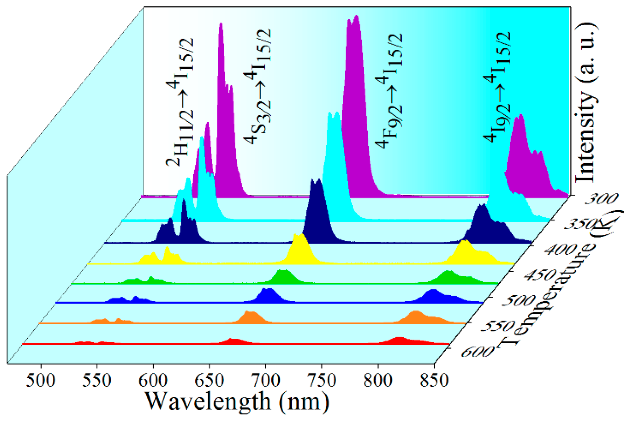
Figure 5.
Typical UC emission spectra of NaErF4 core–shell NCs in the temperature range 303–539 K, stimulated by 1530 nm laser (20 W/cm2).
Figure 5.
Typical UC emission spectra of NaErF4 core–shell NCs in the temperature range 303–539 K, stimulated by 1530 nm laser (20 W/cm2).

Figure 6.
The FIR as a function of temperature based on (a) I520/I540, (b) I806/I540, and (c) I806/I654. Fitting functions are also given; (d–f) the corresponding relative and absolute sensitivity by using various emission bands.
Figure 6.
The FIR as a function of temperature based on (a) I520/I540, (b) I806/I540, and (c) I806/I654. Fitting functions are also given; (d–f) the corresponding relative and absolute sensitivity by using various emission bands.


Table 1.
Lifetimes of NaErF4 core–shell NCs at various temperatures upon 980 or 1530 nm excitation.
| Excitation | Initial Level | Temperature-Dependent Lifetime (μs) | ||||
|---|---|---|---|---|---|---|
| 300 K | 350 K | 400 K | 450 K | 500 K | ||
| 980 nm | 4F9/2 | 130 | 130 | 130 | 140 | 140 |
| 4I9/2 | 230 | 230 | 250 | 260 | 260 | |
| 1530 nm | 4F9/2 | 140 | 130 | 140 | 140 | 140 |
| 4I9/2 | 170 | 170 | 180 | 180 | 180 | |
Table 2.
Comparisons of Sa and Sr based on FIR in different materials.
| Materials | Transitions | Range (K) | Sa-max (K−1) (maximum) | Sr-max (% K−1) | Ref. |
|---|---|---|---|---|---|
| LuVO4:Yb/Er | 2H11/2/4S3/2 | 303–423 | 0.0083 (123 K) | 1.14 | [44] |
| Y2O3:Er | 2H11/2/4S3/2 | 93–613 | 0.0044 (427K) | 0.98 | [45] |
| Gd2O3:Yb/Er | 2H11/2/4S3/2 | 298–573 | 0.0078 (528 K) | 1.16 | [46] |
| NaYF4:Yb/Er@SiO2 | 2H11/2/4S3/2 | 300–900 | N.A. | 1.02 | [47] |
| NaGdF4:Yb/Er | 2H11/2/4S3/2 | 303–363 | 0.0025 (360 K) | 1.12 | [48] |
| NaLuF4:Yb/Er | 4D7/2/4G9/2 | 303–523 | 0.0052 (303 K) | 0.43 | [49] |
| NaYF4:Yb/Er | 2H11/2/4S3/2 | 303–743 | 0.0044 (637 K) | 0.46 | [50] |
| NaErF4@NaGdF4 | 4I9/2/4S3/2 | 303–593 | 0.0149 (593 K) | 1.15 | This work |
| 2H11/2/4S3/2 | 0.0020 (544 K) | 0.48 | |||
| 4I9/2/4F9/2 | 0.0024 (303 K) | 0.59 |
© 2019 by the authors. Licensee MDPI, Basel, Switzerland. This article is an open access article distributed under the terms and conditions of the Creative Commons Attribution (CC BY) license (http://creativecommons.org/licenses/by/4.0/).
Share and Cite
MDPI and ACS Style
Lu, K.; Yi, Y.; Xu, L.; Sun, X.; Liu, L.; Li, H. Temperature-Independent Lifetime and Thermometer Operated in a Biological Window of Upconverting NaErF4 Nanocrystals. Nanomaterials 2020, 10, 24. https://doi.org/10.3390/nano10010024
AMA Style
Lu K, Yi Y, Xu L, Sun X, Liu L, Li H. Temperature-Independent Lifetime and Thermometer Operated in a Biological Window of Upconverting NaErF4 Nanocrystals. Nanomaterials. 2020; 10(1):24. https://doi.org/10.3390/nano10010024
Chicago/Turabian StyleLu, Kailei, Yingxin Yi, Li Xu, Xianhao Sun, Lu Liu, and Hanyang Li. 2020. "Temperature-Independent Lifetime and Thermometer Operated in a Biological Window of Upconverting NaErF4 Nanocrystals" Nanomaterials 10, no. 1: 24. https://doi.org/10.3390/nano10010024
Note that from the first issue of 2016, this journal uses article numbers instead of page numbers. See further details here.