
HIV Expression in Infected T Cell Clones

HIV Dynamics and Replication Program, Center for Cancer Research, National Cancer Institute, Frederick, MD 21702, USA
*
Author to whom correspondence should be addressed.
Viruses 2024, 16(1), 108; https://doi.org/10.3390/v16010108
Submission received: 13 December 2023
/
Revised: 4 January 2024
/
Accepted: 6 January 2024
/
Published: 11 January 2024
(This article belongs to the Special Issue Regulation of HIV-1 Transcription and Latency)
Abstract
:The principal barrier to an HIV-1 cure is the persistence of infected cells harboring replication-competent proviruses despite antiretroviral therapy (ART). HIV-1 transcriptional suppression, referred to as viral latency, is foremost among persistence determinants, as it allows infected cells to evade the cytopathic effects of virion production and killing by cytotoxic T lymphocytes (CTL) and other immune factors. HIV-1 persistence is also governed by cellular proliferation, an innate and essential capacity of CD4+ T cells that both sustains cell populations over time and enables a robust directed response to immunological threats. However, when HIV-1 infects CD4+ T cells, this capacity for proliferation can enable surreptitious HIV-1 propagation without the deleterious effects of viral gene expression in latently infected cells. Over time on ART, the HIV-1 reservoir is shaped by both persistence determinants, with selective forces most often favoring clonally expanded infected cell populations harboring transcriptionally quiescent proviruses. Moreover, if HIV latency is incomplete or sporadically reversed in clonal infected cell populations that are replenished faster than they are depleted, such populations could both persist indefinitely and contribute to low-level persistent viremia during ART and viremic rebound if treatment is withdrawn. In this review, select genetic, epigenetic, cellular, and immunological determinants of viral transcriptional suppression and clonal expansion of HIV-1 reservoir T cells, interdependencies among these determinants, and implications for HIV-1 persistence will be presented and discussed.
1. Introduction
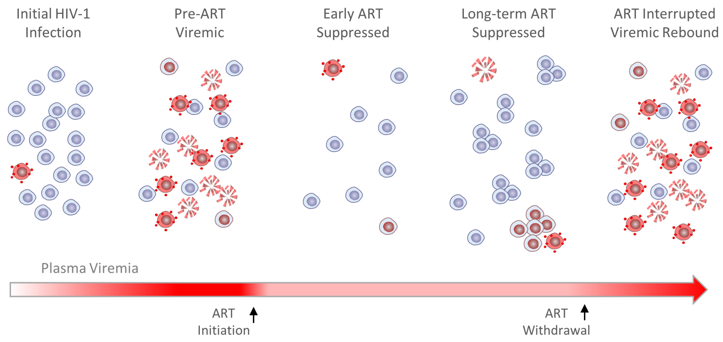
Modern ART virtually eliminates the clinical sequelae of HIV-1 infection and typically reduces plasma viremia to levels undetectable by standard clinical assays in people living with HIV (PLWH) [1,2,3]. Yet ART is not curative, as viremia typically returns to levels at or near the initial pre-ART viral set-point within a few weeks of treatment withdrawal, even after years or decades of uninterrupted treatment [4,5]. This viremic rebound originates from infected cells that harbor genetically intact proviruses capable of templating production of new infectious viral particles, referred to collectively as the HIV reservoir [6,7]. This reservoir is mostly comprised of infected CD4+ T cells [8,9]—long-lived master regulators of innate and adaptive immune responses. Infection of cells can be latent, i.e., viral RNA transcription from HIV-1 proviruses integrated into host genomic DNA in these cells is minimal, unproductive, or does not occur [6]. Silencing of viral transcription renders HIV infection minimally disruptive to normal cellular functions and permits evasion of the cytopathicity associated with viral protein production and CTL killing that would otherwise likely result in infected cell death [10,11,12,13,14]. However, the capacity to seed viremic rebound requires either that some cells in the HIV reservoir persist despite incomplete transcriptional suppression, that latency reversal occurs spontaneously and perhaps arbitrarily in some infected cells, or both.
The native and essential capacity of CD4+ T cells for homeostatic or antigen-driven clonal expansion likewise contributes to HIV-1 persistence when manifest in infected cells [15]. Infected T cell proliferation is also sometimes influenced by HIV-1 integration into select human genes [16]. When these cells are latently infected, their proliferation enables expansion of provirus populations while avoiding the deleterious effects of viral gene expression. Moreover, clonal expansion mitigates the risks of sporadic latency reversal, such as may be required for viremic rebound; i.e., if a clonal population of infected cells is replenished or expands more rapidly than it is attritted by spontaneous provirus activation, it will persist indefinitely. Together, HIV-1 transcriptional suppression and infected cell clonal expansion are the two principal determinants of HIV-1 persistence, the primary barrier to a cure for HIV-1 infection. Regulation of and interplay between these two determinants, with emphasis on epigenetic control and in the context of normal CD4+ T cell function, are the topics of this review.
2. The HIV-1 Reservoir Is Shaped by Viral Transcriptional Suppression and Clonal Expansion
Administration of ART has profound and lasting effects on HIV-1 population dynamics. Often originating from a single founder virus [17], prolific HIV replication spreads the infection quickly and systemically, thus rapidly expanding the size of the virus population. This unchecked expansion is reflected in extreme viremia during acute infection prior to ART administration, with 107 HIV-1 RNA copies per mL of plasma not uncommon [18,19]. Genetic diversity in the virus population expands rapidly during infection, owing primarily to error-prone reverse transcription [20] coupled with high rates of viral recombination and turnover [21,22,23]. Together, these HIV-1 population features early in infection enable facile selection of adaptive forms that evade selective pressures, potentially including those that result in attenuated viral gene expression. Additionally, prior to ART administration, T cells typically harbor genetically intact proviruses that are likely actively transcribed, generating alternatively spliced or unspliced transcripts that are translated into viral gene products or packaged into nascent virions, respectively. Due to the deleterious consequences of viral protein production, T cells infect pre-ART turnover rapidly, with an estimated half-life of 1–2 days [22,23]. Yet even in the absence of ART, infectious proviruses in some cells are transcriptionally suppressed, and recent evidence indicates that the HIV-1 reservoir population after years or decades on ART is descended from these latently infected cells [24,25].
Administration of effective ART rapidly and systemically halts ongoing virus replication [26,27,28,29,30,31]. However, since neither viral RNA transcription nor translation are among the molecular targets of modern ART, some infected cells capable of producing virions continue to do so even after ART is administered and most die rapidly as a result. These conditions produce marked changes in the dynamics of HIV-1 RNA and DNA populations upon ART initiation. Using the single-copy assay (SCA) [32], viral RNA was shown to decay by approximately 4-logs on ART to an average of 0.6 copies/mL of blood plasma in three phases, attributable to the clearance of infected cells of different types and subsets [33,34,35]. A later study provided evidence for a fourth phase of viral decay on very long-term ART, although the slope appeared to be attributable to only a subset of individuals in the study [36].
More recently, decay rates of defective and predicted-intact viral DNA in peripheral blood mononuclear cells (PBMCs) were dissected in parallel using the Intact Proviral DNA Assay (IPDA [37]) and related approaches [38]. Peripheral blood HIV-1 DNA levels, a reflection of the number of infected cells, also decreased on ART, but more slowly, and with kinetics dependent upon whether proviruses harbored by infected CD4+ T cells were intact or defective. In the specified recent study [38], decay of defective proviral populations had estimated half-lives on the order of several months to a few years, in agreement with prior findings [39,40]. Though not a source of rebound viremia if treatment is withdrawn, persistence of defective proviruses may still lead to CTL recognition and killing [41,42,43], contributing to a state of chronic immune activation often observed in otherwise well-controlled infections [44,45]. In contrast, decay of predicted-intact proviruses on ART was found to be biphasic, with average measured half-lives of 12.9 days and 19.0 months, respectively [38]. Seeming correlation between the second phase of plasma RNA decay and the first phase of viral DNA decay suggests that both may be the result of virus-producing infected T cells in the peripheral blood dying without replacement. Others report that the reservoir decays even more slowly (t1/2 = 44 months [46,47,48]) or not at all [49], as forces leading to loss of viral DNA may be almost entirely countered by infected T cell proliferation, increasing their apparent half-life and thereby reducing or preventing clinically meaningful reservoir decay [49].
Taken together, these findings provide important context for the composition of the HIV-1 reservoir upon ART initiation. It is now widely accepted that, in the absence of ongoing rounds of viral replication, HIV-1 population changes are limited almost entirely to dropout of infected cells and skewed by variable rates of cell survival and clonal expansion. Generally, viral DNA populations in PLWH on long-term ART are increasingly dominated by defective proviruses (95–98%) [50,51], and the relatively few persistent intact proviruses are mostly latent [52,53,54] and frequently found in clonally expanded populations [55,56,57]. These viral DNA population characteristics are reflected in plasma viremia typically being reduced to undetectable levels by standard clinical assays (<50 copies HIV RNA/mL). Yet low-level viremia can usually be detected in PLWH on ART using more sensitive laboratory tests [58], and clinically detectable, non-suppressible viremia persists for long periods in some individuals [59]. Moreover, low levels of unspliced intracellular HIV-1 RNA have been detected in variable fractions of clonal infected cell populations across T cell subsets and harboring intact or defective proviruses [60], indicating that at the population level, viral transcriptional suppression is not always complete.
HIV-1 population dynamics in PLWH on long-term ART are ultimately determined by collective viral gene expression, which is itself governed by layers of viral and host genetic, epigenetic, cellular, and immunological determinants, and overarchingly by natural selection; i.e., T cells harboring genetically intact but latent proviruses tend to persist despite ART, while those harboring transcriptionally active intact proviruses typically do not. However, to support viremic rebound upon ART cessation, there must be at least a few cells hidden among latently infected HIV-1 reservoir populations either capable of spontaneous emergence from HIV-1 transcriptional dormancy or within which viral transcription, gene expression, and infectious virion production are not completely silenced—scenarios evidenced by residual low-level viremia and detection of unspliced intracellular RNA [43,51,60]. Escape from this apparent paradox—that some infected cell lineages persist indefinitely despite being capable of producing infectious virions detrimental to their survival—is enabled, in part, by the capacity of HIV-infected cells to clonally expand their populations. More specifically, it is easy to envisage a heterogeneous clonal population of infected T cells wherein, at any given time, most proviruses are latent while a few actively produce virus, the latter group mostly consigned to death by CTL-killing but capable of sparking viremic rebound with cessation of ART. These groups may be distinguishable by epigenetic markers, activation states, or states of T cell differentiation, the determinants of which might be useful therapeutic targets, or perhaps only by the spontaneous emergence of infrequent, stochastically governed intracellular conditions that favor latency reversal. Regardless, identifying the implicated infected cell populations, understanding the bases for their persistence, clonal expansion, and adaptive transcriptional profiles, and how to counter them, are key to curing HIV-1 infection.
3. Viral Genetic Determinants of HIV-1 RNA Transcription
A defining feature of the retrovirus lifecycle is reverse transcription of the viral RNA genome into double-stranded DNA, which is then integrated into the host cell genome [61]. HIV-1 favors integration into actively transcribed genes with an accessible chromatin structure, but this preference is far from absolute [62,63]. Consequently, sites of integration in infected CD4+ T cell populations are neither random nor predetermined and are widely distributed. Once strand discontinuities created by the integration process are repaired by host DNA repair machinery, HIV-1 DNA becomes contiguous with and practically indistinguishable from the host cell genome. Accumulated reverse transcription errors and integration site variability are the largest sources of genetic variation in reservoir proviruses with time on ART, with both potentially affecting viral genetic determinants and the cellular genetic and epigenetic contexts of viral gene expression.
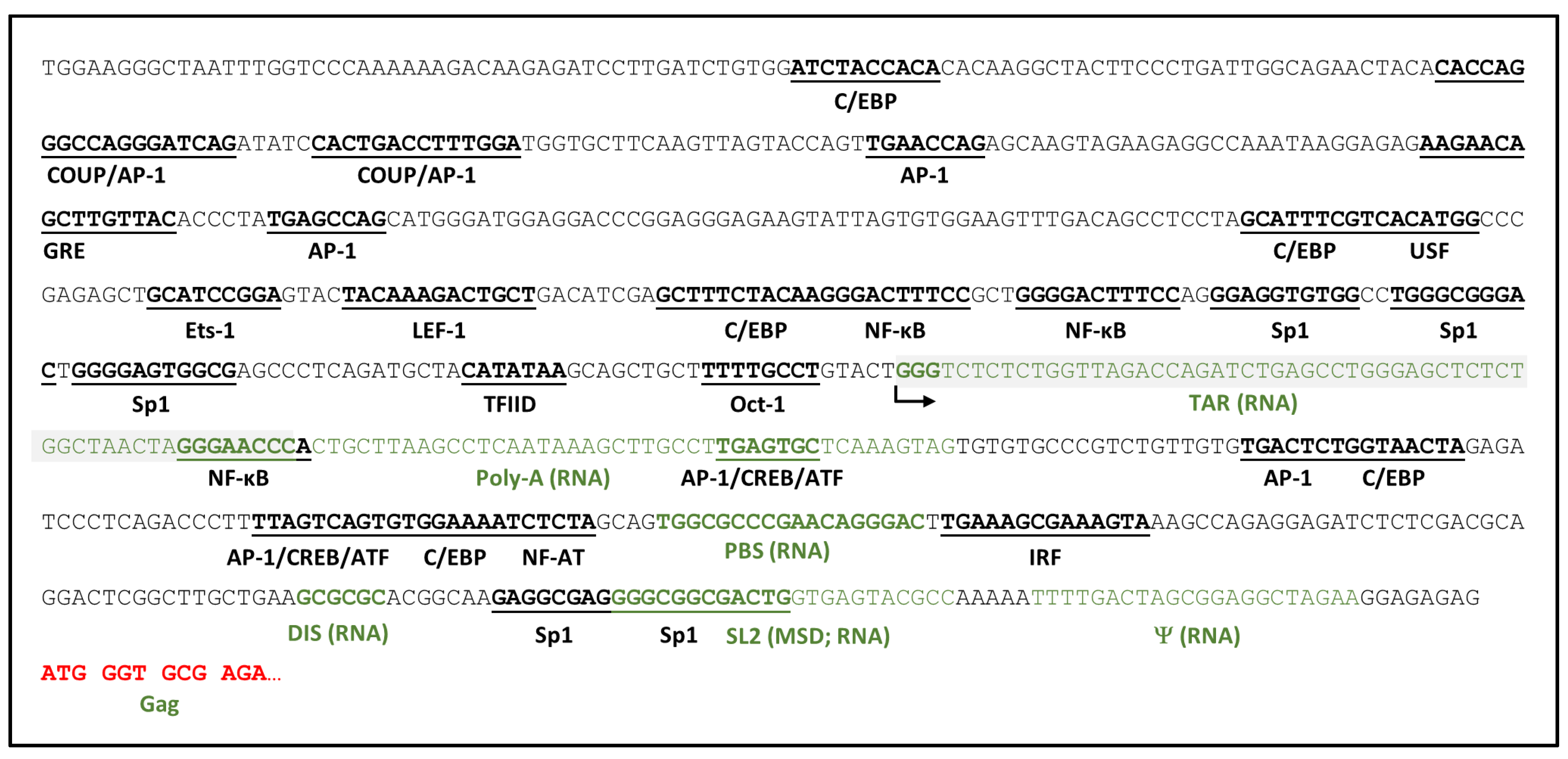
HIV-1 genetic elements directly encoded by the provirus and recognized by cellular and viral transcriptional machinery and regulatory proteins collectively constitute the most basic regulatory level of viral gene expression and are summarized schematically in Figure 1. HIV-1 transcription is catalyzed by the cellular enzyme, RNA polymerase II (RNAPII), initiating from the 5′ long terminal repeat (5′LTR) of the provirus, and is dependent upon host cell and viral transcription factors binding to an array of DNA regulatory elements in the 5′LTR promoter [64,65]. The promoter region can be subdivided into four functional domains: (i) a basal core promoter encompassing the transcription initiation site, a CA/TATAA element, and three Sp1 binding sites; (ii) an upstream enhancer region containing two adjacent binding sites for the inducible transcriptional activator nuclear factor kappa B (NF-κB); (iii) an upstream regulatory region involved in cell type-specific expression, and (iv) a downstream regulatory region containing additional secondary enhancer elements and the nidus for assembly of an unstable nucleosome. The LTR promoter also harbors the trans-activation response (TAR) element immediately downstream of the transcription initiation site. Consequent to this positioning, the 5′ termini of all HIV-1 transcripts contain the RNA version of the TAR element [66], which assumes a stable stem-loop structure recognized by the virus-encoded protein Tat via an arginine-rich motif, forming a complex that enables transition from distributive synthesis of short viral RNAs to efficient synthesis of genome-length viral transcripts [67].
The basic mechanism of Tat trans-activation is through Tat-TAR complex recruitment of positive transcription elongation factor b (pTEFb), a general RNAP II elongation factor comprised of cyclin-dependent kinase 9 (CDK9) and Cyclin T1, into proximity of the RNAP II transcription initiation complex [69]. The CDK9 catalytic component of pTEFb mediates phosphorylation of the carboxyl terminal domain of RNAP II, thus activating its capacity for synthesis of long transcripts. This functional transition is further regulated by binding/dissociation of other cellular proteins and protein complexes to the Tat-TAR and RNAP II transcription complexes, CDK9-mediated phosphorylation of these elements, and phosphorylation or acetylation of Tat itself. Relative activity and availability of many of these components in resting versus activated CD4+ T cells, including and especially Tat, CDK9, and Cyclin T1, are important contributors to the propensities toward latency or active viral transcription in the respective T cell states [70,71,72].
Together with TAR, the core promoter elements are sufficient both to promote basal levels of RNAPII-mediated HIV-1 transcription and for Tat-mediated trans-activation. The rate-limiting step in this process is often formation of the transcription preinitiation complex [73,74], i.e., binding of the CA/TATAA element by the general transcription factor TFIID, a large multiprotein complex comprised of the TATA binding protein (TBP) and several associated factors (TAF) [75]. The initiator element downstream of CA/TATAA determines the transcription start site (TSS) and is comprised of two sequence segments between nucleotide positions −6 and +30 [76,77,78,79]. These HIV-1 elements cannot easily be substituted in genetic experiments, suggesting that they have specifically co-evolved to promote viral transcription in cooperation with Tat trans-activation [80,81]. A GC-rich segment immediately upstream of the CA/TATAA element contains three binding sites for Sp1, a ubiquitous zinc finger transcription factor that, in complex with TFIID and Tat, activates processive basal HIV-1 transcription [82,83]. Other host transcription factors (BTEB, Sp3, and Sp4) can bind the Sp1 sites with differing effects [84,85], but only Sp1 coordinates with NF-κB in modulating viral transcription. Lastly, though it resides within the core promoter region, the Oct-1 site antagonizes HIV-1 transcription. Located between the CA/TATAA and initiator elements, Oct-1 transcription factor binding, alone or in conjunction with YY-1, has been shown to repress both basal and Tat-activated viral transcription [86,87,88,89], with a potential role in mediating HIV-1 quiescence in resting T cells [86,90].
The enhancer element is comprised of tandem adjacent NF-κB binding sites immediately upstream of the Sp1 binding sites that modulate basal viral RNA transcription in response to intracellular signaling. Members of the NF-κB family (e.g., NF-κB1, RelA, c-Rel, NF-κB2, and RelB) bind as dimers to this HIV-1 enhancer and select combinations of subunits favor Tat trans-activation [91]. Though not essential for HIV-1 replication, NF-κB binding to the viral enhancer element has been reported to specifically increase the expression of HIV-1 in macrophages and CD4+ T cells [92,93,94,95] activated by proinflammatory cytokines, including TNFα, IL-1β, and IL-6 [92,93,95,96,97,98,99,100,101], anti-inflammatory cytokines such as TGF-β and IL-10 [102], mitogenic stimuli (e.g., phorbol esters [103]), or mycobacterial invasion [104]. Tat itself can also activate NF-κB to further accelerate and sustain virus transcription [105,106,107,108]. The HIV-1 enhancer can bind other transcription factors as well, including Ets-1 [108] and E2F-1 [109], to inhibit or augment the stimulatory activity of NF-κB [110].
Direct binding of transcription factors C/EBP, AP-1, Ets-1, LEF-1, COUP, and NFAT to binding sites in the regulatory region upstream of the enhancer can also up- or down-regulate HIV-1 gene expression in a cell-type-specific fashion [65,111,112,113,114]. Likewise, additional constitutive (Sp1) and inducible transcription factor binding sites (for AP-1, NF-κB, IRF, and NFAT) have been identified in the regulatory region downstream from the transcription start site (TSS), of which several have been shown to be required for efficient HIV-1 replication in T cells [115,116,117,118,119]. AP-1 sites are specifically responsive to T cell activation signals of the cAMP-dependent protein kinase A (PKA) pathway through binding of CREB/ATF proteins [115,117].
Though not comprehensive, this summary illustrates the complexity of viral genetic elements within the 5′LTR and DNA encoding the 5′UTR that contribute to HIV-1 transcriptional regulation even before considering the epigenetic and cellular context. It is not difficult to envision how a provirus containing a few mutations in these regions might be capable of templating synthesis of nascent virions yet sufficiently attenuated to escape self-destruction by cytopathic gene expression or immune killing. Accordingly, as sophisticated, high-throughput methods for sequencing the proviruses comprising the HIV-1 reservoir in donor samples become more widely utilized, close examination of sequence variation in segments of the viral genome capable of directly influencing viral gene expression should be emphasized.
4. Epigenetic Determinants of HIV RNA Transcription
Unlike assembly of DNA-bound transcription factors into an RNAP II initiation complex, epigenetic regulation of HIV-1 gene expression is often indirect and involves changing the macromolecular structural environment to favor or disfavor transcription initiation and elongation. Human genomic DNA—including integrated proviruses—is organized into a macromolecular nucleoprotein complex called chromatin, the functional repeating unit of which is the nucleosome. Nucleosomes are comprised of 146 base pairs (bp) of DNA wrapped around an octamer of histone proteins—two each of H2A, H2B, H3, and H4—and are separated by 10–80 bp segments of linker DNA stabilized by histone H1. Strings of linked nucleosomes form chromatin fibers, segments of which can assemble into highly compacted heterochromatin or less compacted euchromatin regions that are generally considered to be restrictive or permissive to gene expression, respectively.
Heterochromatin can be further classified as constitutive or facultative, with each class having important structural and functional distinctions [120]. Constitutive heterochromatin is a highly dense and persistent form of chromatin that exists across cell types and is generally non-permissive toward gene expression. It is comprised primarily of highly repetitive, mostly non-genic sequences localized to centromeres, telomeres, and large segments of chromosomes 1, 9, 16, 19, and Y. Constitutive heterochromatin is thought to be more important for regulating nuclear structure and gene spacing than for storing genetic information and is distinguishable by epigenetic characteristics such as frequent cytosine (CpG) methylation, histone hypoacetylation, and histone H3K9 trimethylation (see below), which provides H3K9me3 sites for heterochromatin protein 1 (HP1) binding. In contrast, though also compact and generally not amenable to RNA transcription, facultative heterochromatin is much more responsive to epigenetic signaling and may be dynamically converted to euchromatin during development, differentiation, or as a natural part of adaptive cellular regulation. Enrichment for trimethylated H3K27 (H3K27me3) is characteristic of facultative heterochromatin.
Although HIV-1 integration favors transcriptionally active regions of the human genome, proviruses have been found integrated into heterochromatic regions, though it remains to be determined how the initial chromatin-free state of the pre-integrative HIV DNA intermediate may affect adjacent chromatin structure, and vice versa. However, as will be described, direct and indirect evidence suggest that both genetic elements within the provirus and the chromatin environment into which it is integrated are important determinants of HIV-1 gene expression and latency. Numerous epigenetic factors contributing to governance of the chromatin environment within and proximal to the integrated HIV-1 provirus have been identified, many of which are discussed below and highlighted in Figure 2.
Nucleosome positioning on the HIV-1 provirus is influenced by DNA sequence, DNA binding proteins, and the activity of chromatin remodelers [121]. From studies on HIV-1 infection in cell culture, it was first determined that three nucleosomes (nuc-0, nuc-1, and nuc-2) assemble at specific positions in transcriptionally inactive proviruses, irrespective of their sites of integration [122,123]. One of these, nuc-1, is deposited immediately downstream of the HIV-1 TSS and thus sterically impedes and is highly repressive of viral transcription. Assembly of nuc-1 is mediated by the BAF chromatin remodeling complex, which is also known as SWI/SWF and is recruited to the LTR by BRD4S despite the DNA sequence in this region of the HIV-1 LTR being predicted to be refractory to nucleosome deposition. Conversely, PBAF—a member of the same family of chromatin remodelers—is required for displacement of nuc-1 and activation of viral transcription. Tat has been reported to play a role in recruitment of PBAF to the 5′LTR [124,125], although PBAF-mediated nuc-1 displacement can also occur through a Tat-independent mechanism [126]. BAF/PBAF involvement in nuc-1 establishment is also linked with integration, as the INI-1 subunit of the BAF complex is known to interact with HIV-1 integrase and perhaps also histone chaperone Spt6 [127,128]. For the same reasons, the NuRD/Mi-2/CHD family of chromatin remodelers has also been linked to HIV latency, as have HIRA (Histone Cell Cycle Regulator) and FACT (Facilitates Chromatin Transcription) complexes [129,130].
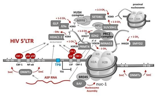
![Viruses 16 00108 g002]()
Figure 2.
Epigenetic suppression of HIV-1 RNA transcription. As described in the text, epigenetic host regulatory machinery can contribute to transcriptional suppression of the HIV-1 provirus in numerous ways. Epigenetic suppressive mechanisms generally involve direct or indirect recruitment of histone-modifying enzymes through sequence-specific binding of host proteins or lncRNAs to promoter or regulatory DNA sequences. Though each of the depicted suppressive mechanisms has been demonstrated in in vitro model systems, their relative importance in maintaining latency in infected CD4+ T cells in PLWH remains to be determined. Adapted from [131].

Nucleosome composition and structure, as determined by post-translational modification of constituent histones, is considered a central player in transcriptional regulation in all human cell types as well as in HIV-1 gene expression in infected CD4+ T cells. Histone amino-terminal tails are disordered, protrude from the nucleosome core [132], and are comprised largely of basic amino acids, the sidechains of which may be acetylated, methylated, phosphorylated, ubiquitinated, or sumoylated by different classes of enzymes. These post-translational modifications can affect local chromatin structure, including the degree to which it is compacted, thus regulating promoter accessibility to transcriptional machinery. Modified chromatin can also serve as a scaffold for recruitment of bifunctional effector proteins with bromo-, chromo-, or PHD domains that each can selectively bind specific post-translational modifications [133]. The governing principles that determine how variations in localized collections of post-translational histone modifications affect RNA transcription in different cellular contexts have suitably been referred to as the “histone code” [134].
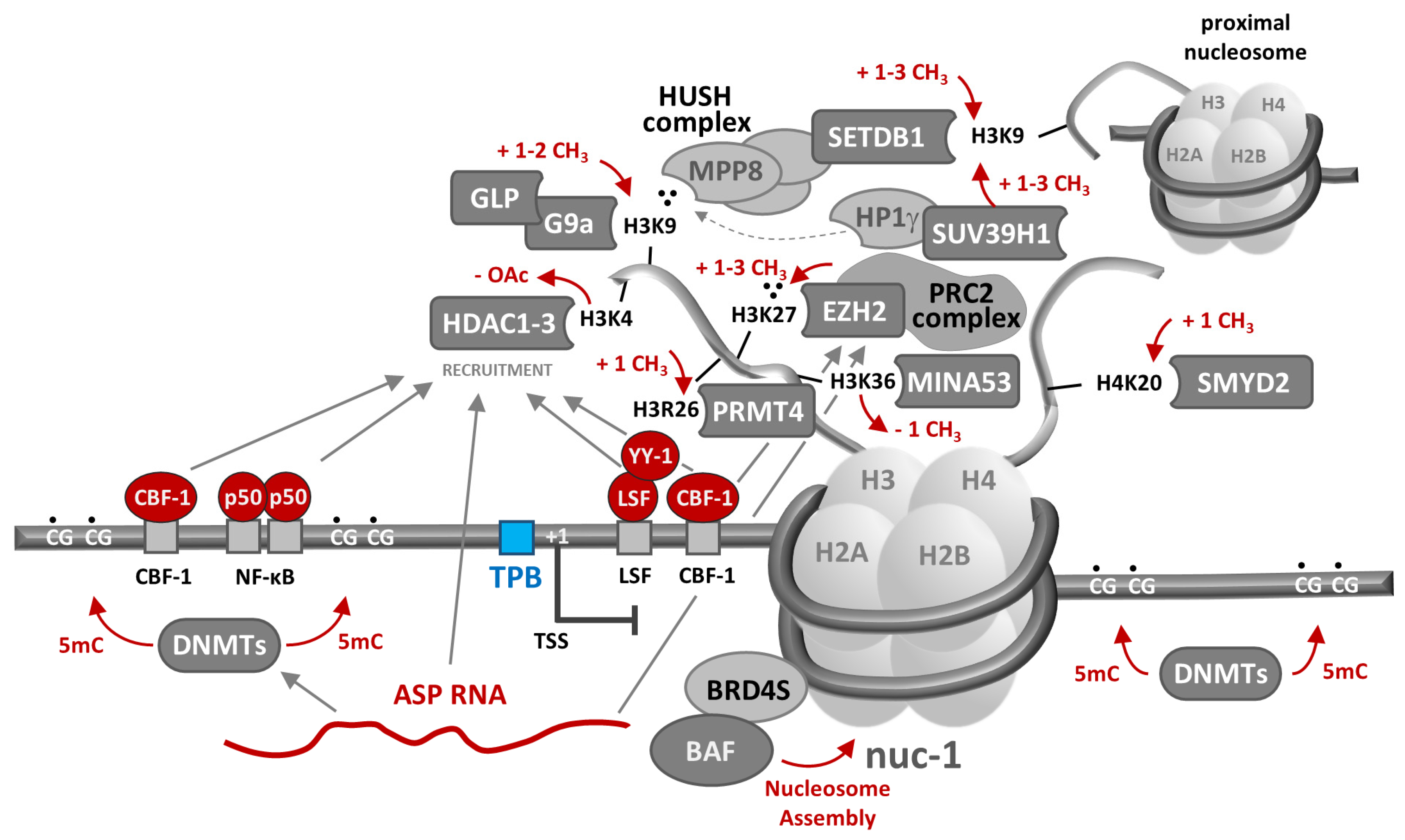
At least 13 of 70 histone modifiers across 12 human gene families from four classes of enzymes have been specifically implicated in HIV-1 latency [135,136,137,138,139,140,141,142,143]. Histone deacetylases (HDACs) erase the acetylation of lysine ε-amino groups on multiple proteins, including histones, with the effects on the latter target generally considered to be repressive to gene expression [144]. HDAC1, HDAC2, and HDAC3 are class I HDACs shown to be recruited to the HIV-1 5′ LTR during viral latency [89,145,146,147,148] by several transcription factors, including the inhibitory p50-p50 NF-κB homodimer [148], YY-1 and LSF [89,149], and CBF-1 [147]. Once brought into proximity, it has been postulated that these HDACs suppress transcription through deacetylation of lysine 9 of histone 3 (H3K4) in LTR-associated histones. In contrast, SIRT1, a class III sirtuin HDAC, mediates HIV-1 transcriptional control by regulating Tat recycling and transactivation feedback [150].
Histone acetyltransferases (KATs broadly, HATs more specifically) catalyze acetylation of lysine side chains, an activity that, with respect to histones, is generally associated with promoting an open chromatin structure and a favorable transcriptional environment, and thus antagonism of HIV-1 latency. However, contrary to this tendency, lysine acetyl transferase 5 (KAT5/TIP60) has been reported to suppress HIV-1 transcription by acetylating H4K20 on provirus-associated nucleosomes, which in turn recruits bromodomain-containing BRD4 to block elongation of HIV-1 transcription [151].
Possible patterns for histone methylation are numerous and complex, even when limited to those demonstrated to play a role in HIV-1 transcriptional suppression. Two classes of histone methyltransferases (HMT) catalyze mono-, di-, or tri-methylation of basic histone side chains: histone lysine methyl transferases (HKMTs), which can methylate histone tail lysine side chains H3K4, H3K9, H3K27, H3K36, H3K79, and/or H4K20, and protein arginine methyl transferases (PRMTs), which can methylate arginines H3R2, H3R8, H3R17, H3R26, and/or H4R3 [152]. HMT EZH2 is an HKMT, is part of the Polycomb Repressive Complex 2 (PRC2), and catalyzes trimethylation of H3K27, which is associated with heterochromatin and promotes transcriptional suppression [153]. In cell lines and primary cell models of HIV-1 latency, HMT EZH2 and its reaction product (H3K27me3) have been found colocalized to nucleosomes proximal to the viral promoter [139]. Euchromatin HMTs EHMT1/GLP and EHMT2/G9a have likewise been shown to colocalize with their H3K9me2 catalytic products on the HIV-1 promoter in latently infected T cell lines [137,140,141]. These methyltransferases can both be recruited to the 5′LTR by CBF-1, together with HDAC1 and HDAC3 [154], suggesting that viral transcriptional suppression by histone methylation and deacetylation may be coordinated. H3K9 trimethylation can also be spread to or from provirus-proximal nucleosomes by the Human Silencing Hub (HUSH) complex, which is composed of the three subunits TASOR, MPP8, and periphilin [136,142]. H3K9me3 is “read” by the chromodomain-containing MPP8 subunit, which recruits the HKMT “writer” SETDB1 to tri-methylate H3K9 in proximal nucleosomes. Chromodomain-containing HP1g and SUV39H1 bind and spread H3K9me3 across provirus-associated nucleosomes in a similar fashion [138]. SMYD2 mono-methylates H4K20 on the 5′LTR, potentially leading to recruitment of PRC1 and chromatin compaction [135], while CARM1/PRMT4 catalyzes H3R26 arginine methylation to mediate HIV-1 transcriptional suppression—the only PRMT so-implicated to date [143].
Histone demethylases (KDMs) remove methyl groups from histone tail basic residues, countering the activities of HMTs. As with histone acetylation catalyzed by KATs and HATs, histone demethylation is usually associated with an open chromatin structure favoring RNA transcription, although one notable exception has been reported with respect to HIV-1 [155]. MINA53, a KDM, was shown to demethylate H3K36me3 at the HIV-1 5′LTR. This action inhibits binding of KAT8, which specifically recognizes H3K36me3 and catalyzes acetylation of K16 in proximal H4 histones, thereby inducing a local chromatin structural change that favors RNA transcription. Hence, in contradistinction to other enzyme family members, the indirect effect of MINA53 demethylase activity is to promote latency by inhibiting a change in chromatin structure that would favor HIV-1 RNA transcription.
Epigenetic transcriptional suppression may also be achieved by direct methylation of deoxy-cytosines (C→5 mC) in clusters of CpG dinucleotides, called CpG islands (CGI), proximal to cellular promoters [156,157]. There are two such CGIs in HIV-1 promoter and adjacent 5′ non-coding regions of the provirus, respectively, methylation of which has been associated with suppression of viral transcription through recruitment or exclusion of transcription factors and histone modifiers. For example, in a latently infected T cell line model, DNA hypermethylation recruits the methyl-binding protein MBD2, a component of the chromatin remodeling complex NuRD that also contains the histone deacetylase HDAC2 [158]. DNA methylation states of cellular CGIs are established and maintained by DNMT3a/DNMT3b and DNMT1 DNA methyltransferases, respectively [159], though their specific roles in regulating HIV-1 DNA methylation and RNA transcription remain unclear. Further investigation is required to assess the importance of CpG methylation in establishing and maintaining latent infection in PLWH, as data on the topic collected from donor samples are mixed and highly condition-specific [51,158,160,161,162,163]. Nevertheless, the evolutionary retention of CpG islands in transcriptional regulatory regions of the HIV-1 genome suggests that they and their susceptibility to methylation contribute significantly to viral fitness.
Piwi-interacting (piRNAs), long non-coding (lncRNAs), and micro-RNAs (miRNAs) add an additional layer of complexity to HIV-1 transcriptional regulation. Although pi-RNAs were originally thought to be expressed only in the germline where they mediate suppression of transposon activity [164,165], there is now evidence that they, together with the PIWIL proteins involved in their synthesis, are expressed in somatic cells [166]. Together with PIWIL4, piRNAs regulate homeostatic and tumorigenic patterns of gene expression [167] and promote HIV-1 promoter heterochromatinization through the recruitment of SETDB1, HP1, and HDAC4 [168]. Two lncRNAs, NRON and NKILA, indirectly restrict HIV-1 gene expression by inducing proteasomal degradation of Tat [169] or by interfering with the NF-κB signaling pathway [170], respectively. In contrast, MALAT1 and HEAL lncRNAs support HIV-1 transcriptional activity, the former by sequestering the EZH2 subunit and inhibiting the HMT activity of PRC2, and the latter by recruiting HATs to the HIV-1 promoter [171,172,173]. ASP RNA (also called AST) is an HIV-1 antisense transcript that, in cell culture studies, has also been shown to possess lncRNA-like activity, suppressing viral transcription through recruitment of PRC2, EZH2, HDAC1, and DNMT3a, thus indirectly inducing H3K27 trimethylation and nuc-1 assembly [174]. Additional studies to establish the importance of ASP RNA as a mediator of latency in PLWH are underway. Finally, five cellular miRNAs (miR-28, miR-125b, miR-150, miR-223, and miR-382) have been shown to mediate transcriptional suppression of HIV-1 by directly targeting the 3′ ends of HIV-1 mRNAs for degradation [175], and two others (miR-17-5p and miR-20a) by indirectly reducing levels of acetylated Tat [176].
5. HIV-1 Transcriptional Suppression and Clonal Expansion in a CD4+ T Cell Functional Context
As regulators of immune responses to diverse and uncertain immunological threats, CD4+ T cells arguably require greater functional plasticity than any other human cell type. This adaptability is manifest in the capacity of these cells to assume any of numerous states of differentiation or effector functionalities in response to immune stimuli, mediated by an exquisitely complex regulatory network of signal transduction. Extracellular environmental signals are transmitted through cell-to-cell contact, cytokine–receptor binding, and antigen recognition complexes to cytoplasmic intermediaries and into the nucleus, where they culminate in transcriptional and epigenetic changes that can persist or be dynamically realigned to fit changing immune conditions. In HIV-1-infected CD4+ T cells in PLWH on ART, the long-term fate of a provirus is inextricably linked to and sometimes dominated by this natural cellular milieu. Accordingly, it is important to consider both viral transcriptional suppression and host cell clonal expansion, the primary determinants of viral persistence, in the context of natural CD4+ T cell phenotypic and regulatory dynamics.
In the thymus early in life and bone marrow later, T cell precursors (thymocytes) differentiate into mature CD4+ T cells expressing unique recombinant T cell receptors (TCRs) on their surfaces. Analogous to antibodies, TCRs recognize specific antigens presented on class II major histocompatibility complexes (MHC II) by antigen-presenting cells (APCs) and must survive positive and negative selection processes to ensure levels of self-antigen recognition sufficient to support homeostatic proliferation yet incapable of precipitating an autoimmune response [177]. Upon maturation, CD4+ T cells are designated naïve because they have yet to encounter sufficient cognate antigen to trigger their activation. Mature naïve CD4+ T cells are released into the circulation, where they migrate via CCR7 homing to secondary lymphoid tissues such as lymph nodes, the tonsils, the spleen, and in various mucous membrane layers in the body, including the gut [178].
Naïve CD4+ T cells are activated when TCR and CD4 co-receptors recognize their cognate antigen in an APC MHC II complex. This activation results in a signaling cascade that drives rapid clonal proliferation and differentiation of activated CD4+ T cells into one of several classes of effector cells depending on the local cytokine milieu, type of APC, concentration of antigen, and co-stimulatory molecules [179,180,181,182]. This process, also called priming to distinguish it from the more rapid secondary activation of memory cells, occurs over 1–2 days and alters the cellular transcription program to endow the T cells with specialized effector functions and a robust proliferative capacity [183]. Polarized CD4+ cell subsets with distinct proliferative capacities and effector functions include T-helper 1 (Th1), T-helper 2 (Th2), T-helper 17 (Th17), follicular helper T cell (TFH), induced T-regulatory cells (iTreg), and the regulatory type 1 cells (Tr1), and potentially distinct classification T-helper 9 (Th9) [181].
Once an immune threat has abated and foreign antigen stimulation subsides, activated CD4+ T cells undergo a contraction phase variably reported to last 1–4 weeks [184,185]. This process is characterized by cessation of proliferation and loss of 90–95% of activated cells to apoptotic or non-apoptotic cell death [184,186,187]; however, a small fraction of these cells survive to become long-lived CD4+ memory T cells capable of responding to subsequent immune challenges more rapidly and robustly than during the primary response. Memory cell subsets include stem cell memory (TSCM), central memory (TCM), transitional memory (TTM), effector memory (TEM), tissue-resident memory (TRM), and recirculating memory (TRCM), and are distinguishable by their relative proliferative potential, capacity for specialized effector functions in a secondary response, surface markers, transcriptional profiles, and tissue residence. Resting memory CD4+ T cell populations can be maintained or restored by intermittent, antigen-independent homeostatic proliferation. Homeostatic proliferation requires both IL-7 and IL-15 and is accelerated by interaction with self-antigen presented on APCs, though self-antigen engagement is not essential [188].
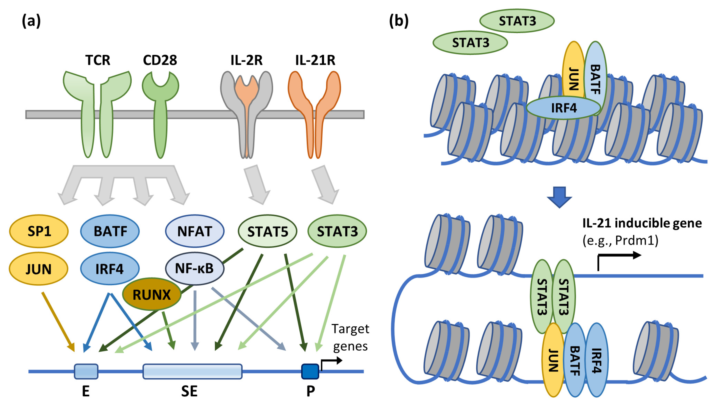
CD4+ T cell lineages are inherently plastic and capable of frequent interconversion among memory and effector CD4+ T cell phenotypes, including capacity for activation-driven clonal expansion [189,190,191]. Both phenotypic maintenance and interconversion are driven by antigen and cytokine environments and manifested through engagement of cell surface receptors and downstream signaling cascades, culminating in institution of specialized, phenotype-specific transcriptional programs and chromatin structural changes [192]. There are numerous examples of how effector cell lineage fidelity and memory cell readiness for secondary immune response are durably altered through epigenetic changes [193,194,195,196,197,198,199], and here we relate both a general mechanism and a specific example of how such signal transduction can activate expression of select CD4+ T cell effector genes, either immediately through engagement of proximal enhancers or super-enhancers or more durably by altering chromatin structure (Figure 3) [200]. Notably, CD4+ T cell phenotypes, including capacity for antigen-driven clonal expansion, are regulated by many of the same factors and mechanisms shown to modulate HIV-1 gene expression, as described in previous sections of this review (e.g., SP1, AP-1, BATF, NF-κB, NFAT, and IRF4). This mechanistic overlap, together with chromatin structure around the provirus, can potentially contribute to conflict or synergy between the two primary determinants of HIV persistence in context-specific fashion.
The capacity of CD4+ T cells to transition between permissive and suppressive transcriptional environments serves as the basis for a fundamental model of HIV-1 latency in which cellular and provirus activation states are linked [201]. In accordance with this paradigm, HIV preferentially infects activated CD4+ T cells [202,203], the intracellular environment of which also supports active virus replication that usually results in rapid infected cell turnover. However, when the immune threat that precipitated T cell activation recedes, a few infected cells can survive both contraction and the potentially deleterious effects of HIV-1 infection and persist as infected resting memory cells with transcriptionally suppressive environments that favor viral latency. The relative abundance of cellular transcription factors that enhance viral gene expression (e.g., NF-κB, NFAT, and pTEFb) in activated and resting CD4+ T cells also supports this paradigm [92,204,205,206,207,208,209,210], as does the consistent finding that latently infected cells are most commonly resting memory T cells [8,9,52,53,54], even though these cells are relatively resistant to infection. More precisely, among CD4+ T cell memory subtypes, most infected cells in PLWH on ART are TSCM and TCM [8,211,212], which have the greatest proliferative capacities, while sequence-intact and potentially replication-competent proviruses are more often found in TEM cells [213,214].
Yet the activation states of infected CD4+ memory T cells and the HIV-1 proviruses they harbor are not always correlated [215], and the mechanistic basis for this functional uncoupling remains incompletely understood. Perhaps the most notable example of this disjunction in donor samples is derived from analysis of a very large CD4+ T cell clone harboring a replication-competent provirus [216]. Despite chronic exposure to cognate antigen sufficient to keep the clonal population in a chronic state of activation, only 2.3% of these infected cells were found to express unspliced HIV-1 RNA [60], indicative of uncoupling between cellular and provirus activation states. This notion is further supported by a subsequent study reporting additional instances of extensive antigen-driven expansion of infected cell clones without corresponding high levels of latency reversal [217]. Moreover, in a longitudinal analysis of samples from PLWH on ART, intact HIV-1 proviruses were frequently found in activated CD4+ T cells (i.e., those expressing the HLA-DR activation marker) [218]. One possible explanation for these observations is that some provirus integration sites are not subject to modulation by natural epigenetic regulation of CD4+ T cell activation or differentiation states. This would be consistent with reports of low levels of HIV-1 transcription in infected cells harboring reported-intact proviruses in non-genic regions [219,220].
Like HIV-1 transcriptional suppression, the natural CD4+ memory T cell capacities for homeostatic and antigen-driven proliferation are important enablers of HIV-1 persistence. In homeostatic proliferation, IL-7 expression is upregulated and promotes clonal expansion in uninfected and HIV-1-infected cells alike [221,222]. Moreover, because this mode of infected cell proliferation does not likely induce reactivation of latent proviruses, latently infected clonal T cell populations can expand without being recognized by immune surveillance. However, because the immune function of homeostatic proliferation is to establish and maintain the T cell repertoire, clonal expansion by this mechanism is relatively slow and gradual, particularly in individuals whose HIV infection is well controlled on ART and who do not have other active infections or cancers.
Conversely, antigen-driven clonal expansion of infected CD4+ T cells during activation is a consequence of specific immune challenges, the occurrence and intensity of which are more variable [223,224]. Since cellular proliferation and transition to a more robust transcriptional program are natural components of this activation response, the infected cell transcriptional environment likewise is believed to generally become more favorable for provirus reactivation—an effective reversal of the induced latency model presented above. Pitting the primary determinants of HIV-1 persistence—viral transcriptional silencing and infected cell clonal expansion—against each other in this manner can significantly affect infected cell clonal population dynamics, perhaps best exemplified in studies of HIV-infected CD4+ T cells with HIV-1 antigen specificities (see below). Cells induced to proliferate by immune challenge can become the predominant plasma clones in the infected cell population either intermittently, waxing and waning in concert with escalation and resolution of acute challenges [223,225,226], or remaining elevated when the challenges are chronic [59,216]. Plasma viremia may likewise either transiently spike, producing viremic “blips”, or persist in accordance with this pattern [59].
Clonal expansion dynamics can also vary among infected CD4+ T cells specific for different pathogens. T cells with HIV-1 antigen specificities can become HIV-infected in untreated individuals, though these cells are relatively few and dysfunctional, including impairments in proliferative capacity. Contributors to this dysfunction include provirus activation [227], upregulation of inhibitory molecules [228,229,230], chronic immune activation [231], and loss of lymphoid structure supporting CD4 homeostasis [232,233,234]. These impairments, coupled with the cytopathicity often caused by HIV-1 protein production in these cells [235], can result in clonal depletion, although proliferative responses can be restored by initiation of ART [236]. Proliferation dynamics of CD4+ T cells specific to antigens expressed on other pathogens are likewise distinctive, owing to differing surface receptor and cytokine synthesis and response profiles. Perhaps most notably, and in contrast to HIV-specific CD4+ T cells, cells recognizing cytomegalovirus (CMV) antigens are generally preserved in function, quantity, and proliferative capacity during HIV-1 infection [237,238,239].
6. Site of Provirus Integration Can Affect Both Viral Gene Expression and Clonal Expansion in HIV-1-Infected CD4+ T Cells
With improved methodologies [219,220,240,241,242,243], evidence is accumulating that sites of provirus integration can shape HIV-1 reservoir dynamics by influencing both viral gene expression and the propensity of infected cells to clonally expand. Nearly a century ago, it was first demonstrated that DNA translocations within and among chromosomes can have a critical influence on cellular phenotype, an influence dubbed ‘position effects’ [244]. Much more recently, the mechanistic basis for position effects was linked to translocated genes adopting the local chromatin structural environment and predisposition toward gene expression thereof [245]. It should therefore perhaps not be surprising if gene expression from integrated HIV-1 proviruses is determined to have a similar position dependence [246].
HIV-1 integration favors active genes [63], particularly at loci with open, accessible chromatin structures [247,248] associated with the histone marker H3K36me3. Following integration, long-term persistence of the infected CD4+ T cells requires mutual compatibility between provirus and proximal gene expression, and this congruence is likely at least partly enabled by adaptations in local chromatin structure. Human chromosomes can be structurally subdivided into topologically associated domains (TADs) flanked by CCCTC-binding factor (CTCF) binding sites that restrict chromatin spread outside domain boundaries. The borders of smaller compartments within these domains are demarcated both by transcriptional activity and 3D interactions, and these environments are preserved by the structural maintenance of chromosome (SMC) complex SMC5/6 [249]. An important component of this maintenance function is resolution of transcriptionally induced topological stress [250,251], such as can occur when HIV-1 provirus transcriptional orientation opposes that of proximal host genes. Though SMC5/6 has not been specifically implicated in HIV-1 transcriptional silencing, this and other epigenetic mechanisms described in this review contribute to the evolution or preservation of provirus chromatin structure over time, perhaps explaining accumulation of silencing histone modifications correlating with provirus quiescence in cell culture [252]. Conversely, though the commonly accepted paradigm is that the chromatin structure of the provirus conforms to the local structural environment into which it has been integrated, it is conceivable that the reverse may also occur, i.e., that provirus chromatin and transcriptional states impose themselves on proximal host genes via SMC5/6, HUSH complexes, or other mechanisms—an intriguing possibility that might confer a survival advantage to select HIV-1 reservoir cells and thus merits further investigation.
A system of analysis based on the proximity ligation (PLA) recently revealed associations among local histone modifications, Tat recruitment, proviral transcriptional activity, and capacity for latent proviral activation by stimulation in individual cells in vitro [240,253]. When HIV-1 integration occurs in stimulated CD4+ T cells, cell and provirus activation states are typically aligned, though some uncoupling has been observed [254,255]. More specifically, short HIV-1 transcripts are frequently detected irrespective of cell activation, consistent with RNAP II stalling 50 bp after initiation [252,256,257], and their presence is correlated with more rapid viremic rebound after cessation of ART [258,259]. Using PLA-based analysis of HIV-1-infected cells induced to activation ex vivo, it was determined that provirus-proximal histone marks K3K4me1 and H3K27ac were often associated with short viral transcripts prior to activation and Tat-mediated latency reversal afterwards [240,252], providing additional chromatin structural context to related observations [246,247,257,260]. These histone marks are characteristic of the nearly one million enhancer and super-enhancer regions of the human genome, shown to be favored targets of HIV-1 integration [247,260] and capable of templating RNAs that—like the short viral transcripts—are neither spliced nor polyadenylated. In contrast, proviruses harboring heterochromatin marks H3K9me3 and H3K27me3 were not easily induced to transcribe viral RNA, in accordance with other studies [138,158,247], while the H3K4me3 histone modification was associated with transcriptionally active proviruses capable of generating full-length viral transcripts in stimulated infected cells. From these collective observations, it has been postulated that, in contrast to histone marks typically associated with heterochromatin, “enhancer” chromatin modifications may prevent compartmentalization of a provirus into transcriptionally repressive heterochromatin and yet limit viral RNA synthesis to short transcripts in reservoir cells, thus avoiding the deleterious effects of HIV-1 gene expression while remaining “primed” for intermittent provirus activation [252,261].
Although persistent infected cells harboring proviruses in an enhancer chromatin structural context is a plausible source of rebound viremia once ART is halted, recent evidence instead suggests a long-term evolutionary trend for HIV-1 reservoir populations in PLWH toward selection of cells harboring proviruses in non-genic, transcriptionally dormant regions of heterochromatin [219,220]. More specifically, analyses of individual proviruses reported to be intact from both PLWH on ART for more than 20 years and elite controllers, i.e., those few individuals who maintain undetectable levels of plasma viremia without ART, revealed that most were integrated into pericentromeric or repetitive satellite DNA regions of transcriptionally inert constitutive heterochromatin and thus predicted to be highly refractory to reactivation. Such proviruses have been referred to as ‘deeply latent’, and it has been postulated that elite control may be a consequence of an HIV-1 reservoir comprised exclusively of cells harboring ‘deeply latent’ proviruses [262].
In these and other studies [59,219,220,262,263,264,265,266], Krüppel-associated box (KRAB) zinc finger (ZNF) genes have also been shown to be highly represented among genic integration sites of persistent demonstrated- or reported-intact proviruses in PLWH on long-term ART. Acquired over the course of evolution to suppress endogenous transposable elements [267], KRAB-ZNF genes encode bifunctional proteins usually comprised of C-terminal arrays of C2H2-type zinc finger motifs that bind select DNA targets and at least one N-terminal KRAB effector domain, the most common variety of which recruits KAP1/TRIM28 and SETDB1 to locally suppress target gene expression through H3K9 trimethylation [268,269]. Most KRAB-ZNF genes (60%) reside in six gene clusters distributed throughout heterochromatin-enriched chromosome 19, and KRAB-ZNF genes have generally been identified as targets for the HUSH complex, which likewise suppresses gene expression through H3K9 trimethylation [270,271,272]. For these reasons, the chromatin structural environment around KRAB-ZNF genes has been considered generally repressive, and HIV-1 proviruses integrated into KRAB-ZNF genes refractory to induction to a degree comparable to those integrated into pericentromeric constitutive heterochromatin.
There is ample evidence, however, that KRAB-ZNF genes are not constitutively dormant, and both the original and evolved functions of KRAB-ZNF genes suggest a potentially unique niche as a safe harbor for latent replication-competent proviruses capable of re-activation. For instance, in a sample donated by a PLWH on ART for more than 21 years, an infected T cell clone harboring a confirmed-intact provirus integrated into ZNF268 was shown to contribute to that individual’s non-suppressible viremia—a clear indication that this integrant is not ‘deeply latent’ [59]. More recently, two T cell clones harboring confirmed-intact proviruses integrated into ZNF470 and ZNF721 (dubbed ZNF470i and ZNF721i, respectively) were identified in samples donated by an elite controller with metastatic lung cancer, with the latter clone supporting low-level viral transcription when infected cells were induced to activation by cognate antigen ex vivo [266]. Moreover, all three clones harboring intact proviruses in KRAB-ZNF genes were very large relative to the respective infected CD4+ T cell populations of the two respective donors, suggesting that provirus inducibility, infected T cell proliferative potential, and sites of provirus integration are potentially interrelated.
In a comparative study of HIV-1 integration sites identified in primary CD4+ T cells infected ex vivo and samples from PLWH on ART, integration into any of seven genes was found to confer a highly statistically significant proliferative and/or survival advantage to infected cell clonal populations [16]. These genes, i.e., STAT5B, BACH2, MKL2, MKL1, IL2RB, MYB, and POU2F1, are all cancer-associated, and proviruses were found integrated into intronic or non-coding regions of these genes upstream of some or all exons and most often in the same orientation as gene transcription, in contradistinction to the pattern generally observed in persistent infected cell populations in PLWH on ART [216,273,274]. Together, these observations suggest that a survival advantage may be conferred through insertion of an exogenous HIV-1 LTR promoter(s) in non-coding regions of select cancer-related genes, resulting in their overexpression or dysregulation. This, in turn, may reduce or increase infected cell responsiveness to apoptotic or proliferative signals, respectively. Reported associations between HIV-1 integration into STAT3 and LCK genes and development of AIDS-related T cell lymphomas represent perhaps the most extreme example of such HIV-1 integration-induced cellular dysregulation [275].
Yet HIV-1 integration into select cancer-related genes is a relatively minor contributor to clonal expansion and persistence of infected CD4+ T cells in PLWH on long-term ART [16]. Moreover, all implicated proviruses in the cited examples were defective, and thus incapable of contributing to viremic rebound upon ART cessation. In contrast, KRAB-ZNF genes not only appear to be specifically overrepresented among genic integration sites in CD4+ T cells harboring intact proviruses in PLWH on long-term ART, but anecdotal evidence suggests that cells harboring such proviruses in KRAB-ZNF genes may be more clonally expanded than is typically observed for other genic targets [264]. The question of whether enrichment and propensity toward clonal expansion of CD4+ T cells harboring intact proviruses in KRAB-ZNF genes is related to dysregulation of target gene function, a distinct chromatin structural environment, or both, is actively being investigated in the field.
Finally, powerful new single-cell multiomic methodologies have enabled profiling of infected cells in multiple dimensions simultaneously. For instance, using an approach called PheP-seq, it was recently demonstrated that in large, clonally expanded populations, infected T cells harboring intact proviruses frequently express both ensemble signatures of surface markers conferring increased resistance to immune-mediated killing (i.e., CD44, CD28, CD127, and the IL-21 receptor) and increased levels of immune checkpoint markers likely to limit viral RNA transcription [243]. Single-cell DOGMA-seq [276] was used to demonstrate specific epigenetic programs driving four distinct cellular states of HIV-1-infected cells, as well as elevated levels of IKZF3 expression in cells harboring both latent and transcriptionally active proviruses [276]. IKZF3 encodes the transcription factor Aiolos, a driver of Bcl-2 expression [277] and NF-κB signaling [278] and shown to promote survival and proliferation of HIV-infected cells [279]. DOGMA-seq was also used to show that an HIV-1 provirus can act as an ectopic enhancer that increases chromatin accessibility and recruitment of ETS, RUNT, and ZNF family host transcription factors to regions of chromatin spatially proximal to sites of integration [280]. These and other studies that exploit advanced single-cell technologies will be instrumental in developing a comprehensive yet granular model of HIV persistence.
7. Conclusions
Because ART halts ongoing cycles of viral replication, comparing HIV-infected CD4+ T cell populations shortly after ART is initiated to those that persist after decades on ART is highly revelatory of determinants most critical to HIV-1 persistence. Foremost among these is HIV-1 transcriptional latency, governed immediately by availability of transcription factors and engagement with viral genetic elements and more durably by local chromatin structural changes and histone modifications in nucleosomes within and proximal to the proviruses. The proviral transcriptional environment can also be influenced by the activation state of the CD4+ T cell host, though recent advances indicate a selective persistence of reservoir cells with time on ART in which cellular and provirus activation states are uncoupled, a phenomenon that may be related to sites of HIV-1 integration and provirus-proximal chromatin environment. With T cell activation comes antigen-driven clonal expansion, a formidable contributor to HIV-1 persistence, particularly when activation is chronic and provirus quiescence is widely—but incompletely—maintained. Such a condition might be characterized by low basal levels or bursts of stochastic provirus activation in clonal infected cell populations, resulting in low-level systemic virion production and occasional infected cell death, the latter effect counterbalanced by cellular proliferation (Figure 4). These observations collectively profile the dynamics of HIV-1 transcriptional regulation and clonal expansion of infected CD4+ T cells, understanding of which is essential if we are to overcome viral persistence despite ART, the principal barrier to a cure for HIV-1 infection.
Author Contributions
Conceptualization, M.F.K. and J.W.R.; writing—original draft preparation, J.W.R., S.P. (Shadab Parvez) and S.P. (Sachi Pathak); writing—review and editing, J.W.R., A.A.C. and M.F.K.; visualization, J.W.R.; supervision, M.F.K.; project administration, J.W.R. and M.F.K.; funding acquisition, M.F.K. All authors have read and agreed to the published version of the manuscript.
Funding
The M.F.K. laboratory is funded by the National Cancer Institute and the Office of AIDS Research.
Institutional Review Board Statement
Not applicable.
Informed Consent Statement
Not applicable.
Data Availability Statement
Data sharing is not applicable.
Acknowledgments
We sincerely thank our funding agencies and all study participants, without whom much of the work described herein would not have been possible.
Conflicts of Interest
The authors declare no conflicts of interest. The funders had no role in the design of the study; in the collection, analyses, or interpretation of data; in the writing of the manuscript, or in the decision to publish the results.
References
- Gulick, R.M.; Mellors, J.W.; Havlir, D.; Eron, J.J.; Gonzalez, C.; McMahon, D.; Richman, D.D.; Valentine, F.T.; Jonas, L.; Meibohm, A.; et al. Treatment with indinavir, zidovudine, and lamivudine in adults with human immunodeficiency virus infection and prior antiretroviral therapy. N. Engl. J. Med. 1997, 337, 734–739. [Google Scholar] [CrossRef] [PubMed]
- Hammer, S.M.; Squires, K.E.; Hughes, M.D.; Grimes, J.M.; Demeter, L.M.; Currier, J.S.; Eron, J.J., Jr.; Feinberg, J.E.; Balfour, H.H., Jr.; Deyton, L.R.; et al. A controlled trial of two nucleoside analogues plus indinavir in persons with human immunodeficiency virus infection and CD4 cell counts of 200 per cubic millimeter or less. AIDS Clinical Trials Group 320 Study Team. N. Engl. J. Med. 1997, 337, 725–733. [Google Scholar] [CrossRef] [PubMed]
- Perelson, A.S.; Essunger, P.; Cao, Y.; Vesanen, M.; Hurley, A.; Saksela, K.; Markowitz, M.; Ho, D.D. Decay characteristics of HIV-1-infected compartments during combination therapy. Nature 1997, 387, 188–191. [Google Scholar] [CrossRef]
- Davey, R.T., Jr.; Bhat, N.; Yoder, C.; Chun, T.W.; Metcalf, J.A.; Dewar, R.; Natarajan, V.; Lempicki, R.A.; Adelsberger, J.W.; Miller, K.D.; et al. HIV-1 and T cell dynamics after interruption of highly active antiretroviral therapy (HAART) in patients with a history of sustained viral suppression. Proc. Natl. Acad. Sci. USA 1999, 96, 15109–15114. [Google Scholar] [CrossRef] [PubMed]
- Rothenberger, M.K.; Keele, B.F.; Wietgrefe, S.W.; Fletcher, C.V.; Beilman, G.J.; Chipman, J.G.; Khoruts, A.; Estes, J.D.; Anderson, J.; Callisto, S.P.; et al. Large number of rebounding/founder HIV variants emerge from multifocal infection in lymphatic tissues after treatment interruption. Proc. Natl. Acad. Sci. USA 2015, 112, E1126–E1134. [Google Scholar] [CrossRef]
- Blankson, J.N.; Persaud, D.; Siliciano, R.F. The challenge of viral reservoirs in HIV-1 infection. Annu. Rev. Med. 2002, 53, 557–593. [Google Scholar] [CrossRef] [PubMed]
- Eisele, E.; Siliciano, R.F. Redefining the viral reservoirs that prevent HIV-1 eradication. Immunity 2012, 37, 377–388. [Google Scholar] [CrossRef] [PubMed]
- Chomont, N.; El-Far, M.; Ancuta, P.; Trautmann, L.; Procopio, F.A.; Yassine-Diab, B.; Boucher, G.; Boulassel, M.R.; Ghattas, G.; Brenchley, J.M.; et al. HIV reservoir size and persistence are driven by T cell survival and homeostatic proliferation. Nat. Med. 2009, 15, 893–900. [Google Scholar] [CrossRef]
- Chun, T.W.; Carruth, L.; Finzi, D.; Shen, X.; DiGiuseppe, J.A.; Taylor, H.; Hermankova, M.; Chadwick, K.; Margolick, J.; Quinn, T.C.; et al. Quantification of latent tissue reservoirs and total body viral load in HIV-1 infection. Nature 1997, 387, 183–188. [Google Scholar] [CrossRef]
- Borrow, P.; Lewicki, H.; Wei, X.; Horwitz, M.S.; Peffer, N.; Meyers, H.; Nelson, J.A.; Gairin, J.E.; Hahn, B.H.; Oldstone, M.B.; et al. Antiviral pressure exerted by HIV-1-specific cytotoxic T lymphocytes (CTLs) during primary infection demonstrated by rapid selection of CTL escape virus. Nat. Med. 1997, 3, 205–211. [Google Scholar] [CrossRef]
- Cooper, A.; Garcia, M.; Petrovas, C.; Yamamoto, T.; Koup, R.A.; Nabel, G.J. HIV-1 causes CD4 cell death through DNA-dependent protein kinase during viral integration. Nature 2013, 498, 376–379. [Google Scholar] [CrossRef] [PubMed]
- Koup, R.A.; Safrit, J.T.; Cao, Y.; Andrews, C.A.; McLeod, G.; Borkowsky, W.; Farthing, C.; Ho, D.D. Temporal association of cellular immune responses with the initial control of viremia in primary human immunodeficiency virus type 1 syndrome. J. Virol. 1994, 68, 4650–4655. [Google Scholar] [CrossRef] [PubMed]
- Sakai, K.; Dimas, J.; Lenardo, M.J. The Vif and Vpr accessory proteins independently cause HIV-1-induced T cell cytopathicity and cell cycle arrest. Proc. Natl. Acad. Sci. USA 2006, 103, 3369–3374. [Google Scholar] [CrossRef] [PubMed]
- Walker, B.D.; Chakrabarti, S.; Moss, B.; Paradis, T.J.; Flynn, T.; Durno, A.G.; Blumberg, R.S.; Kaplan, J.C.; Hirsch, M.S.; Schooley, R.T. HIV-specific cytotoxic T lymphocytes in seropositive individuals. Nature 1987, 328, 345–348. [Google Scholar] [CrossRef]
- Liu, R.; Simonetti, F.R.; Ho, Y.C. The forces driving clonal expansion of the HIV-1 latent reservoir. Virol. J. 2020, 17, 4. [Google Scholar] [CrossRef] [PubMed]
- Coffin, J.M.; Bale, M.J.; Wells, D.; Guo, S.; Luke, B.; Zerbato, J.M.; Sobolewski, M.D.; Sia, T.; Shao, W.; Wu, X.; et al. Integration in oncogenes plays only a minor role in determining the in vivo distribution of HIV integration sites before or during suppressive antiretroviral therapy. PLoS Pathog. 2021, 17, e1009141. [Google Scholar] [CrossRef] [PubMed]
- Kearney, M.; Maldarelli, F.; Shao, W.; Margolick, J.B.; Daar, E.S.; Mellors, J.W.; Rao, V.; Coffin, J.M.; Palmer, S. Human immunodeficiency virus type 1 population genetics and adaptation in newly infected individuals. J. Virol. 2009, 83, 2715–2727. [Google Scholar] [CrossRef]
- Clark, S.J.; Saag, M.S.; Decker, W.D.; Campbell-Hill, S.; Roberson, J.L.; Veldkamp, P.J.; Kappes, J.C.; Hahn, B.H.; Shaw, G.M. High titers of cytopathic virus in plasma of patients with symptomatic primary HIV-1 infection. N. Engl. J. Med. 1991, 324, 954–960. [Google Scholar] [CrossRef]
- Daar, E.S.; Moudgil, T.; Meyer, R.D.; Ho, D.D. Transient high levels of viremia in patients with primary human immunodeficiency virus type 1 infection. N. Engl. J. Med. 1991, 324, 961–964. [Google Scholar] [CrossRef]
- Mansky, L.M.; Temin, H.M. Lower in vivo mutation rate of human immunodeficiency virus type 1 than that predicted from the fidelity of purified reverse transcriptase. J. Virol. 1995, 69, 5087–5094. [Google Scholar] [CrossRef]
- Ho, D.D.; Neumann, A.U.; Perelson, A.S.; Chen, W.; Leonard, J.M.; Markowitz, M. Rapid turnover of plasma virions and CD4 lymphocytes in HIV-1 infection. Nature 1995, 373, 123–126. [Google Scholar] [CrossRef]
- Perelson, A.S.; Neumann, A.U.; Markowitz, M.; Leonard, J.M.; Ho, D.D. HIV-1 dynamics in vivo: Virion clearance rate, infected cell life-span, and viral generation time. Science 1996, 271, 1582–1586. [Google Scholar] [CrossRef] [PubMed]
- Wei, X.; Ghosh, S.K.; Taylor, M.E.; Johnson, V.A.; Emini, E.A.; Deutsch, P.; Lifson, J.D.; Bonhoeffer, S.; Nowak, M.A.; Hahn, B.H.; et al. Viral dynamics in human immunodeficiency virus type 1 infection. Nature 1995, 373, 117–122. [Google Scholar] [CrossRef] [PubMed]
- Gantner, P.; Buranapraditkun, S.; Pagliuzza, A.; Dufour, C.; Pardons, M.; Mitchell, J.L.; Kroon, E.; Sacdalan, C.; Tulmethakaan, N.; Pinyakorn, S.; et al. HIV rapidly targets a diverse pool of CD4(+) T cells to establish productive and latent infections. Immunity 2023, 56, 653–668.e5. [Google Scholar] [CrossRef] [PubMed]
- Shahid, A.; MacLennan, S.; Jones, B.R.; Sudderuddin, H.; Dang, Z.; Cobamibias, K.; Duncan, M.C.; Kinloch, N.N.; Dapp, M.J.; Archin, N.M.; et al. The replication-competent HIV reservoir is a genetically restricted, younger subset of the overall pool of HIV proviruses persisting during therapy, which is highly genetically stable over time. Res. Sq. 2023. [Google Scholar] [CrossRef]
- Dinoso, J.B.; Kim, S.Y.; Wiegand, A.M.; Palmer, S.E.; Gange, S.J.; Cranmer, L.; O’Shea, A.; Callender, M.; Spivak, A.; Brennan, T.; et al. Treatment intensification does not reduce residual HIV-1 viremia in patients on highly active antiretroviral therapy. Proc. Natl. Acad. Sci. USA 2009, 106, 9403–9408. [Google Scholar] [CrossRef] [PubMed]
- Gandhi, R.T.; Zheng, L.; Bosch, R.J.; Chan, E.S.; Margolis, D.M.; Read, S.; Kallungal, B.; Palmer, S.; Medvik, K.; Lederman, M.M.; et al. The effect of raltegravir intensification on low-level residual viremia in HIV-infected patients on antiretroviral therapy: A randomized controlled trial. PLoS Med. 2010, 7, e1000321. [Google Scholar] [CrossRef]
- Hatano, H.; Hayes, T.L.; Dahl, V.; Sinclair, E.; Lee, T.H.; Hoh, R.; Lampiris, H.; Hunt, P.W.; Palmer, S.; McCune, J.M.; et al. A randomized, controlled trial of raltegravir intensification in antiretroviral-treated, HIV-infected patients with a suboptimal CD4+ T cell response. J. Infect. Dis. 2011, 203, 960–968. [Google Scholar] [CrossRef]
- Kearney, M.F.; Wiegand, A.; Shao, W.; McManus, W.R.; Bale, M.J.; Luke, B.; Maldarelli, F.; Mellors, J.W.; Coffin, J.M. Ongoing HIV Replication During ART Reconsidered. Open Forum Infect. Dis. 2017, 4, ofx173. [Google Scholar] [CrossRef]
- McMahon, D.; Jones, J.; Wiegand, A.; Gange, S.J.; Kearney, M.; Palmer, S.; McNulty, S.; Metcalf, J.A.; Acosta, E.; Rehm, C.; et al. Short-course raltegravir intensification does not reduce persistent low-level viremia in patients with HIV-1 suppression during receipt of combination antiretroviral therapy. Clin. Infect. Dis. 2010, 50, 912–919. [Google Scholar] [CrossRef]
- Kearney, M.F.; Spindler, J.; Shao, W.; Yu, S.; Anderson, E.M.; O’Shea, A.; Rehm, C.; Poethke, C.; Kovacs, N.; Mellors, J.W.; et al. Lack of detectable HIV-1 molecular evolution during suppressive antiretroviral therapy. PLoS Pathog. 2014, 10, e1004010. [Google Scholar] [CrossRef] [PubMed]
- Palmer, S.; Wiegand, A.P.; Maldarelli, F.; Bazmi, H.; Mican, J.M.; Polis, M.; Dewar, R.L.; Planta, A.; Liu, S.; Metcalf, J.A.; et al. New real-time reverse transcriptase-initiated PCR assay with single-copy sensitivity for human immunodeficiency virus type 1 RNA in plasma. J. Clin. Microbiol. 2003, 41, 4531–4536. [Google Scholar] [CrossRef] [PubMed]
- Andrade, A.; Rosenkranz, S.L.; Cillo, A.R.; Lu, D.; Daar, E.S.; Jacobson, J.M.; Lederman, M.; Acosta, E.P.; Campbell, T.; Feinberg, J.; et al. Three distinct phases of HIV-1 RNA decay in treatment-naive patients receiving raltegravir-based antiretroviral therapy: ACTG A5248. J. Infect. Dis. 2013, 208, 884–891. [Google Scholar] [CrossRef] [PubMed]
- Maldarelli, F.; Palmer, S.; King, M.S.; Wiegand, A.; Polis, M.A.; Mican, J.; Kovacs, J.A.; Davey, R.T.; Rock-Kress, D.; Dewar, R.; et al. ART suppresses plasma HIV-1 RNA to a stable set point predicted by pretherapy viremia. PLoS Pathog. 2007, 3, e46. [Google Scholar] [CrossRef] [PubMed]
- Palmer, S.; Maldarelli, F.; Wiegand, A.; Bernstein, B.; Hanna, G.J.; Brun, S.C.; Kempf, D.J.; Mellors, J.W.; Coffin, J.M.; King, M.S. Low-level viremia persists for at least 7 years in patients on suppressive antiretroviral therapy. Proc. Natl. Acad. Sci. USA 2008, 105, 3879–3884. [Google Scholar] [CrossRef] [PubMed]
- Riddler, S.A.; Aga, E.; Bosch, R.J.; Bastow, B.; Bedison, M.; Vagratian, D.; Vaida, F.; Eron, J.J.; Gandhi, R.T.; Mellors, J.W.; et al. Continued Slow Decay of the Residual Plasma Viremia Level in HIV-1-Infected Adults Receiving Long-term Antiretroviral Therapy. J. Infect. Dis. 2016, 213, 556–560. [Google Scholar] [CrossRef] [PubMed]
- Bruner, K.M.; Wang, Z.; Simonetti, F.R.; Bender, A.M.; Kwon, K.J.; Sengupta, S.; Fray, E.J.; Beg, S.A.; Antar, A.A.R.; Jenike, K.M.; et al. A quantitative approach for measuring the reservoir of latent HIV-1 proviruses. Nature 2019, 566, 120–125. [Google Scholar] [CrossRef] [PubMed]
- White, J.A.; Simonetti, F.R.; Beg, S.; McMyn, N.F.; Dai, W.; Bachmann, N.; Lai, J.; Ford, W.C.; Bunch, C.; Jones, J.L.; et al. Complex decay dynamics of HIV virions, intact and defective proviruses, and 2LTR circles following initiation of antiretroviral therapy. Proc. Natl. Acad. Sci. USA 2022, 119, e2120326119. [Google Scholar] [CrossRef]
- Anderson, E.M.; Simonetti, F.R.; Gorelick, R.J.; Hill, S.; Gouzoulis, M.A.; Bell, J.; Rehm, C.; Perez, L.; Boritz, E.; Wu, X.; et al. Dynamic Shifts in the HIV Proviral Landscape During Long Term Combination Antiretroviral Therapy: Implications for Persistence and Control of HIV Infections. Viruses 2020, 12, 136. [Google Scholar] [CrossRef]
- Peluso, M.J.; Bacchetti, P.; Ritter, K.D.; Beg, S.; Lai, J.; Martin, J.N.; Hunt, P.W.; Henrich, T.J.; Siliciano, J.D.; Siliciano, R.F.; et al. Differential decay of intact and defective proviral DNA in HIV-1-infected individuals on suppressive antiretroviral therapy. JCI Insight 2020, 5, e132997. [Google Scholar] [CrossRef]
- Imamichi, H.; Dewar, R.L.; Adelsberger, J.W.; Rehm, C.A.; O’Doherty, U.; Paxinos, E.E.; Fauci, A.S.; Lane, H.C. Defective HIV-1 proviruses produce novel protein-coding RNA species in HIV-infected patients on combination antiretroviral therapy. Proc. Natl. Acad. Sci. USA 2016, 113, 8783–8788. [Google Scholar] [CrossRef]
- Imamichi, H.; Smith, M.; Adelsberger, J.W.; Izumi, T.; Scrimieri, F.; Sherman, B.T.; Rehm, C.A.; Imamichi, T.; Pau, A.; Catalfamo, M.; et al. Defective HIV-1 proviruses produce viral proteins. Proc. Natl. Acad. Sci. USA 2020, 117, 3704–3710.e4. [Google Scholar] [CrossRef] [PubMed]
- Pollack, R.A.; Jones, R.B.; Pertea, M.; Bruner, K.M.; Martin, A.R.; Thomas, A.S.; Capoferri, A.A.; Beg, S.A.; Huang, S.H.; Karandish, S.; et al. Defective HIV-1 Proviruses Are Expressed and Can Be Recognized by Cytotoxic T Lymphocytes, which Shape the Proviral Landscape. Cell Host Microbe 2017, 21, 494–506. [Google Scholar] [CrossRef] [PubMed]
- Hatano, H.; Jain, V.; Hunt, P.W.; Lee, T.H.; Sinclair, E.; Do, T.D.; Hoh, R.; Martin, J.N.; McCune, J.M.; Hecht, F.; et al. Cell-based measures of viral persistence are associated with immune activation and programmed cell death protein 1 (PD-1)-expressing CD4+ T cells. J. Infect. Dis. 2013, 208, 50–56. [Google Scholar] [CrossRef] [PubMed]
- Stunnenberg, M.; Sprokholt, J.K.; van Hamme, J.L.; Kaptein, T.M.; Zijlstra-Willems, E.M.; Gringhuis, S.I.; Geijtenbeek, T.B.H. Synthetic Abortive HIV-1 RNAs Induce Potent Antiviral Immunity. Front. Immunol. 2020, 11, 8. [Google Scholar] [CrossRef] [PubMed]
- Crooks, A.M.; Bateson, R.; Cope, A.B.; Dahl, N.P.; Griggs, M.K.; Kuruc, J.D.; Gay, C.L.; Eron, J.J.; Margolis, D.M.; Bosch, R.J.; et al. Precise Quantitation of the Latent HIV-1 Reservoir: Implications for Eradication Strategies. J. Infect. Dis. 2015, 212, 1361–1365. [Google Scholar] [CrossRef] [PubMed]
- Finzi, D.; Blankson, J.; Siliciano, J.D.; Margolick, J.B.; Chadwick, K.; Pierson, T.; Smith, K.; Lisziewicz, J.; Lori, F.; Flexner, C.; et al. Latent infection of CD4+ T cells provides a mechanism for lifelong persistence of HIV-1, even in patients on effective combination therapy. Nat. Med. 1999, 5, 512–517. [Google Scholar] [CrossRef]
- Siliciano, J.D.; Kajdas, J.; Finzi, D.; Quinn, T.C.; Chadwick, K.; Margolick, J.B.; Kovacs, C.; Gange, S.J.; Siliciano, R.F. Long-term follow-up studies confirm the stability of the latent reservoir for HIV-1 in resting CD4+ T cells. Nat. Med. 2003, 9, 727–728. [Google Scholar] [CrossRef]
- McMyn, N.F.; Varriale, J.; Fray, E.J.; Zitzmann, C.; MacLeod, H.; Lai, J.; Singhal, A.; Moskovljevic, M.; Garcia, M.A.; Lopez, B.M.; et al. The latent reservoir of inducible, infectious HIV-1 does not decrease despite decades of antiretroviral therapy. J. Clin. Investig. 2023, 133, e171554. [Google Scholar] [CrossRef]
- Bruner, K.M.; Murray, A.J.; Pollack, R.A.; Soliman, M.G.; Laskey, S.B.; Capoferri, A.A.; Lai, J.; Strain, M.C.; Lada, S.M.; Hoh, R.; et al. Defective proviruses rapidly accumulate during acute HIV-1 infection. Nat. Med. 2016, 22, 1043–1049. [Google Scholar] [CrossRef]
- Ho, Y.C.; Shan, L.; Hosmane, N.N.; Wang, J.; Laskey, S.B.; Rosenbloom, D.I.; Lai, J.; Blankson, J.N.; Siliciano, J.D.; Siliciano, R.F. Replication-competent noninduced proviruses in the latent reservoir increase barrier to HIV-1 cure. Cell 2013, 155, 540–551. [Google Scholar] [CrossRef]
- Chun, T.W.; Stuyver, L.; Mizell, S.B.; Ehler, L.A.; Mican, J.A.; Baseler, M.; Lloyd, A.L.; Nowak, M.A.; Fauci, A.S. Presence of an inducible HIV-1 latent reservoir during highly active antiretroviral therapy. Proc. Natl. Acad. Sci. USA 1997, 94, 13193–13197. [Google Scholar] [CrossRef] [PubMed]
- Finzi, D.; Hermankova, M.; Pierson, T.; Carruth, L.M.; Buck, C.; Chaisson, R.E.; Quinn, T.C.; Chadwick, K.; Margolick, J.; Brookmeyer, R.; et al. Identification of a reservoir for HIV-1 in patients on highly active antiretroviral therapy. Science 1997, 278, 1295–1300. [Google Scholar] [CrossRef] [PubMed]
- Wong, J.K.; Hezareh, M.; Gunthard, H.F.; Havlir, D.V.; Ignacio, C.C.; Spina, C.A.; Richman, D.D. Recovery of replication-competent HIV despite prolonged suppression of plasma viremia. Science 1997, 278, 1291–1295. [Google Scholar] [CrossRef]
- Bui, J.K.; Sobolewski, M.D.; Keele, B.F.; Spindler, J.; Musick, A.; Wiegand, A.; Luke, B.T.; Shao, W.; Hughes, S.H.; Coffin, J.M.; et al. Proviruses with identical sequences comprise a large fraction of the replication-competent HIV reservoir. PLoS Pathog. 2017, 13, e1006283. [Google Scholar] [CrossRef]
- Hosmane, N.N.; Kwon, K.J.; Bruner, K.M.; Capoferri, A.A.; Beg, S.; Rosenbloom, D.I.; Keele, B.F.; Ho, Y.C.; Siliciano, J.D.; Siliciano, R.F. Proliferation of latently infected CD4(+) T cells carrying replication-competent HIV-1: Potential role in latent reservoir dynamics. J. Exp. Med. 2017, 214, 959–972. [Google Scholar] [CrossRef]
- Lorenzi, J.C.; Cohen, Y.Z.; Cohn, L.B.; Kreider, E.F.; Barton, J.P.; Learn, G.H.; Oliveira, T.; Lavine, C.L.; Horwitz, J.A.; Settler, A.; et al. Paired quantitative and qualitative assessment of the replication-competent HIV-1 reservoir and comparison with integrated proviral DNA. Proc. Natl. Acad. Sci. USA 2016, 113, E7908–E7916. [Google Scholar] [CrossRef] [PubMed]
- Malisa, J.; Manak, M.; Michelo, C.; Imami, N.; Kibirige, C.N. Use of laboratory-developed assays in global HIV-1 treatment-monitoring and research. Sci. Rep. 2023, 13, 4578. [Google Scholar] [CrossRef]
- Halvas, E.K.; Joseph, K.W.; Brandt, L.D.; Guo, S.; Sobolewski, M.D.; Jacobs, J.L.; Tumiotto, C.; Bui, J.K.; Cyktor, J.C.; Keele, B.F.; et al. HIV-1 viremia not suppressible by antiretroviral therapy can originate from large T cell clones producing infectious virus. J. Clin. Investig. 2020, 130, 5847–5857. [Google Scholar] [CrossRef]
- Musick, A.; Spindler, J.; Boritz, E.; Perez, L.; Crespo-Velez, D.; Patro, S.C.; Sobolewski, M.D.; Bale, M.J.; Reid, C.; Keele, B.F.; et al. HIV Infected T Cells Can Proliferate in vivo Without Inducing Expression of the Integrated Provirus. Front. Microbiol. 2019, 10, 2204. [Google Scholar] [CrossRef]
- Brown, P.O. Integration. In Retroviruses; Coffin, J.M., Hughes, S.H., Varmus, H.E., Eds.; Cold Spring Harbor Laboratory Press: Cold Spring Harbor, NY, USA, 1997. [Google Scholar]
- Mitchell, R.S.; Beitzel, B.F.; Schroder, A.R.; Shinn, P.; Chen, H.; Berry, C.C.; Ecker, J.R.; Bushman, F.D. Retroviral DNA integration: ASLV, HIV, and MLV show distinct target site preferences. PLoS Biol. 2004, 2, E234. [Google Scholar] [CrossRef] [PubMed]
- Schroder, A.R.; Shinn, P.; Chen, H.; Berry, C.; Ecker, J.R.; Bushman, F. HIV-1 integration in the human genome favors active genes and local hotspots. Cell 2002, 110, 521–529. [Google Scholar] [CrossRef] [PubMed]
- Gaynor, R. Cellular transcription factors involved in the regulation of HIV-1 gene expression. AIDS 1992, 6, 347–363. [Google Scholar] [CrossRef] [PubMed]
- Jones, K.A.; Peterlin, B.M. Control of RNA initiation and elongation at the HIV-1 promoter. Annu. Rev. Biochem. 1994, 63, 717–743. [Google Scholar] [CrossRef] [PubMed]
- Berkhout, B.; Silverman, R.H.; Jeang, K.T. Tat trans-activates the human immunodeficiency virus through a nascent RNA target. Cell 1989, 59, 273–282. [Google Scholar] [CrossRef]
- Jones, K.A. Tat and the HIV-1 promoter. Curr. Opin. Cell Biol. 1993, 5, 461–468. [Google Scholar] [CrossRef] [PubMed]
- Roebuck, K.A.; Saifuddin, M. Regulation of HIV-1 transcription. Gene Expr. 1999, 8, 67–84. [Google Scholar]
- Rice, A.P. The HIV-1 Tat Protein: Mechanism of Action and Target for HIV-1 Cure Strategies. Curr. Pharm. Des. 2017, 23, 4098–4102. [Google Scholar] [CrossRef]
- Ghose, R.; Liou, L.Y.; Herrmann, C.H.; Rice, A.P. Induction of TAK (cyclin T1/P-TEFb) in purified resting CD4(+) T lymphocytes by combination of cytokines. J. Virol. 2001, 75, 11336–11343. [Google Scholar] [CrossRef]
- Herrmann, C.H.; Carroll, R.G.; Wei, P.; Jones, K.A.; Rice, A.P. Tat-associated kinase, TAK, activity is regulated by distinct mechanisms in peripheral blood lymphocytes and promonocytic cell lines. J. Virol. 1998, 72, 9881–9888. [Google Scholar] [CrossRef]
- Sung, T.L.; Rice, A.P. Effects of prostratin on Cyclin T1/P-TEFb function and the gene expression profile in primary resting CD4+ T cells. Retrovirology 2006, 3, 66. [Google Scholar] [CrossRef] [PubMed]
- Majello, B.; Napolitano, G.; De Luca, P.; Lania, L. Recruitment of human TBP selectively activates RNA polymerase II TATA-dependent promoters. J. Biol. Chem. 1998, 273, 16509–16516. [Google Scholar] [CrossRef] [PubMed]
- Pendergrast, P.S.; Morrison, D.; Tansey, W.P.; Hernandez, N. Mutations in the carboxy-terminal domain of TBP affect the synthesis of human immunodeficiency virus type 1 full-length and short transcripts similarly. J. Virol. 1996, 70, 5025–5034. [Google Scholar] [CrossRef] [PubMed]
- Verrijzer, C.P.; Tjian, R. TAFs mediate transcriptional activation and promoter selectivity. Trends Biochem. Sci. 1996, 21, 338–342. [Google Scholar] [CrossRef]
- Bielinska, A.; Krasnow, S.; Nabel, G.J. NF-kappa B-mediated activation of the human immunodeficiency virus enhancer: Site of transcriptional initiation is independent of the TATA box. J. Virol. 1989, 63, 4097–4100. [Google Scholar] [CrossRef] [PubMed]
- Margolis, D.M.; Ostrove, J.M.; Straus, S.E. HSV-1 activation of HIV-1 transcription is augmented by a cellular protein that binds near the initiator element. Virology 1993, 192, 370–374. [Google Scholar] [CrossRef] [PubMed]
- Rittner, K.; Churcher, M.J.; Gait, M.J.; Karn, J. The human immunodeficiency virus long terminal repeat includes a specialised initiator element which is required for Tat-responsive transcription. J. Mol. Biol. 1995, 248, 562–580. [Google Scholar] [CrossRef] [PubMed]
- Zenzie-Gregory, B.; Sheridan, P.; Jones, K.A.; Smale, S.T. HIV-1 core promoter lacks a simple initiator element but contains a bipartite activator at the transcription start site. J. Biol. Chem. 1993, 268, 15823–15832. [Google Scholar] [CrossRef]
- Berkhout, B.; Jeang, K.T. Functional roles for the TATA promoter and enhancers in basal and Tat-induced expression of the human immunodeficiency virus type 1 long terminal repeat. J. Virol. 1992, 66, 139–149. [Google Scholar] [CrossRef]
- Olsen, H.S.; Rosen, C.A. Contribution of the TATA motif to Tat-mediated transcriptional activation of human immunodeficiency virus gene expression. J. Virol. 1992, 66, 5594–5597. [Google Scholar] [CrossRef]
- Kamine, J.; Chinnadurai, G. Synergistic activation of the human immunodeficiency virus type 1 promoter by the viral Tat protein and cellular transcription factor Sp1. J. Virol. 1992, 66, 3932–3936. [Google Scholar] [CrossRef] [PubMed]
- Sune, C.; Garcia-Blanco, M.A. Transcriptional trans activation by human immunodeficiency virus type 1 Tat requires specific coactivators that are not basal factors. J. Virol. 1995, 69, 3098–3107. [Google Scholar] [CrossRef] [PubMed]
- Majello, B.; De Luca, P.; Hagen, G.; Suske, G.; Lania, L. Different members of the Sp1 multigene family exert opposite transcriptional regulation of the long terminal repeat of HIV-1. Nucleic Acids Res. 1994, 22, 4914–4921. [Google Scholar] [CrossRef] [PubMed]
- Jones, K.A.; Luciw, P.A.; Duchange, N. Structural arrangements of transcription control domains within the 5′-untranslated leader regions of the HIV-1 and HIV-2 promoters. Genes Dev. 1988, 2, 1101–1114. [Google Scholar] [CrossRef] [PubMed]
- Kato, H.; Horikoshi, M.; Roeder, R.G. Repression of HIV-1 transcription by a cellular protein. Science 1991, 251, 1476–1479. [Google Scholar] [CrossRef] [PubMed]
- Liu, Y.Z.; Latchman, D.S. The octamer-binding proteins Oct-1 and Oct-2 repress the HIV long terminal repeat promoter and its transactivation by Tat. Biochem. J. 1997, 322 Pt 1, 155–158. [Google Scholar] [CrossRef] [PubMed]
- Margolis, D.M.; Somasundaran, M.; Green, M.R. Human transcription factor YY1 represses human immunodeficiency virus type 1 transcription and virion production. J. Virol. 1994, 68, 905–910. [Google Scholar] [CrossRef]
- Romerio, F.; Gabriel, M.N.; Margolis, D.M. Repression of human immunodeficiency virus type 1 through the novel cooperation of human factors YY1 and LSF. J. Virol. 1997, 71, 9375–9382. [Google Scholar] [CrossRef]
- Parada, C.A.; Yoon, J.B.; Roeder, R.G. A novel LBP-1-mediated restriction of HIV-1 transcription at the level of elongation in vitro. J. Biol. Chem. 1995, 270, 2274–2283. [Google Scholar] [CrossRef]
- Liu, J.; Perkins, N.D.; Schmid, R.M.; Nabel, G.J. Specific NF-kappa B subunits act in concert with Tat to stimulate human immunodeficiency virus type 1 transcription. J. Virol. 1992, 66, 3883–3887. [Google Scholar] [CrossRef]
- Duh, E.J.; Maury, W.J.; Folks, T.M.; Fauci, A.S.; Rabson, A.B. Tumor necrosis factor alpha activates human immunodeficiency virus type 1 through induction of nuclear factor binding to the NF-kappa B sites in the long terminal repeat. Proc. Natl. Acad. Sci. USA 1989, 86, 5974–5978. [Google Scholar] [CrossRef] [PubMed]
- Israel, N.; Hazan, U.; Alcami, J.; Munier, A.; Arenzana-Seisdedos, F.; Bachelerie, F.; Israel, A.; Virelizier, J.L. Tumor necrosis factor stimulates transcription of HIV-1 in human T lymphocytes, independently and synergistically with mitogens. J. Immunol. 1989, 143, 3956–3960. [Google Scholar] [CrossRef] [PubMed]
- Krasnow, S.W.; Zhang, L.Q.; Leung, K.Y.; Osborn, L.; Kunkel, S.; Nabel, G.J. Tumor necrosis factor-alpha, interleukin 1, and phorbol myristate acetate are independent activators of NF-kappa B which differentially activate T cells. Cytokine 1991, 3, 372–379. [Google Scholar] [CrossRef]
- Mellors, J.W.; Griffith, B.P.; Ortiz, M.A.; Landry, M.L.; Ryan, J.L. Tumor necrosis factor-alpha/cachectin enhances human immunodeficiency virus type 1 replication in primary macrophages. J. Infect. Dis. 1991, 163, 78–82. [Google Scholar] [CrossRef] [PubMed]
- Atwood, W.J.; Tornatore, C.S.; Traub, R.; Conant, K.; Drew, P.D.; Major, E.O. Stimulation of HIV type 1 gene expression and induction of NF-kappa B (p50/p65)-binding activity in tumor necrosis factor alpha-treated human fetal glial cells. AIDS Res. Hum. Retroviruses 1994, 10, 1207–1211. [Google Scholar] [CrossRef] [PubMed]
- Folks, T.M.; Justement, J.; Kinter, A.; Dinarello, C.A.; Fauci, A.S. Cytokine-induced expression of HIV-1 in a chronically infected promonocyte cell line. Science 1987, 238, 800–802. [Google Scholar] [CrossRef] [PubMed]
- Goldman, P.S.; Tran, V.K.; Goodman, R.H. The multifunctional role of the co-activator CBP in transcriptional regulation. Recent. Prog. Horm. Res. 1997, 52, 103–119; discussion 119–120. [Google Scholar]
- Li, Q.; Gebhard, K.; Schacker, T.; Henry, K.; Haase, A.T. The relationship between tumor necrosis factor and human immunodeficiency virus gene expression in lymphoid tissue. J. Virol. 1997, 71, 7080–7082. [Google Scholar] [CrossRef]
- Okamoto, T.; Matsuyama, T.; Mori, S.; Hamamoto, Y.; Kobayashi, N.; Yamamoto, N.; Josephs, S.F.; Wong-Staal, F.; Shimotohno, K. Augmentation of human immunodeficiency virus type 1 gene expression by tumor necrosis factor alpha. AIDS Res. Hum. Retroviruses 1989, 5, 131–138. [Google Scholar] [CrossRef]
- Poli, G.; Bressler, P.; Kinter, A.; Duh, E.; Timmer, W.C.; Rabson, A.; Justement, J.S.; Stanley, S.; Fauci, A.S. Interleukin 6 induces human immunodeficiency virus expression in infected monocytic cells alone and in synergy with tumor necrosis factor alpha by transcriptional and post-transcriptional mechanisms. J. Exp. Med. 1990, 172, 151–158. [Google Scholar] [CrossRef]
- Li, J.M.; Shen, X.; Hu, P.P.; Wang, X.F. Transforming growth factor beta stimulates the human immunodeficiency virus 1 enhancer and requires NF-kappaB activity. Mol. Cell Biol. 1998, 18, 110–121. [Google Scholar] [CrossRef] [PubMed]
- Dinter, H.; Chiu, R.; Imagawa, M.; Karin, M.; Jones, K.A. In vitro activation of the HIV-1 enhancer in extracts from cells treated with a phorbol ester tumor promoter. EMBO J. 1987, 6, 4067–4071. [Google Scholar] [CrossRef]
- Ghassemi, M.; Andersen, B.R.; Roebuck, K.A.; Rabbi, M.F.; Plate, J.M.; Novak, R.M. Mycobacterium avium complex activates nuclear factor kappaB via induction of inflammatory cytokines. Cell Immunol. 1999, 191, 117–123. [Google Scholar] [CrossRef]
- Biswas, D.K.; Salas, T.R.; Wang, F.; Ahlers, C.M.; Dezube, B.J.; Pardee, A.B. A Tat-induced auto-up-regulatory loop for superactivation of the human immunodeficiency virus type 1 promoter. J. Virol. 1995, 69, 7437–7444. [Google Scholar] [CrossRef] [PubMed]
- Conant, K.; Ma, M.; Nath, A.; Major, E.O. Extracellular human immunodeficiency virus type 1 Tat protein is associated with an increase in both NF-kappa B binding and protein kinase C activity in primary human astrocytes. J. Virol. 1996, 70, 1384–1389. [Google Scholar] [CrossRef] [PubMed]
- Cujec, T.P.; Cho, H.; Maldonado, E.; Meyer, J.; Reinberg, D.; Peterlin, B.M. The human immunodeficiency virus transactivator Tat interacts with the RNA polymerase II holoenzyme. Mol. Cell Biol. 1997, 17, 1817–1823. [Google Scholar] [CrossRef]
- Bassuk, A.G.; Anandappa, R.T.; Leiden, J.M. Physical interactions between Ets and NF-kappaB/NFAT proteins play an important role in their cooperative activation of the human immunodeficiency virus enhancer in T cells. J. Virol. 1997, 71, 3563–3573. [Google Scholar] [CrossRef]
- Kundu, M.; Srinivasan, A.; Pomerantz, R.J.; Khalili, K. Evidence that a cell cycle regulator, E2F1, down-regulates transcriptional activity of the human immunodeficiency virus type 1 promoter. J. Virol. 1995, 69, 6940–6946. [Google Scholar] [CrossRef]
- Lodie, T.A.; Reiner, M.; Coniglio, S.; Viglianti, G.; Fenton, M.J. Both PU.1 and nuclear factor-kappa B mediate lipopolysaccharide- induced HIV-1 long terminal repeat transcription in macrophages. J. Immunol. 1998, 161, 268–276. [Google Scholar] [CrossRef]
- Canonne-Hergaux, F.; Aunis, D.; Schaeffer, E. Interactions of the transcription factor AP-1 with the long terminal repeat of different human immunodeficiency virus type 1 strains in Jurkat, glial, and neuronal cells. J. Virol. 1995, 69, 6634–6642. [Google Scholar] [CrossRef]
- Henderson, A.J.; Calame, K.L. CCAAT/enhancer binding protein (C/EBP) sites are required for HIV-1 replication in primary macrophages but not CD4(+) T cells. Proc. Natl. Acad. Sci. USA 1997, 94, 8714–8719. [Google Scholar] [CrossRef] [PubMed]
- Henderson, A.J.; Connor, R.I.; Calame, K.L. C/EBP activators are required for HIV-1 replication and proviral induction in monocytic cell lines. Immunity 1996, 5, 91–101. [Google Scholar] [CrossRef] [PubMed]
- Henderson, A.J.; Zou, X.; Calame, K.L. C/EBP proteins activate transcription from the human immunodeficiency virus type 1 long terminal repeat in macrophages/monocytes. J. Virol. 1995, 69, 5337–5344. [Google Scholar] [CrossRef] [PubMed]
- Rabbi, M.F.; Al-Harthi, L.; Roebuck, K.A. TNFalpha cooperates with the protein kinase A pathway to synergistically increase HIV-1 LTR transcription via downstream TRE-like cAMP response elements. Virology 1997, 237, 422–429. [Google Scholar] [CrossRef] [PubMed]
- Rabbi, M.F.; al-Harthi, L.; Saifuddin, M.; Roebuck, K.A. The cAMP-dependent protein kinase A and protein kinase C-beta pathways synergistically interact to activate HIV-1 transcription in latently infected cells of monocyte/macrophage lineage. Virology 1998, 245, 257–269. [Google Scholar] [CrossRef] [PubMed]
- Rabbi, M.F.; Saifuddin, M.; Gu, D.S.; Kagnoff, M.F.; Roebuck, K.A. U5 region of the human immunodeficiency virus type 1 long terminal repeat contains TRE-like cAMP-responsive elements that bind both AP-1 and CREB/ATF proteins. Virology 1997, 233, 235–245. [Google Scholar] [CrossRef] [PubMed]
- Roebuck, K.A.; Gu, D.S.; Kagnoff, M.F. Activating protein-1 cooperates with phorbol ester activation signals to increase HIV-1 expression. AIDS 1996, 10, 819–826. [Google Scholar] [CrossRef] [PubMed]
- Van Lint, C.; Amella, C.A.; Emiliani, S.; John, M.; Jie, T.; Verdin, E. Transcription factor binding sites downstream of the human immunodeficiency virus type 1 transcription start site are important for virus infectivity. J. Virol. 1997, 71, 6113–6127. [Google Scholar] [CrossRef]
- Trojer, P.; Reinberg, D. Facultative heterochromatin: Is there a distinctive molecular signature? Mol. Cell 2007, 28, 1–13. [Google Scholar] [CrossRef]
- Chereji, R.V.; Clark, D.J. Major Determinants of Nucleosome Positioning. Biophys. J. 2018, 114, 2279–2289. [Google Scholar] [CrossRef]
- Rafati, H.; Parra, M.; Hakre, S.; Moshkin, Y.; Verdin, E.; Mahmoudi, T. Repressive LTR nucleosome positioning by the BAF complex is required for HIV latency. PLoS Biol. 2011, 9, e1001206. [Google Scholar] [CrossRef] [PubMed]
- Verdin, E. DNase I-hypersensitive sites are associated with both long terminal repeats and with the intragenic enhancer of integrated human immunodeficiency virus type 1. J. Virol. 1991, 65, 6790–6799. [Google Scholar] [CrossRef] [PubMed]
- Mahmoudi, T.; Parra, M.; Vries, R.G.; Kauder, S.E.; Verrijzer, C.P.; Ott, M.; Verdin, E. The SWI/SNF chromatin-remodeling complex is a cofactor for Tat transactivation of the HIV promoter. J. Biol. Chem. 2006, 281, 19960–19968. [Google Scholar] [CrossRef] [PubMed]
- Treand, C.; du Chene, I.; Bres, V.; Kiernan, R.; Benarous, R.; Benkirane, M.; Emiliani, S. Requirement for SWI/SNF chromatin-remodeling complex in Tat-mediated activation of the HIV-1 promoter. EMBO J. 2006, 25, 1690–1699. [Google Scholar] [CrossRef] [PubMed]
- Mizutani, T.; Ishizaka, A.; Tomizawa, M.; Okazaki, T.; Yamamichi, N.; Kawana-Tachikawa, A.; Iwamoto, A.; Iba, H. Loss of the Brm-type SWI/SNF chromatin remodeling complex is a strong barrier to the Tat-independent transcriptional elongation of human immunodeficiency virus type 1 transcripts. J. Virol. 2009, 83, 11569–11580. [Google Scholar] [CrossRef] [PubMed]
- Gerard, A.; Segeral, E.; Naughtin, M.; Abdouni, A.; Charmeteau, B.; Cheynier, R.; Rain, J.C.; Emiliani, S. The integrase cofactor LEDGF/p75 associates with Iws1 and Spt6 for postintegration silencing of HIV-1 gene expression in latently infected cells. Cell Host Microbe 2015, 17, 107–117. [Google Scholar] [CrossRef] [PubMed]
- Venkatesh, S.; Workman, J.L. Histone exchange, chromatin structure and the regulation of transcription. Nat. Rev. Mol. Cell Biol. 2015, 16, 178–189. [Google Scholar] [CrossRef]
- Gallastegui, E.; Millan-Zambrano, G.; Terme, J.M.; Chavez, S.; Jordan, A. Chromatin reassembly factors are involved in transcriptional interference promoting HIV latency. J. Virol. 2011, 85, 3187–3202. [Google Scholar] [CrossRef]
- Rodgers, M.J.; Banks, D.J.; Bradley, K.A.; Young, J.A. CHD1 and CHD2 are positive regulators of HIV-1 gene expression. Virol. J. 2014, 11, 180. [Google Scholar] [CrossRef]
- Verdikt, R.; Hernalsteens, O.; Van Lint, C. Epigenetic Mechanisms of HIV-1 Persistence. Vaccines 2021, 9, 514. [Google Scholar] [CrossRef]
- Bannister, A.J.; Kouzarides, T. Regulation of chromatin by histone modifications. Cell Res. 2011, 21, 381–395. [Google Scholar] [CrossRef]
- Tan, M.; Luo, H.; Lee, S.; Jin, F.; Yang, J.S.; Montellier, E.; Buchou, T.; Cheng, Z.; Rousseaux, S.; Rajagopal, N.; et al. Identification of 67 histone marks and histone lysine crotonylation as a new type of histone modification. Cell 2011, 146, 1016–1028. [Google Scholar] [CrossRef]
- Strahl, B.D.; Allis, C.D. The language of covalent histone modifications. Nature 2000, 403, 41–45. [Google Scholar] [CrossRef] [PubMed]
- Boehm, D.; Jeng, M.; Camus, G.; Gramatica, A.; Schwarzer, R.; Johnson, J.R.; Hull, P.A.; Montano, M.; Sakane, N.; Pagans, S.; et al. SMYD2-Mediated Histone Methylation Contributes to HIV-1 Latency. Cell Host Microbe 2017, 21, 569–579.e6. [Google Scholar] [CrossRef] [PubMed]
- Chougui, G.; Munir-Matloob, S.; Matkovic, R.; Martin, M.M.; Morel, M.; Lahouassa, H.; Leduc, M.; Ramirez, B.C.; Etienne, L.; Margottin-Goguet, F. HIV-2/SIV viral protein X counteracts HUSH repressor complex. Nat. Microbiol. 2018, 3, 891–897. [Google Scholar] [CrossRef]
- Ding, D.; Qu, X.; Li, L.; Zhou, X.; Liu, S.; Lin, S.; Wang, P.; Liu, S.; Kong, C.; Wang, X.; et al. Involvement of histone methyltransferase GLP in HIV-1 latency through catalysis of H3K9 dimethylation. Virology 2013, 440, 182–189. [Google Scholar] [CrossRef]
- Du Chene, I.; Basyuk, E.; Lin, Y.L.; Triboulet, R.; Knezevich, A.; Chable-Bessia, C.; Mettling, C.; Baillat, V.; Reynes, J.; Corbeau, P.; et al. Suv39H1 and HP1gamma are responsible for chromatin-mediated HIV-1 transcriptional silencing and post-integration latency. EMBO J. 2007, 26, 424–435. [Google Scholar] [CrossRef] [PubMed]
- Friedman, J.; Cho, W.K.; Chu, C.K.; Keedy, K.S.; Archin, N.M.; Margolis, D.M.; Karn, J. Epigenetic silencing of HIV-1 by the histone H3 lysine 27 methyltransferase enhancer of Zeste 2. J. Virol. 2011, 85, 9078–9089. [Google Scholar] [CrossRef]
- Imai, K.; Togami, H.; Okamoto, T. Involvement of histone H3 lysine 9 (H3K9) methyltransferase G9a in the maintenance of HIV-1 latency and its reactivation by BIX01294. J. Biol. Chem. 2010, 285, 16538–16545. [Google Scholar] [CrossRef]
- Nguyen, K.; Das, B.; Dobrowolski, C.; Karn, J. Multiple Histone Lysine Methyltransferases Are Required for the Establishment and Maintenance of HIV-1 Latency. mBio 2017, 8, 10–1128. [Google Scholar] [CrossRef]
- Yurkovetskiy, L.; Guney, M.H.; Kim, K.; Goh, S.L.; McCauley, S.; Dauphin, A.; Diehl, W.E.; Luban, J. Primate immunodeficiency virus proteins Vpx and Vpr counteract transcriptional repression of proviruses by the HUSH complex. Nat. Microbiol. 2018, 3, 1354–1361. [Google Scholar] [CrossRef] [PubMed]
- Zhang, Z.; Nikolai, B.C.; Gates, L.A.; Jung, S.Y.; Siwak, E.B.; He, B.; Rice, A.P.; O’Malley, B.W.; Feng, Q. Crosstalk between histone modifications indicates that inhibition of arginine methyltransferase CARM1 activity reverses HIV latency. Nucleic Acids Res. 2017, 45, 9348–9360. [Google Scholar] [CrossRef] [PubMed]
- Gregoretti, I.V.; Lee, Y.M.; Goodson, H.V. Molecular evolution of the histone deacetylase family: Functional implications of phylogenetic analysis. J. Mol. Biol. 2004, 338, 17–31. [Google Scholar] [CrossRef] [PubMed]
- Huber, K.; Doyon, G.; Plaks, J.; Fyne, E.; Mellors, J.W.; Sluis-Cremer, N. Inhibitors of histone deacetylases: Correlation between isoform specificity and reactivation of HIV type 1 (HIV-1) from latently infected cells. J. Biol. Chem. 2011, 286, 22211–22218. [Google Scholar] [CrossRef] [PubMed]
- Keedy, K.S.; Archin, N.M.; Gates, A.T.; Espeseth, A.; Hazuda, D.J.; Margolis, D.M. A limited group of class I histone deacetylases acts to repress human immunodeficiency virus type 1 expression. J. Virol. 2009, 83, 4749–4756. [Google Scholar] [CrossRef] [PubMed]
- Tyagi, M.; Karn, J. CBF-1 promotes transcriptional silencing during the establishment of HIV-1 latency. EMBO J. 2007, 26, 4985–4995. [Google Scholar] [CrossRef]
- Williams, S.A.; Chen, L.F.; Kwon, H.; Ruiz-Jarabo, C.M.; Verdin, E.; Greene, W.C. NF-kappaB p50 promotes HIV latency through HDAC recruitment and repression of transcriptional initiation. EMBO J. 2006, 25, 139–149. [Google Scholar] [CrossRef] [PubMed]
- He, G.; Margolis, D.M. Counterregulation of chromatin deacetylation and histone deacetylase occupancy at the integrated promoter of human immunodeficiency virus type 1 (HIV-1) by the HIV-1 repressor YY1 and HIV-1 activator Tat. Mol. Cell Biol. 2002, 22, 2965–2973. [Google Scholar] [CrossRef]
- Karn, J. The molecular biology of HIV latency: Breaking and restoring the Tat-dependent transcriptional circuit. Curr. Opin. HIV AIDS 2011, 6, 4–11. [Google Scholar] [CrossRef]
- Li, Z.; Mbonye, U.; Feng, Z.; Wang, X.; Gao, X.; Karn, J.; Zhou, Q. The KAT5-Acetyl-Histone4-Brd4 axis silences HIV-1 transcription and promotes viral latency. PLoS Pathog. 2018, 14, e1007012. [Google Scholar] [CrossRef]
- Greer, E.L.; Shi, Y. Histone methylation: A dynamic mark in health, disease and inheritance. Nat. Rev. Genet. 2012, 13, 343–357. [Google Scholar] [CrossRef] [PubMed]
- Chammas, P.; Mocavini, I.; Di Croce, L. Engaging chromatin: PRC2 structure meets function. Br. J. Cancer 2020, 122, 315–328. [Google Scholar] [CrossRef] [PubMed]
- Zhang, T.; Cooper, S.; Brockdorff, N. The interplay of histone modifications—Writers that read. EMBO Rep. 2015, 16, 1467–1481. [Google Scholar] [CrossRef] [PubMed]
- Huang, H.; Kong, W.; Jean, M.; Fiches, G.; Zhou, D.; Hayashi, T.; Que, J.; Santoso, N.; Zhu, J. A CRISPR/Cas9 screen identifies the histone demethylase MINA53 as a novel HIV-1 latency-promoting gene (LPG). Nucleic Acids Res. 2019, 47, 7333–7347. [Google Scholar] [CrossRef]
- Bird, A. DNA methylation patterns and epigenetic memory. Genes Dev. 2002, 16, 6–21. [Google Scholar] [CrossRef]
- Goll, M.G.; Bestor, T.H. Eukaryotic cytosine methyltransferases. Annu. Rev. Biochem. 2005, 74, 481–514. [Google Scholar] [CrossRef]
- Kauder, S.E.; Bosque, A.; Lindqvist, A.; Planelles, V.; Verdin, E. Epigenetic regulation of HIV-1 latency by cytosine methylation. PLoS Pathog. 2009, 5, e1000495. [Google Scholar] [CrossRef]
- Lyko, F. The DNA methyltransferase family: A versatile toolkit for epigenetic regulation. Nat. Rev. Genet. 2018, 19, 81–92. [Google Scholar] [CrossRef]
- Blazkova, J.; Murray, D.; Justement, J.S.; Funk, E.K.; Nelson, A.K.; Moir, S.; Chun, T.W.; Fauci, A.S. Paucity of HIV DNA methylation in latently infected, resting CD4+ T cells from infected individuals receiving antiretroviral therapy. J. Virol. 2012, 86, 5390–5392. [Google Scholar] [CrossRef]
- Blazkova, J.; Trejbalova, K.; Gondois-Rey, F.; Halfon, P.; Philibert, P.; Guiguen, A.; Verdin, E.; Olive, D.; Van Lint, C.; Hejnar, J.; et al. CpG methylation controls reactivation of HIV from latency. PLoS Pathog. 2009, 5, e1000554. [Google Scholar] [CrossRef]
- Boltz, V.F.; Ceriani, C.; Rausch, J.W.; Shao, W.; Bale, M.J.; Keele, B.F.; Hoh, R.; Milush, J.M.; Deeks, S.G.; Maldarelli, F.; et al. CpG Methylation Profiles of HIV-1 Pro-Viral DNA in Individuals on ART. Viruses 2021, 13, 799. [Google Scholar] [CrossRef] [PubMed]
- Weber, S.; Weiser, B.; Kemal, K.S.; Burger, H.; Ramirez, C.M.; Korn, K.; Anastos, K.; Kaul, R.; Kovacs, C.; Doerfler, W. Epigenetic analysis of HIV-1 proviral genomes from infected individuals: Predominance of unmethylated CpG’s. Virology 2014, 449, 181–189. [Google Scholar] [CrossRef] [PubMed]
- Holoch, D.; Moazed, D. RNA-mediated epigenetic regulation of gene expression. Nat. Rev. Genet. 2015, 16, 71–84. [Google Scholar] [CrossRef]
- Kaikkonen, M.U.; Lam, M.T.; Glass, C.K. Non-coding RNAs as regulators of gene expression and epigenetics. Cardiovasc. Res. 2011, 90, 430–440. [Google Scholar] [CrossRef] [PubMed]
- Li, D.; Taylor, D.H.; van Wolfswinkel, J.C. PIWI-mediated control of tissue-specific transposons is essential for somatic cell differentiation. Cell Rep. 2021, 37, 109776. [Google Scholar] [CrossRef] [PubMed]
- Moyano, M.; Stefani, G. piRNA involvement in genome stability and human cancer. J. Hematol. Oncol. 2015, 8, 38. [Google Scholar] [CrossRef] [PubMed]
- He, Z.; Jing, S.; Yang, T.; Chen, J.; Huang, F.; Zhang, W.; Peng, Z.; Liu, B.; Ma, X.; Wu, L.; et al. PIWIL4 Maintains HIV-1 Latency by Enforcing Epigenetically Suppressive Modifications on the 5′ Long Terminal Repeat. J. Virol. 2020, 94, e01923-19. [Google Scholar] [CrossRef] [PubMed]
- Li, J.; Chen, C.; Ma, X.; Geng, G.; Liu, B.; Zhang, Y.; Zhang, S.; Zhong, F.; Liu, C.; Yin, Y.; et al. Long noncoding RNA NRON contributes to HIV-1 latency by specifically inducing tat protein degradation. Nat. Commun. 2016, 7, 11730. [Google Scholar] [CrossRef]
- Wang, H.; Liu, Y.; Huan, C.; Yang, J.; Li, Z.; Zheng, B.; Wang, Y.; Zhang, W. NF-kappaB-Interacting Long Noncoding RNA Regulates HIV-1 Replication and Latency by Repressing NF-kappaB Signaling. J. Virol. 2020, 94, 10–1128. [Google Scholar] [CrossRef]
- Bignami, F.; Pilotti, E.; Bertoncelli, L.; Ronzi, P.; Gulli, M.; Marmiroli, N.; Magnani, G.; Pinti, M.; Lopalco, L.; Mussini, C.; et al. Stable changes in CD4+ T lymphocyte miRNA expression after exposure to HIV-1. Blood 2012, 119, 6259–6267. [Google Scholar] [CrossRef]
- Houzet, L.; Yeung, M.L.; de Lame, V.; Desai, D.; Smith, S.M.; Jeang, K.T. MicroRNA profile changes in human immunodeficiency virus type 1 (HIV-1) seropositive individuals. Retrovirology 2008, 5, 118. [Google Scholar] [CrossRef]
- Qu, D.; Sun, W.W.; Li, L.; Ma, L.; Sun, L.; Jin, X.; Li, T.; Hou, W.; Wang, J.H. Long noncoding RNA MALAT1 releases epigenetic silencing of HIV-1 replication by displacing the polycomb repressive complex 2 from binding to the LTR promoter. Nucleic Acids Res. 2019, 47, 3013–3027. [Google Scholar] [CrossRef] [PubMed]
- Zapata, J.C.; Campilongo, F.; Barclay, R.A.; DeMarino, C.; Iglesias-Ussel, M.D.; Kashanchi, F.; Romerio, F. The Human Immunodeficiency Virus 1 ASP RNA promotes viral latency by recruiting the Polycomb Repressor Complex 2 and promoting nucleosome assembly. Virology 2017, 506, 34–44. [Google Scholar] [CrossRef] [PubMed]
- Huang, J.; Wang, F.; Argyris, E.; Chen, K.; Liang, Z.; Tian, H.; Huang, W.; Squires, K.; Verlinghieri, G.; Zhang, H. Cellular microRNAs contribute to HIV-1 latency in resting primary CD4+ T lymphocytes. Nat. Med. 2007, 13, 1241–1247. [Google Scholar] [CrossRef]
- Triboulet, R.; Mari, B.; Lin, Y.L.; Chable-Bessia, C.; Bennasser, Y.; Lebrigand, K.; Cardinaud, B.; Maurin, T.; Barbry, P.; Baillat, V.; et al. Suppression of microRNA-silencing pathway by HIV-1 during virus replication. Science 2007, 315, 1579–1582. [Google Scholar] [CrossRef] [PubMed]
- Klein, L.; Hinterberger, M.; Wirnsberger, G.; Kyewski, B. Antigen presentation in the thymus for positive selection and central tolerance induction. Nat. Rev. Immunol. 2009, 9, 833–844. [Google Scholar] [CrossRef]
- Drayton, D.L.; Liao, S.; Mounzer, R.H.; Ruddle, N.H. Lymphoid organ development: From ontogeny to neogenesis. Nat. Immunol. 2006, 7, 344–353. [Google Scholar] [CrossRef] [PubMed]
- Ashkar, S.; Weber, G.F.; Panoutsakopoulou, V.; Sanchirico, M.E.; Jansson, M.; Zawaideh, S.; Rittling, S.R.; Denhardt, D.T.; Glimcher, M.J.; Cantor, H. Eta-1 (osteopontin): An early component of type-1 (cell-mediated) immunity. Science 2000, 287, 860–864. [Google Scholar] [CrossRef]
- Jenkins, M.K.; Khoruts, A.; Ingulli, E.; Mueller, D.L.; McSorley, S.J.; Reinhardt, R.L.; Itano, A.; Pape, K.A. In vivo activation of antigen-specific CD4 T cells. Annu. Rev. Immunol. 2001, 19, 23–45. [Google Scholar] [CrossRef]
- Luckheeram, R.V.; Zhou, R.; Verma, A.D.; Xia, B. CD4(+)T cells: Differentiation and functions. Clin. Dev. Immunol. 2012, 2012, 925135. [Google Scholar] [CrossRef]
- Tao, X.; Constant, S.; Jorritsma, P.; Bottomly, K. Strength of TCR signal determines the costimulatory requirements for Th1 and Th2 CD4+ T cell differentiation. J. Immunol. 1997, 159, 5956–5963. [Google Scholar] [CrossRef] [PubMed]
- Jelley-Gibbs, D.M.; Lepak, N.M.; Yen, M.; Swain, S.L. Two distinct stages in the transition from naive CD4 T cells to effectors, early antigen-dependent and late cytokine-driven expansion and differentiation. J. Immunol. 2000, 165, 5017–5026. [Google Scholar] [CrossRef] [PubMed]
- Pepper, M.; Jenkins, M.K. Origins of CD4(+) effector and central memory T cells. Nat. Immunol. 2011, 12, 467–471. [Google Scholar] [CrossRef]
- Whitmire, J.K.; Asano, M.S.; Kaech, S.M.; Sarkar, S.; Hannum, L.G.; Shlomchik, M.J.; Ahmed, R. Requirement of B cells for generating CD4+ T cell memory. J. Immunol. 2009, 182, 1868–1876. [Google Scholar] [CrossRef] [PubMed]
- Ahmed, R.; Gray, D. Immunological memory and protective immunity: Understanding their relation. Science 1996, 272, 54–60. [Google Scholar] [CrossRef]
- Swain, S.L.; Croft, M.; Dubey, C.; Haynes, L.; Rogers, P.; Zhang, X.; Bradley, L.M. From naive to memory T cells. Immunol. Rev. 1996, 150, 143–167. [Google Scholar] [CrossRef] [PubMed]
- Surh, C.D.; Sprent, J. Homeostasis of naive and memory T cells. Immunity 2008, 29, 848–862. [Google Scholar] [CrossRef]
- MacLeod, M.K.; Kappler, J.W.; Marrack, P. Memory CD4 T cells: Generation, reactivation and re-assignment. Immunology 2010, 130, 10–15. [Google Scholar] [CrossRef]
- Nakayamada, S.; Takahashi, H.; Kanno, Y.; O’Shea, J.J. Helper T cell diversity and plasticity. Curr. Opin. Immunol. 2012, 24, 297–302. [Google Scholar] [CrossRef]
- Yamane, H.; Paul, W.E. Memory CD4+ T cells: Fate determination, positive feedback and plasticity. Cell Mol. Life Sci. 2012, 69, 1577–1583. [Google Scholar] [CrossRef]
- Gasper, D.J.; Tejera, M.M.; Suresh, M. CD4 T-cell memory generation and maintenance. Crit. Rev. Immunol. 2014, 34, 121–146. [Google Scholar] [CrossRef] [PubMed]
- Avni, O.; Lee, D.; Macian, F.; Szabo, S.J.; Glimcher, L.H.; Rao, A. T(H) cell differentiation is accompanied by dynamic changes in histone acetylation of cytokine genes. Nat. Immunol. 2002, 3, 643–651. [Google Scholar] [CrossRef]
- Ohkura, N.; Hamaguchi, M.; Morikawa, H.; Sugimura, K.; Tanaka, A.; Ito, Y.; Osaki, M.; Tanaka, Y.; Yamashita, R.; Nakano, N.; et al. T cell receptor stimulation-induced epigenetic changes and Foxp3 expression are independent and complementary events required for Treg cell development. Immunity 2012, 37, 785–799. [Google Scholar] [CrossRef] [PubMed]
- Onrust-van Schoonhoven, A.; de Bruijn, M.J.W.; Stikker, B.; Brouwer, R.W.W.; Braunstahl, G.J.; van IJcken, W.F.; Graf, T.; Huylebroeck, D.; Hendriks, R.W.; Stik, G.; et al. 3D chromatin reprogramming primes human memory T(H)2 cells for rapid recall and pathogenic dysfunction. Sci. Immunol. 2023, 8, eadg3917. [Google Scholar] [CrossRef] [PubMed]
- Wei, G.; Wei, L.; Zhu, J.; Zang, C.; Hu-Li, J.; Yao, Z.; Cui, K.; Kanno, Y.; Roh, T.Y.; Watford, W.T.; et al. Global mapping of H3K4me3 and H3K27me3 reveals specificity and plasticity in lineage fate determination of differentiating CD4+ T cells. Immunity 2009, 30, 155–167. [Google Scholar] [CrossRef] [PubMed]
- Wilson, C.B.; Rowell, E.; Sekimata, M. Epigenetic control of T-helper-cell differentiation. Nat. Rev. Immunol. 2009, 9, 91–105. [Google Scholar] [CrossRef]
- Youngblood, B.; Hale, J.S.; Ahmed, R. T-cell memory differentiation: Insights from transcriptional signatures and epigenetics. Immunology 2013, 139, 277–284. [Google Scholar] [CrossRef]
- Youngblood, B.; Hale, J.S.; Akondy, R. Using epigenetics to define vaccine-induced memory T cells. Curr. Opin. Virol. 2013, 3, 371–376. [Google Scholar] [CrossRef]
- Li, P.; Leonard, W.J. Chromatin Accessibility and Interactions in the Transcriptional Regulation of T Cells. Front. Immunol. 2018, 9, 2738. [Google Scholar] [CrossRef]
- Murray, A.J.; Kwon, K.J.; Farber, D.L.; Siliciano, R.F. The Latent Reservoir for HIV-1: How Immunologic Memory and Clonal Expansion Contribute to HIV-1 Persistence. J. Immunol. 2016, 197, 407–417. [Google Scholar] [CrossRef]
- Margolick, J.B.; Volkman, D.J.; Folks, T.M.; Fauci, A.S. Amplification of HTLV-III/LAV infection by antigen-induced activation of T cells and direct suppression by virus of lymphocyte blastogenic responses. J. Immunol. 1987, 138, 1719–1723. [Google Scholar] [CrossRef]
- Zhang, Z.; Schuler, T.; Zupancic, M.; Wietgrefe, S.; Staskus, K.A.; Reimann, K.A.; Reinhart, T.A.; Rogan, M.; Cavert, W.; Miller, C.J.; et al. Sexual transmission and propagation of SIV and HIV in resting and activated CD4+ T cells. Science 1999, 286, 1353–1357. [Google Scholar] [CrossRef]
- Adams, M.; Sharmeen, L.; Kimpton, J.; Romeo, J.M.; Garcia, J.V.; Peterlin, B.M.; Groudine, M.; Emerman, M. Cellular latency in human immunodeficiency virus-infected individuals with high CD4 levels can be detected by the presence of promoter-proximal transcripts. Proc. Natl. Acad. Sci. USA 1994, 91, 3862–3866. [Google Scholar] [CrossRef]
- Bohnlein, E.; Lowenthal, J.W.; Siekevitz, M.; Ballard, D.W.; Franza, B.R.; Greene, W.C. The same inducible nuclear proteins regulates mitogen activation of both the interleukin-2 receptor-alpha gene and type 1 HIV. Cell 1988, 53, 827–836. [Google Scholar] [CrossRef] [PubMed]
- Kinoshita, S.; Chen, B.K.; Kaneshima, H.; Nolan, G.P. Host control of HIV-1 parasitism in T cells by the nuclear factor of activated T cells. Cell 1998, 95, 595–604. [Google Scholar] [CrossRef]
- Lin, X.; Irwin, D.; Kanazawa, S.; Huang, L.; Romeo, J.; Yen, T.S.; Peterlin, B.M. Transcriptional profiles of latent human immunodeficiency virus in infected individuals: Effects of Tat on the host and reservoir. J. Virol. 2003, 77, 8227–8236. [Google Scholar] [CrossRef] [PubMed]
- Nabel, G.; Baltimore, D. An inducible transcription factor activates expression of human immunodeficiency virus in T cells. Nature 1987, 326, 711–713. [Google Scholar] [CrossRef]
- Pessler, F.; Cron, R.Q. Reciprocal regulation of the nuclear factor of activated T cells and HIV-1. Genes Immun. 2004, 5, 158–167. [Google Scholar] [CrossRef] [PubMed]
- Rice, A.P.; Herrmann, C.H. Regulation of TAK/P-TEFb in CD4+ T lymphocytes and macrophages. Curr. HIV Res. 2003, 1, 395–404. [Google Scholar] [CrossRef]
- Buzon, M.J.; Sun, H.; Li, C.; Shaw, A.; Seiss, K.; Ouyang, Z.; Martin-Gayo, E.; Leng, J.; Henrich, T.J.; Li, J.Z.; et al. HIV-1 persistence in CD4+ T cells with stem cell-like properties. Nat. Med. 2014, 20, 139–142. [Google Scholar] [CrossRef]
- Jaafoura, S.; de Goer de Herve, M.G.; Hernandez-Vargas, E.A.; Hendel-Chavez, H.; Abdoh, M.; Mateo, M.C.; Krzysiek, R.; Merad, M.; Seng, R.; Tardieu, M.; et al. Progressive contraction of the latent HIV reservoir around a core of less-differentiated CD4(+) memory T Cells. Nat. Commun. 2014, 5, 5407. [Google Scholar] [CrossRef] [PubMed]
- Kulpa, D.A.; Chomont, N. HIV persistence in the setting of antiretroviral therapy: When, where and how does HIV hide? J. Virus Erad. 2015, 1, 59–66. [Google Scholar] [CrossRef] [PubMed]
- Kulpa, D.A.; Talla, A.; Brehm, J.H.; Ribeiro, S.P.; Yuan, S.; Bebin-Blackwell, A.G.; Miller, M.; Barnard, R.; Deeks, S.G.; Hazuda, D.; et al. Differentiation into an Effector Memory Phenotype Potentiates HIV-1 Latency Reversal in CD4(+) T Cells. J. Virol. 2019, 93, e00969-19. [Google Scholar] [CrossRef] [PubMed]
- Razooky, B.S.; Pai, A.; Aull, K.; Rouzine, I.M.; Weinberger, L.S. A hardwired HIV latency program. Cell 2015, 160, 990–1001. [Google Scholar] [CrossRef]
- Maldarelli, F.; Wu, X.; Su, L.; Simonetti, F.R.; Shao, W.; Hill, S.; Spindler, J.; Ferris, A.L.; Mellors, J.W.; Kearney, M.F.; et al. HIV latency. Specific HIV integration sites are linked to clonal expansion and persistence of infected cells. Science 2014, 345, 179–183. [Google Scholar] [CrossRef] [PubMed]
- Simonetti, F.R.; Zhang, H.; Soroosh, G.P.; Duan, J.; Rhodehouse, K.; Hill, A.L.; Beg, S.A.; McCormick, K.; Raymond, H.E.; Nobles, C.L.; et al. Antigen-driven clonal selection shapes the persistence of HIV-1-infected CD4+ T cells in vivo. J. Clin. Invest. 2021, 131, e145254. [Google Scholar] [CrossRef] [PubMed]
- Horsburgh, B.A.; Hiener, B.; Fisher, K.; Lee, E.; Morgan, H.; Eden, J.S.; von Stockenstrom, S.; Odevall, L.; Milush, J.M.; Hoh, R.; et al. Cellular Activation, Differentiation, and Proliferation Influence the Dynamics of Genetically Intact Proviruses Over Time. J. Infect. Dis. 2022, 225, 1168–1178. [Google Scholar] [CrossRef]
- Einkauf, K.B.; Lee, G.Q.; Gao, C.; Sharaf, R.; Sun, X.; Hua, S.; Chen, S.M.; Jiang, C.; Lian, X.; Chowdhury, F.Z.; et al. Intact HIV-1 proviruses accumulate at distinct chromosomal positions during prolonged antiretroviral therapy. J. Clin. Investig. 2019, 129, 988–998. [Google Scholar] [CrossRef]
- Einkauf, K.B.; Osborn, M.R.; Gao, C.; Sun, W.; Sun, X.; Lian, X.; Parsons, E.M.; Gladkov, G.T.; Seiger, K.W.; Blackmer, J.E.; et al. Parallel analysis of transcription, integration, and sequence of single HIV-1 proviruses. Cell 2022, 185, 266–282.e15. [Google Scholar] [CrossRef]
- Katlama, C.; Lambert-Niclot, S.; Assoumou, L.; Papagno, L.; Lecardonnel, F.; Zoorob, R.; Tambussi, G.; Clotet, B.; Youle, M.; Achenbach, C.J.; et al. Treatment intensification followed by interleukin-7 reactivates HIV without reducing total HIV DNA: A randomized trial. AIDS 2016, 30, 221–230. [Google Scholar] [CrossRef]
- Vandergeeten, C.; Fromentin, R.; DaFonseca, S.; Lawani, M.B.; Sereti, I.; Lederman, M.M.; Ramgopal, M.; Routy, J.P.; Sekaly, R.P.; Chomont, N. Interleukin-7 promotes HIV persistence during antiretroviral therapy. Blood 2013, 121, 4321–4329. [Google Scholar] [CrossRef]
- Douek, D.C.; Brenchley, J.M.; Betts, M.R.; Ambrozak, D.R.; Hill, B.J.; Okamoto, Y.; Casazza, J.P.; Kuruppu, J.; Kunstman, K.; Wolinsky, S.; et al. HIV preferentially infects HIV-specific CD4+ T cells. Nature 2002, 417, 95–98. [Google Scholar] [CrossRef]
- Simonetti, F.R.; Sobolewski, M.D.; Fyne, E.; Shao, W.; Spindler, J.; Hattori, J.; Anderson, E.M.; Watters, S.A.; Hill, S.; Wu, X.; et al. Clonally expanded CD4+ T cells can produce infectious HIV-1 in vivo. Proc. Natl. Acad. Sci. USA 2016, 113, 1883–1888. [Google Scholar] [CrossRef] [PubMed]
- Bailey, J.R.; Sedaghat, A.R.; Kieffer, T.; Brennan, T.; Lee, P.K.; Wind-Rotolo, M.; Haggerty, C.M.; Kamireddi, A.R.; Liu, Y.; Lee, J.; et al. Residual human immunodeficiency virus type 1 viremia in some patients on antiretroviral therapy is dominated by a small number of invariant clones rarely found in circulating CD4+ T cells. J. Virol. 2006, 80, 6441–6457. [Google Scholar] [CrossRef] [PubMed]
- Wagner, T.A.; McKernan, J.L.; Tobin, N.H.; Tapia, K.A.; Mullins, J.I.; Frenkel, L.M. An increasing proportion of monotypic HIV-1 DNA sequences during antiretroviral treatment suggests proliferation of HIV-infected cells. J. Virol. 2013, 87, 1770–1778. [Google Scholar] [CrossRef] [PubMed]
- Meyaard, L.; Otto, S.A.; Jonker, R.R.; Mijnster, M.J.; Keet, R.P.; Miedema, F. Programmed death of T cells in HIV-1 infection. Science 1992, 257, 217–219. [Google Scholar] [CrossRef] [PubMed]
- Day, C.L.; Kaufmann, D.E.; Kiepiela, P.; Brown, J.A.; Moodley, E.S.; Reddy, S.; Mackey, E.W.; Miller, J.D.; Leslie, A.J.; DePierres, C.; et al. PD-1 expression on HIV-specific T cells is associated with T-cell exhaustion and disease progression. Nature 2006, 443, 350–354. [Google Scholar] [CrossRef] [PubMed]
- Porichis, F.; Hart, M.G.; Massa, A.; Everett, H.L.; Morou, A.; Richard, J.; Brassard, N.; Veillette, M.; Hassan, M.; Ly, N.L.; et al. Immune Checkpoint Blockade Restores HIV-Specific CD4 T Cell Help for NK Cells. J. Immunol. 2018, 201, 971–981. [Google Scholar] [CrossRef]
- Porichis, F.; Kwon, D.S.; Zupkosky, J.; Tighe, D.P.; McMullen, A.; Brockman, M.A.; Pavlik, D.F.; Rodriguez-Garcia, M.; Pereyra, F.; Freeman, G.J.; et al. Responsiveness of HIV-specific CD4 T cells to PD-1 blockade. Blood 2011, 118, 965–974. [Google Scholar] [CrossRef]
- Sousa, A.E.; Carneiro, J.; Meier-Schellersheim, M.; Grossman, Z.; Victorino, R.M. CD4 T cell depletion is linked directly to immune activation in the pathogenesis of HIV-1 and HIV-2 but only indirectly to the viral load. J. Immunol. 2002, 169, 3400–3406. [Google Scholar] [CrossRef]
- Brenchley, J.M.; Schacker, T.W.; Ruff, L.E.; Price, D.A.; Taylor, J.H.; Beilman, G.J.; Nguyen, P.L.; Khoruts, A.; Larson, M.; Haase, A.T.; et al. CD4+ T cell depletion during all stages of HIV disease occurs predominantly in the gastrointestinal tract. J. Exp. Med. 2004, 200, 749–759. [Google Scholar] [CrossRef]
- Estes, J.; Baker, J.V.; Brenchley, J.M.; Khoruts, A.; Barthold, J.L.; Bantle, A.; Reilly, C.S.; Beilman, G.J.; George, M.E.; Douek, D.C.; et al. Collagen deposition limits immune reconstitution in the gut. J. Infect. Dis. 2008, 198, 456–464. [Google Scholar] [CrossRef] [PubMed]
- Schacker, T.W.; Nguyen, P.L.; Beilman, G.J.; Wolinsky, S.; Larson, M.; Reilly, C.; Haase, A.T. Collagen deposition in HIV-1 infected lymphatic tissues and T cell homeostasis. J. Clin. Investig. 2002, 110, 1133–1139. [Google Scholar] [CrossRef] [PubMed]
- Laurent-Crawford, A.G.; Krust, B.; Muller, S.; Riviere, Y.; Rey-Cuille, M.A.; Bechet, J.M.; Montagnier, L.; Hovanessian, A.G. The cytopathic effect of HIV is associated with apoptosis. Virology 1991, 185, 829–839. [Google Scholar] [CrossRef] [PubMed]
- Ndhlovu, Z.M.; Kazer, S.W.; Nkosi, T.; Ogunshola, F.; Muema, D.M.; Anmole, G.; Swann, S.A.; Moodley, A.; Dong, K.; Reddy, T.; et al. Augmentation of HIV-specific T cell function by immediate treatment of hyperacute HIV-1 infection. Sci. Transl. Med. 2019, 11, eaau0528. [Google Scholar] [CrossRef] [PubMed]
- Gomez-Mora, E.; Garcia, E.; Urrea, V.; Massanella, M.; Puig, J.; Negredo, E.; Clotet, B.; Blanco, J.; Cabrera, C. Preserved immune functionality and high CMV-specific T-cell responses in HIV-infected individuals with poor CD4(+) T-cell immune recovery. Sci. Rep. 2017, 7, 11711. [Google Scholar] [CrossRef] [PubMed]
- Hu, H.; Nau, M.; Ehrenberg, P.; Chenine, A.L.; Macedo, C.; Zhou, Y.; Daye, Z.J.; Wei, Z.; Vahey, M.; Michael, N.L.; et al. Distinct gene-expression profiles associated with the susceptibility of pathogen-specific CD4 T cells to HIV-1 infection. Blood 2013, 121, 1136–1144. [Google Scholar] [CrossRef] [PubMed]
- Papagno, L.; Appay, V.; Sutton, J.; Rostron, T.; Gillespie, G.M.; Ogg, G.S.; King, A.; Makadzanhge, A.T.; Waters, A.; Balotta, C.; et al. Comparison between HIV- and CMV-specific T cell responses in long-term HIV infected donors. Clin. Exp. Immunol. 2002, 130, 509–517. [Google Scholar] [CrossRef] [PubMed]
- Lindqvist, B.; Jutte, B.B.; Love, L.; Assi, W.; Roux, J.; Sonnerborg, A.; Tezil, T.; Verdin, E.; Svensson, J.P. T cell stimulation remodels the latently HIV-1 infected cell population by differential activation of proviral chromatin. PLoS Pathog. 2022, 18, e1010555. [Google Scholar] [CrossRef]
- Patro, S.C.; Brandt, L.D.; Bale, M.J.; Halvas, E.K.; Joseph, K.W.; Shao, W.; Wu, X.; Guo, S.; Murrell, B.; Wiegand, A.; et al. Combined HIV-1 sequence and integration site analysis informs viral dynamics and allows reconstruction of replicating viral ancestors. Proc. Natl. Acad. Sci. USA 2019, 116, 25891–25899. [Google Scholar] [CrossRef]
- Sun, C.; Liu, L.; Perez, L.; Li, X.; Liu, Y.; Xu, P.; Boritz, E.A.; Mullins, J.I.; Abate, A.R. Droplet-microfluidics-assisted sequencing of HIV proviruses and their integration sites in cells from people on antiretroviral therapy. Nat. Biomed. Eng. 2022, 6, 1004–1012. [Google Scholar] [CrossRef] [PubMed]
- Sun, W.; Gao, C.; Hartana, C.A.; Osborn, M.R.; Einkauf, K.B.; Lian, X.; Bone, B.; Bonheur, N.; Chun, T.W.; Rosenberg, E.S.; et al. Phenotypic signatures of immune selection in HIV-1 reservoir cells. Nature 2023, 614, 309–317. [Google Scholar] [CrossRef] [PubMed]
- Muller, H.J. Types of visible variaitons induced by X-rays in Drosophila. J. Genet. 1930, 22, 299–334. [Google Scholar] [CrossRef]
- Rea, S.; Eisenhaber, F.; O’Carroll, D.; Strahl, B.D.; Sun, Z.W.; Schmid, M.; Opravil, S.; Mechtler, K.; Ponting, C.P.; Allis, C.D.; et al. Regulation of chromatin structure by site-specific histone H3 methyltransferases. Nature 2000, 406, 593–599. [Google Scholar] [CrossRef] [PubMed]
- Chen, H.C.; Martinez, J.P.; Zorita, E.; Meyerhans, A.; Filion, G.J. Position effects influence HIV latency reversal. Nat. Struct. Mol. Biol. 2017, 24, 47–54. [Google Scholar] [CrossRef] [PubMed]
- Battivelli, E.; Dahabieh, M.S.; Abdel-Mohsen, M.; Svensson, J.P.; Tojal Da Silva, I.; Cohn, L.B.; Gramatica, A.; Deeks, S.; Greene, W.C.; Pillai, S.K.; et al. Distinct chromatin functional states correlate with HIV latency reactivation in infected primary CD4(+) T cells. Elife 2018, 7, e34655. [Google Scholar] [CrossRef] [PubMed]
- Wang, G.P.; Ciuffi, A.; Leipzig, J.; Berry, C.C.; Bushman, F.D. HIV integration site selection: Analysis by massively parallel pyrosequencing reveals association with epigenetic modifications. Genome Res. 2007, 17, 1186–1194. [Google Scholar] [CrossRef] [PubMed]
- Pradhan, B.; Kanno, T.; Umeda Igarashi, M.; Loke, M.S.; Baaske, M.D.; Wong, J.S.K.; Jeppsson, K.; Bjorkegren, C.; Kim, E. The Smc5/6 complex is a DNA loop-extruding motor. Nature 2023, 616, 843–848. [Google Scholar] [CrossRef]
- Betts Lindroos, H.; Strom, L.; Itoh, T.; Katou, Y.; Shirahige, K.; Sjogren, C. Chromosomal association of the Smc5/6 complex reveals that it functions in differently regulated pathways. Mol. Cell 2006, 22, 755–767. [Google Scholar] [CrossRef] [PubMed]
- Kegel, A.; Betts-Lindroos, H.; Kanno, T.; Jeppsson, K.; Strom, L.; Katou, Y.; Itoh, T.; Shirahige, K.; Sjogren, C. Chromosome length influences replication-induced topological stress. Nature 2011, 471, 392–396. [Google Scholar] [CrossRef]
- Lindqvist, B.; Svensson Akusjarvi, S.; Sonnerborg, A.; Dimitriou, M.; Svensson, J.P. Chromatin maturation of the HIV-1 provirus in primary resting CD4+ T cells. PLoS Pathog. 2020, 16, e1008264. [Google Scholar] [CrossRef]
- Wang, P.; Qu, X.; Wang, X.; Zhu, X.; Zeng, H.; Chen, H.; Zhu, H. Specific reactivation of latent HIV-1 with designer zinc-finger transcription factors targeting the HIV-1 5′-LTR promoter. Gene Ther. 2014, 21, 490–495. [Google Scholar] [CrossRef]
- Razooky, B.S.; Cao, Y.; Hansen, M.M.K.; Perelson, A.S.; Simpson, M.L.; Weinberger, L.S. Nonlatching positive feedback enables robust bimodality by decoupling expression noise from the mean. PLoS Biol. 2017, 15, e2000841. [Google Scholar] [CrossRef]
- Weinberger, L.S.; Dar, R.D.; Simpson, M.L. Transient-mediated fate determination in a transcriptional circuit of HIV. Nat. Genet. 2008, 40, 466–470. [Google Scholar] [CrossRef]
- Wietgrefe, S.W.; Duan, L.; Anderson, J.; Marques, G.; Sanders, M.; Cummins, N.W.; Badley, A.D.; Dobrowolski, C.; Karn, J.; Pagliuzza, A.; et al. Detecting Sources of Immune Activation and Viral Rebound in HIV Infection. J. Virol. 2022, 96, e0088522. [Google Scholar] [CrossRef] [PubMed]
- Yukl, S.A.; Kaiser, P.; Kim, P.; Telwatte, S.; Joshi, S.K.; Vu, M.; Lampiris, H.; Wong, J.K. HIV latency in isolated patient CD4(+) T cells may be due to blocks in HIV transcriptional elongation, completion, and splicing. Sci. Transl. Med. 2018, 10, eaap9927. [Google Scholar] [CrossRef]
- Moron-Lopez, S.; Kim, P.; Sogaard, O.S.; Tolstrup, M.; Wong, J.K.; Yukl, S.A. Characterization of the HIV-1 transcription profile after romidepsin administration in ART-suppressed individuals. AIDS 2019, 33, 425–431. [Google Scholar] [CrossRef]
- Pasternak, A.O.; Grijsen, M.L.; Wit, F.W.; Bakker, M.; Jurriaans, S.; Prins, J.M.; Berkhout, B. Cell-associated HIV-1 RNA predicts viral rebound and disease progression after discontinuation of temporary early ART. JCI Insight 2020, 5, e134196. [Google Scholar] [CrossRef] [PubMed]
- Lucic, B.; Chen, H.C.; Kuzman, M.; Zorita, E.; Wegner, J.; Minneker, V.; Wang, W.; Fronza, R.; Laufs, S.; Schmidt, M.; et al. Spatially clustered loci with multiple enhancers are frequent targets of HIV-1 integration. Nat. Commun. 2019, 10, 4059. [Google Scholar] [CrossRef] [PubMed]
- Jutte, B.B.; Love, L.; Svensson, J.P. Molecular Mechanisms of HIV-1 Latency from a Chromatin and Epigenetic Perspective. In Current Clinical Microbiology Reports; Springer: Berlin/Heidelberg, Germany, 2023. [Google Scholar] [CrossRef]
- Jiang, C.; Lian, X.; Gao, C.; Sun, X.; Einkauf, K.B.; Chevalier, J.M.; Chen, S.M.Y.; Hua, S.; Rhee, B.; Chang, K.; et al. Distinct viral reservoirs in individuals with spontaneous control of HIV-1. Nature 2020, 585, 261–267. [Google Scholar] [CrossRef]
- Cole, B.; Lambrechts, L.; Gantner, P.; Noppe, Y.; Bonine, N.; Witkowski, W.; Chen, L.; Palmer, S.; Mullins, J.I.; Chomont, N.; et al. In-depth single-cell analysis of translation-competent HIV-1 reservoirs identifies cellular sources of plasma viremia. Nat. Commun. 2021, 12, 3727. [Google Scholar] [CrossRef] [PubMed]
- Huang, A.S.; Ramos, V.; Oliveira, T.Y.; Gaebler, C.; Jankovic, M.; Nussenzweig, M.C.; Cohn, L.B. Integration features of intact latent HIV-1 in CD4+ T cell clones contribute to viral persistence. J. Exp. Med. 2021, 218, e20211427. [Google Scholar] [CrossRef] [PubMed]
- Lian, X.; Seiger, K.W.; Parsons, E.M.; Gao, C.; Sun, W.; Gladkov, G.T.; Roseto, I.C.; Einkauf, K.B.; Osborn, M.R.; Chevalier, J.M.; et al. Progressive transformation of the HIV-1 reservoir cell profile over two decades of antiviral therapy. Cell Host Microbe 2023, 31, 83–96.e5. [Google Scholar] [CrossRef] [PubMed]
- Dragoni, F.; Kwaa, A.K.; Traut, C.C.; Veenhuis, R.T.; Woldemeskel, B.A.; Camilo-Contreras, A.; Raymond, H.E.; Dykema, A.G.; Scully, E.P.; Rosecrans, A.M.; et al. Proviral location affects cognate peptide-induced virus production and immune recognition of HIV-1-infected T cell clones. J. Clin. Investig. 2023, 133, e171097. [Google Scholar] [CrossRef] [PubMed]
- Rosspopoff, O.; Trono, D. Take a walk on the KRAB side. Trends Genet. 2023, 39, 844–857. [Google Scholar] [CrossRef] [PubMed]
- Matsui, T.; Leung, D.; Miyashita, H.; Maksakova, I.A.; Miyachi, H.; Kimura, H.; Tachibana, M.; Lorincz, M.C.; Shinkai, Y. Proviral silencing in embryonic stem cells requires the histone methyltransferase ESET. Nature 2010, 464, 927–931. [Google Scholar] [CrossRef] [PubMed]
- Schultz, D.C.; Ayyanathan, K.; Negorev, D.; Maul, G.G.; Rauscher, F.J., 3rd. SETDB1: A novel KAP-1-associated histone H3, lysine 9-specific methyltransferase that contributes to HP1-mediated silencing of euchromatic genes by KRAB zinc-finger proteins. Genes Dev. 2002, 16, 919–932. [Google Scholar] [CrossRef] [PubMed]
- Matkovic, R.; Morel, M.; Lanciano, S.; Larrous, P.; Martin, B.; Bejjani, F.; Vauthier, V.; Hansen, M.M.K.; Emiliani, S.; Cristofari, G.; et al. TASOR epigenetic repressor cooperates with a CNOT1 RNA degradation pathway to repress HIV. Nat. Commun. 2022, 13, 66. [Google Scholar] [CrossRef]
- Seczynska, M.; Bloor, S.; Cuesta, S.M.; Lehner, P.J. Genome surveillance by HUSH-mediated silencing of intronless mobile elements. Nature 2022, 601, 440–445. [Google Scholar] [CrossRef]
- Tchasovnikarova, I.A.; Timms, R.T.; Matheson, N.J.; Wals, K.; Antrobus, R.; Gottgens, B.; Dougan, G.; Dawson, M.A.; Lehner, P.J. GENE SILENCING. Epigenetic silencing by the HUSH complex mediates position-effect variegation in human cells. Science 2015, 348, 1481–1485. [Google Scholar] [CrossRef]
- Ferris, A.L.; Wells, D.W.; Guo, S.; Del Prete, G.Q.; Swanstrom, A.E.; Coffin, J.M.; Wu, X.; Lifson, J.D.; Hughes, S.H. Clonal expansion of SIV-infected cells in macaques on antiretroviral therapy is similar to that of HIV-infected cells in humans. PLoS Pathog. 2019, 15, e1007869. [Google Scholar] [CrossRef]
- Wagner, T.A.; McLaughlin, S.; Garg, K.; Cheung, C.Y.; Larsen, B.B.; Styrchak, S.; Huang, H.C.; Edlefsen, P.T.; Mullins, J.I.; Frenkel, L.M. HIV latency. Proliferation of cells with HIV integrated into cancer genes contributes to persistent infection. Science 2014, 345, 570–573. [Google Scholar] [CrossRef]
- Mellors, J.W.; Guo, S.; Naqvi, A.; Brandt, L.D.; Su, L.; Sun, Z.; Joseph, K.W.; Demirov, D.; Halvas, E.K.; Butcher, D.; et al. Insertional activation of STAT3 and LCK by HIV-1 proviruses in T cell lymphomas. Sci. Adv. 2021, 7, eabi8795. [Google Scholar] [CrossRef] [PubMed]
- Mimitou, E.P.; Lareau, C.A.; Chen, K.Y.; Zorzetto-Fernandes, A.L.; Hao, Y.; Takeshima, Y.; Luo, W.; Huang, T.S.; Yeung, B.Z.; Papalexi, E.; et al. Scalable, multimodal profiling of chromatin accessibility, gene expression and protein levels in single cells. Nat. Biotechnol. 2021, 39, 1246–1258. [Google Scholar] [CrossRef] [PubMed]
- Romero, F.; Martinez, A.C.; Camonis, J.; Rebollo, A. Aiolos transcription factor controls cell death in T cells by regulating Bcl-2 expression and its cellular localization. EMBO J. 1999, 18, 3419–3430. [Google Scholar] [CrossRef] [PubMed]
- Lazarian, G.; Yin, S.; Ten Hacken, E.; Sewastianik, T.; Uduman, M.; Font-Tello, A.; Gohil, S.H.; Li, S.; Kim, E.; Joyal, H.; et al. A hotspot mutation in transcription factor IKZF3 drives B cell neoplasia via transcriptional dysregulation. Cancer Cell 2021, 39, 380–393.e388. [Google Scholar] [CrossRef]
- Cohn, L.B.; da Silva, I.T.; Valieris, R.; Huang, A.S.; Lorenzi, J.C.C.; Cohen, Y.Z.; Pai, J.A.; Butler, A.L.; Caskey, M.; Jankovic, M.; et al. Clonal CD4(+) T cells in the HIV-1 latent reservoir display a distinct gene profile upon reactivation. Nat. Med. 2018, 24, 604–609. [Google Scholar] [CrossRef] [PubMed]
- Collora, J.A.; Ho, Y.C. Integration site-dependent HIV-1 promoter activity shapes host chromatin conformation. Genome Res. 2023, 33, 891–906. [Google Scholar] [CrossRef] [PubMed]
Figure 1.
HIV-1 genetic determinants of viral RNA transcription. HIV-1 5′LTR to the initial Gag-coding sequence with transcription factor binding sites and DNA equivalents of select viral RNA elements—TAR, Poly-A signal, PBS, dimerization initiation sequence (DIS), SL2, within which the major splice donor (MSD) is embedded, and packaging signal (Ψ) are highlighted. Arrow denotes the transcription start site. Functions of most elements important for HIV-1 transcriptional control are described in the text. Adapted from [68].

Figure 3.
Signal transduction and epigenetic regulation of T cell activation and differentiation. (a) Engagement of TCR and co-stimulatory receptor CD28 or cytokine-specific receptors IL-2R and IL-21R activates signaling cascades that determine the genome-wide transcriptional programs that define T cell activation and differentiation states. These effects can be mediated through transcription factor binding to enhancer, super-enhancer, or promoter elements, thereby upregulating target gene expression. (b) Select inducible factors can also affect a more durable change in the transcriptional landscape through chromatin remodeling. In this example, the effect is mediated through cooperative binding of AP-1 (a BATF-JUN heterodimer) and IRF-4, which also recruits transcriptional activator STAT3—perhaps via a specific STAT3–JUN interaction. This regulatory mechanism has been specifically demonstrated for upregulation of the IL-21 inducible gene prdm1. Notably, several of these transcription factors (e.g., SP1, AP-1, BATF, NF-κB, NFAT, and IRF4) have also been shown to directly or epigenetically regulate HIV-1 RNA transcription. Adapted from [200].

Figure 4.
Dynamics of HIV-1 infection and persistence in PLWH on ART. The model depicts the dynamics of uninfected (blue), latently infected (red nucleus, blue cytoplasm), virus-producing (red, with virions), killed (red, fragmented), and clonally expanded (clustered) CD4+ T cells pre-ART, on ART, and after ART is withdrawn. The model considers only infection with replication-competent proviruses. HIV-1 infection typically begins with a burst of virus replication from a single founder virus and spreads rapidly to other CD4+ T cells. Most infected T cells produce virus and are rapidly killed by the cytopathic effects of viral gene expression, a CD8+ T cell immune response, or other immune-killing mechanisms, while the few harboring latent proviruses selectively persist on ART. Likelihood of persistence is further enabled by homeostatic or antigen-driven proliferation, whereby infected T cell populations can expand without the deleterious effects of viral gene expression. Infrequent, sporadic provirus activation in clonally expanded infected CD4+ T cell populations may contribute to rebound viremia within a few weeks after ART is withdrawn.

Disclaimer/Publisher’s Note: The statements, opinions and data contained in all publications are solely those of the individual author(s) and contributor(s) and not of MDPI and/or the editor(s). MDPI and/or the editor(s) disclaim responsibility for any injury to people or property resulting from any ideas, methods, instructions or products referred to in the content. |
© 2024 by the authors. Licensee MDPI, Basel, Switzerland. This article is an open access article distributed under the terms and conditions of the Creative Commons Attribution (CC BY) license (https://creativecommons.org/licenses/by/4.0/).
Share and Cite
MDPI and ACS Style
Rausch, J.W.; Parvez, S.; Pathak, S.; Capoferri, A.A.; Kearney, M.F. HIV Expression in Infected T Cell Clones. Viruses 2024, 16, 108. https://doi.org/10.3390/v16010108
AMA Style
Rausch JW, Parvez S, Pathak S, Capoferri AA, Kearney MF. HIV Expression in Infected T Cell Clones. Viruses. 2024; 16(1):108. https://doi.org/10.3390/v16010108
Chicago/Turabian StyleRausch, Jason W., Shadab Parvez, Sachi Pathak, Adam A. Capoferri, and Mary F. Kearney. 2024. "HIV Expression in Infected T Cell Clones" Viruses 16, no. 1: 108. https://doi.org/10.3390/v16010108
Note that from the first issue of 2016, this journal uses article numbers instead of page numbers. See further details here.