Terahertz Spectroscopy and Imaging: A Cutting-Edge Method for Diagnosing Digestive Cancers

 , , , , , ,
Abstract
:1. Introduction
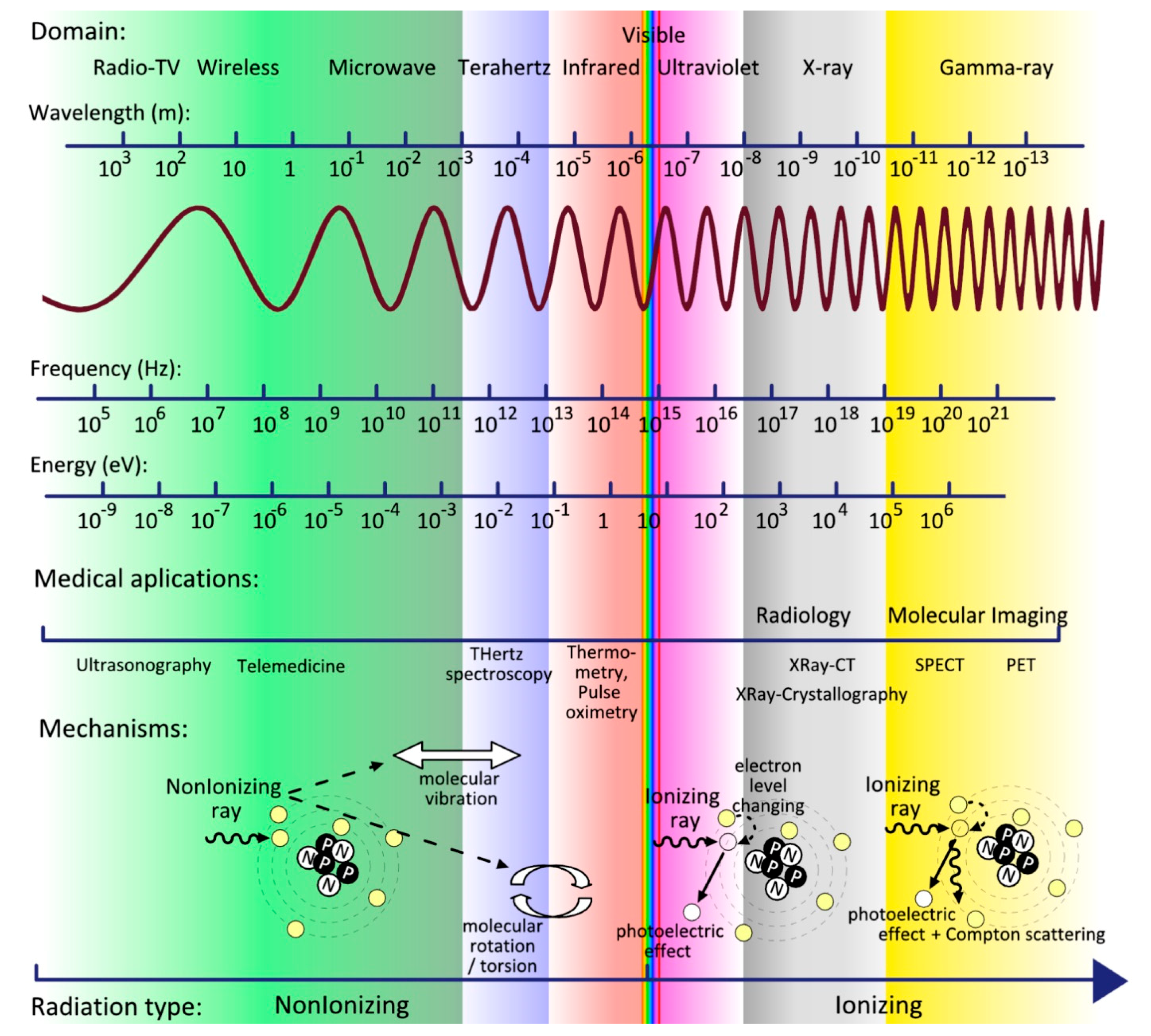
2. THz Attractive Characteristics and Interactions with Tissues
3. Integrating THz-Based Technologies with Current Diagnosis Techniques
3.1. THz Spectroscopy
3.2. THz-Tomography
3.3. THZ-Endoscope
3.4. THz Sensors Metamaterial Based
3.5. Enhancing THz Contrast
4. Applications of THz Imaging and THz Spectroscopy in Digestive Cancers
4.1. Oral Carcinoma
4.2. Esophageal Carcinoma
4.3. Gastric Carcinoma
4.4. Colorectal Tumors
4.5. Hepatocarcinoma
4.6. Pancreatic Cancer
5. Team’s Preliminary Results in THz Field
6. Conclusions
Author Contributions
Funding
Conflicts of Interest
References
- Mantsch, H.H.; Naumann, D. Terahertz spectroscopy: The renaissance of far infrared spectroscopy. J. Mol. Struct. 2010, 964, 1–4. [Google Scholar] [CrossRef] [Green Version]
- Parrott, E.P.J.; Sun, Y.; Pickwell-MacPherson, E. Terahertz spectroscopy: Its future role in medical diagnoses. J. Mol. Struct. 2011, 1006, 66–76. [Google Scholar] [CrossRef]
- Ren, A.; Zahid, A.; Fan, D.; Yang, X.; Imran, M.A.; Alomainy, A.; Abbasi, Q.H. State-of-the-art in terahertz sensing for food and water security—A comprehensive review. Trends Food Sci. Technol. 2019, 85, 241–251. [Google Scholar] [CrossRef]
- Hu, B.B.; Nuss, M.C. Imaging with terahertz waves. Opt. Lett. 1995, 20, 1716–1718. [Google Scholar] [CrossRef] [PubMed]
- Wietzke, S.; Jansen, C.; Reuter, M.; Jung, T.; Kraft, D.; Chatterjee, S.; Fischer, B.M.; Koch, M. Terahertz spectroscopy on polymers: A review of morphological studies. J. Mol. Struct. 2011, 1006, 41–51. [Google Scholar] [CrossRef]
- Yang, X.; Zhao, X.; Yang, K.; Liu, Y.; Liu, Y.; Fu, W.; Luo, Y. Biomedical Applications of Terahertz Spectroscopy and Imaging. Trends Biotechnol. 2016, 34, 810–824. [Google Scholar] [CrossRef] [PubMed]
- Woolard, D.L.; Brown, R.; Pepper, M.; Kemp, M. Terahertz Frequency Sensing and Imaging: A Time of Reckoning Future Applications? Proc. IEEE 2005, 93, 1722–1743. [Google Scholar] [CrossRef]
- Clothier, R.H.; Bourne, N. Effects of THz exposure on human primary keratinocyte differentiation and viability. J. Biol. Phys. 2003, 29, 179–185. [Google Scholar] [CrossRef]
- Zeni, O.; Gallerano, G.P.; Perrotta, A.; Romanò, M.; Sannino, A.; Sarti, M.; D’Arienzo, M.; Doria, A.; Giovenale, E.; Lai, A.; Messina, G.; Scarfì, M.R. Cytogenetic observations in human peripheral blood leukocytes following in vitro exposure to THz radiation: A pilot study. Health Phys. 2007, 92, 349–357. [Google Scholar] [CrossRef]
- Wilmink, G.J.; Rivest, B.D.; Ibey, B.L.; Roth, C.L.; Bernhard, J.; Roach, W.P. Quantitative investigation of the bioeffects associated with terahertz radiation. Proc. SPIE 2010, 7562, 75620L. [Google Scholar] [CrossRef]
- Borovkova, M.; Serebriakova, M.; Fedorov, V.; Sedykh, E.; Vaks, V.; Lichutin, A.; Salnikova, A.; Khodzitsky, M. Investigation of terahertz radiation influence on rat glial cells. Biomed. Opt. Express 2017, 8, 273–280. [Google Scholar] [CrossRef]
- Pereira, M.F.; Zubelli, J.P.; Winge, D.; Wacker, A.; Rodrigues, A.S.; Anfertev, V.; Vaks, V. Theory and measurements of harmonic generation in semiconductor superlattices with applications in the 100 GHz to 1 THz range. Phys. Rev. B 2017, 96, 045306. [Google Scholar] [CrossRef] [Green Version]
- Apostolakis, A.; Pereira, M.F. Controlling the harmonic conversion efficiency in semiconductor superlattices by interface roughness design. AIP Adv. 2019, 9, 015022. [Google Scholar] [CrossRef]
- He, M.; Azad, A.K.; Ye, S.; Zhang, W. Far-infrared signature of animal tissues characterized by terahertz time-domain spectroscopy. Opt. Commun. 2006, 259, 389–392. [Google Scholar] [CrossRef]
- Pickwell-MacPherson, E.; Wallace, V.P. Terahertz pulsed imaging-A potential medical imaging modality? Photodiagnosis Photodyn. Ther. 2009, 6, 128–134. [Google Scholar] [CrossRef]
- Kagadis, G.C.; Ford, N.L.; Karnabatidis, D.N.; Loudos, G.K. Handbook of Small Animal Imaging: Preclinical Imaging, Therapy, and Applications, 1st ed.; CRC Press: Boca Raton, FL, USA, 2009; pp. 1–602. [Google Scholar]
- Ashworth, P.C.; Pickwell-MacPherson, E.; Provenzano, E.; Pinder, S.E.; Purushotham, A.D.; Pepper, M.; Wallace, V.P. Terahertz pulsed spectroscopy of freshly excised human breast cancer. Opt. Express 2009, 17, 12444–12454. [Google Scholar] [CrossRef]
- Ahi, K. A method and system for enhancing the resolution of terahertz imaging. Measurement 2019, 138, 614–619. [Google Scholar] [CrossRef]
- Yu, C.; Fan, S.; Sun, Y.; Pickwell-Macpherson, E. The potential of terahertz imaging for cancer diagnosis: A review of investigations to date. Quant. Imaging Med. Surg. 2012, 2, 33–45. [Google Scholar]
- Guillet, J.P.; Recur, B.; Frederique, L.; Bousquet, B.; Canioni, L.; Manek-Hönninger, I.; Desbarats, P.; Mounaix, P. Review of Terahertz Tomography Techniques. J. Infrared. Millim. Terahertz Waves 2014, 35, 382–411. [Google Scholar] [CrossRef] [Green Version]
- Sun, Y.; Sy, M.Y.; Wang, Y.-X.J.; Ahuja, A.T.; Zhang, Y.-T. Pickwell-Macpherson E. A promising diagnostic method: Terahertz pulsed imaging and spectroscopy. World J. Radiol. 2011, 3, 55–65. [Google Scholar] [CrossRef]
- Chiriac, A.P.; Balan, V.; Asandulesa, M.; Butnaru, E.; Tudorachi, N.; Stoleru, E.; Nita, L.E.; Neamtu, I.; Diaconu, A. Investigation on thermal, rheological, dielectric and spectroscopic properties of a polymer containing pendant spiroacetal moieties. Mater. Chem. Phys. 2016, 180, 291–300. [Google Scholar] [CrossRef]
- Born, B.; Kim, S.J.; Ebbinghaus, S.; Gruebele, M.; Havenith, M. The terahertz dance of water with the proteins: The effect of protein flexibility on the dynamical hydration shell of ubiquitin. Faraday Discuss. 2009, 141, 161–173. [Google Scholar] [CrossRef]
- Wahaia, F.; Valusis, G.; Bernardo, L.M.; Almeida, A.; Moreira, J.A.; Lopes, P.C.; Macutkevic, J.; Kasalynas, I.; Seliuta, D.; Adomavicius, R.; et al. Detection of colon cancer by terahertz techniques. J. Mol. Struct. 2011, 1006, 77–82. [Google Scholar] [CrossRef]
- Eadie, L.H.; Reid, C.B.; Fitzgerald, A.J.; Wallace, V.P. Optimizing multi-dimensional terahertz imaging analysis for colon cancer diagnosis. Expert Syst. Appl. 2013, 40, 2043–2050. [Google Scholar] [CrossRef] [Green Version]
- Doradla, P.; Joseph, C.; Giles, R.H. Terahertz endoscopic imaging for colorectal cancer detection: Current status and future perspectives. World J. Gastrointest. Endosc. 2017, 9, 346–358. [Google Scholar] [CrossRef]
- Reid, C.B.; Fitzgerald, A.; Reese, G.; Goldin, R.; Tekkis, P.; O’Kelly, P.S.; Pickwell-MacPherson, E.; Gibson, A.P.; Wallace, V.P. Terahertz pulsed imaging of freshly excised human colonic tissues. Phys. Med. Biol. 2011, 56, 4333–4353. [Google Scholar] [CrossRef] [Green Version]
- Ji, Y.B.; Kim, S.-H.; Jeong, K.; Choi, Y.; Son, J.-H.; Park, D.W.; Noh, S.K.; Jeon, T.-I.; Huh, Y.-M.; Haam, S.; et al. Terahertz spectroscopic imaging and properties of gastrointestinal tract in a rat model. Biomed. Opt. Express 2014, 5, 4162. [Google Scholar] [CrossRef]
- Goryachuk, A.; Simonova, A.; Khodzitsky, M.; Borovkova, M.; Khamid, A. Gastrointestinal cancer diagnostics by terahertz time domain spectroscopy. In Proceedings of the 2017 IEEE International Symposium on Medical Measurements and Applications (MeMeA), Rochester, MN, USA, 7–10 May 2017; pp. 134–137. [Google Scholar]
- Wahaia, F.; Kasalynas, I.; Venckevicius, R.; Seliuta, D.; Valusis, G.; Urbanowicz, A.; Molis, G.; Carneiro, F.; Carvalho Silva, C.D.; Granja, P.L. Terahertz absorption and reflection imaging of carcinoma-affected colon tissues embedded in paraffin. J. Mol. Struct. 2016, 1107, 214–219. [Google Scholar] [CrossRef]
- Fischer, B.M.; Walther, M.; Jepsen, P.U. Far-infrared vibrational modes of DNA components studied by terahertz time-domain spectroscopy. Phys. Med. Biol. 2002, 47, 3807–3814. [Google Scholar] [CrossRef]
- Globus, T.; Woolard, D.; Crowe, T.W.; Khromova, T.; Gelmont, B.; Hesler, J. Terahertz Fourier transform characterization of biological materials in a liquid phase. J. Phys. D Appl. Phys. 2006, 39, 3405–3413. [Google Scholar]
- Tang, M.; Huang, Q.; Wei, D.; Zhao, G.; Chang, T.; Kou, K.; Wang, M.; Du, C.; Fu, W.; Cui, H.-L. Terahertz spectroscopy of oligonucleotides in aqueous solutions. J. Biomed. Opt. 2015, 20, 095009. [Google Scholar] [CrossRef] [PubMed]
- Brown, E.R.; Mendoza, E.A.; Xia, D.; Brueck, S.R.J. Narrow THz Spectral signatures through an RNA solution in nanofluidic channels. IEEE Sens. J. 2010, 10, 755–759. [Google Scholar] [CrossRef]
- Zhang, W.; Brown, E.R.; Rahman, M.; Norton, M.L. Observation of terahertz absorption signatures in microliter DNA solutions. Appl. Phys. Lett. 2013, 102, 023701. [Google Scholar] [CrossRef]
- Hasebe, T.; Kawabe, S.; Matsui, H.; Tabata, H. Metallic mesh-based terahertz biosensing of single- and double-stranded DNA. J. Appl. Phys. 2012, 112, 094702. [Google Scholar] [CrossRef]
- Brucherseifer, M.; Nagel, M.; Haring Bolivar, P.; Kurz, H. Label-free probing of the binding state of DNA by time-domain terahertz sensing. Appl. Phys. Lett. 2000, 77, 4049–4051. [Google Scholar] [CrossRef]
- Kutteruf, M.; Brown, C.M.; Iwaki, L.K.; Campbell, M.B.; Korter, T.M.; Heilweil, E.J. Terahertz spectroscopy of short-chain polypeptides. Chem. Phys. Lett. 2003, 375, 337–343. [Google Scholar] [CrossRef]
- Korter, T.; Balu, R.; Campbell, M.; Beard, M.; Gregurick, S.; Heilweil, E. Terahertz spectroscopy of solid serine and cysteine. Chem. Phys. Lett. 2006, 418, 65–70. [Google Scholar] [CrossRef]
- Rungsawang, R.; Ueno, Y.; Tomita, I.; Ajito, K. Angle-Dependent Terahertz Time-Domain Spectroscopy of Amino Acid Single Crystals. J. Phys. Chem. B 2006, 110, 21259–21263. [Google Scholar] [CrossRef]
- Yamamoto, K.; Tominaga, K.; Sasakawa, H.; Tamura, A.; Murakami, H.; Ohtake, H.; Sarukura, N. Terahertz Time-Domain Spectroscopy of Amino Acids and Polypeptides. Biophys. J. 2005, 89, L22–L24. [Google Scholar] [CrossRef] [Green Version]
- Ueno, Y.; Rungsawang, R.; Tomita, I.; Ajito, K. Quantitative measurements of amino acids by terahertz time-domain transmission spectroscopy. Anal. Chem. 2006, 78, 5424–5428. [Google Scholar] [CrossRef]
- Fuglewicz, B.; Pliński, E.F.; Jarzab, P.P.; Plińska, S.; Cebrat, M.; Nowak, K.; Augustyn, L.; Walczakowski, M.J.; Mikulics, M.; Pałka, N.; et al. Selected nonapeptides in terahertz light. Opt. Appl. 2014, 44, 159–171. [Google Scholar]
- Ding, T.; Li, R.; Zeitler, J.A.; Huber, T.L.; Gladden, L.F.; Middelberg, A.P.J.; Falconer, R.J. Terahertz and far infrared Spectroscopy of alanine-rich peptides having variable ellipticity. Opt. Express 2010, 18, 27431–27444. [Google Scholar] [CrossRef] [PubMed]
- Kikuchi, N.; Tanno, T.; Watanabe, M.; Kurabayashi, T. A membrane method for terahertz spectroscopy of amino acids. Anal. Sci. 2009, 25, 457–459. [Google Scholar] [CrossRef]
- Whitmire, S.E.; Wolpert, D.; Markelz, A.G.; Hillebrecht, J.R.; Galan, J.; Birge, R.R. Protein Flexibility and Conformational State: A Comparison of Collective Vibrational Modes of Wild-Type and D96N Bacteriorhodopsin. Biophys. J. 2003, 85, 1269–1277. [Google Scholar] [CrossRef] [Green Version]
- Balu, R.; Zhang, H.; Zukowski, E.; Chen, J.-Y.; Markelz, A.G.; Gregurick, S.K. Terahertz Spectroscopy of Bacteriorhodopsin and Rhodopsin: Similarities and Differences. Biophys. J. 2008, 94, 3217–3226. [Google Scholar] [CrossRef] [PubMed] [Green Version]
- Knab, J.; Chen, J.-Y.; Markelz, A. Hydration Dependence of Conformational Dielectric Relaxation of Lysozyme. Biophys. J. 2006, 90, 2576–2581. [Google Scholar] [CrossRef] [Green Version]
- Sun, Y.; Zhang, Y.-T.; Pickwell-Macpherson, E. Investigating Antibody Interactions with a Polar Liquid Using Terahertz Pulsed Spectroscopy. Biophys. J. 2011, 100, 225–231. [Google Scholar] [CrossRef] [Green Version]
- Wilmink, G.J.; Ibey, B.L.; Rivest, B.D.; Grundt, J.E.; Roach, W.P.; Tongue, T.D.; Schulkin, B.J.; Laman, N.; Peralta, X.G.; Roth, C.C.; Cerna, C.Z. Development of a compact terahertz time-domain spectrometer for the measurement of the optical properties of biological tissues. J. Biomed. Opt. 2011, 16, 047006. [Google Scholar] [CrossRef] [Green Version]
- Chen, J.Y.; Knab, J.R.; Ye, S.; He, Y.; Markelz, A.G. Terahertz dielectric assay of solution phase protein binding. Appl. Phys. Lett. 2007, 90, 243901. [Google Scholar] [CrossRef]
- Lee, J.W.; Lee, Y.-S.; Park, J.-M.; Shin, D.-C.; Jung, G.B.; Shin, J.-H.; Kim, S.; Kee, C.-S.; Kang, C. Terahertz spectroscopy of human sclera. Curr. Appl. Phys. 2015, 15, 1156–1159. [Google Scholar] [CrossRef]
- Liu, W.-Q.; Lu, Y.-F.; Jiao, G.-H.; Chen, X.-F.; Li, J.-Y.; Chen, S.-H.; Dong, Y.-M.; Lv, J.-C. Terahertz optical properties of the cornea. Opt. Commun. 2016, 359, 344–348. [Google Scholar] [CrossRef]
- Wallace, V.P.; Fitzgerald, A.J.; Pickwell, E.; Pye, R.J.; Taday, P.F.; Flanagan, N.; Ha, T. Terahertz Pulsed Spectroscopy of Human Basal Cell Carcinoma. Appl. Spectroscopy 2006, 60, 1127–1133. [Google Scholar] [CrossRef] [PubMed]
- Stringer, M.R.; Lund, D.N.; Foulds, A.P.; Uddin, A.; Berry, E.; Miles, R.E.; Davies, A.G. The analysis of human cortical bone by terahertz time-domain spectroscopy. Phys. Med. Biol. 2005, 3211, 3211–3219. [Google Scholar] [CrossRef]
- Png, G.M.; Flook, R.; Ng, B.W.-H.; Abbott, D. Terahertz spectroscopy of snap-frozen human brain tissue: An initial study. Electron. Lett. 2009, 45, 343–344. [Google Scholar] [CrossRef]
- Zhang, R.; Wu, T.; Zhao, Y. Terahertz signal analysis for biological tissues based on empirical mode decomposition. Optik 2019, 183, 906–911. [Google Scholar] [CrossRef]
- Huang, S.Y.; Wang, Y.X.J.; Yeung, D.K.W.; Ahuja, A.T.; Zhang, Y.-T.; Pickwell-MacPherson, E. Tissue characterization using terahertz pulsed imaging in reflection geometry. Phys. Med. Biol. 2009, 54, 149–160. [Google Scholar] [CrossRef]
- Berry, E.; Handley, J.W.; Fitzgerald, A.J.; Merchant, W.J.; Boyle, R.D.; Zinov’ev, N.N.; Miles, R.E.; Chamberlain, J.M.; Smith, M.A. Multispectral classification techniques for terahertz pulsed imaging: An example in histopathology. Med. Eng. Phys. 2004, 26, 423–430. [Google Scholar] [CrossRef] [PubMed]
- Taylor, Z.D.; Singh, R.S.; Culjat, M.O.; Suen, J.Y.; Grundfest, W.S.; Lee, H.; Brown, E.R. Reflective terahertz imaging of porcine skin burns. Opt. Lett. 2008, 33, 1258–1260. [Google Scholar] [CrossRef]
- Cole, B.E.; Woodward, R.M.; Crawley, D.A.; Wallace, V.P.; Arnone, D.D.; Pepper, M. Terahertz imaging and spectroscopy of human skin in vivo. In Proceedings of the Commercial and Biomedical Applications of Ultrashort Pulse Lasers—Laser Plasma Generation and Diagnostics (2011), San Jose, CA, USA, 20–26 January 2001; p. 4276. [Google Scholar]
- Crawley, D.; Longbottom, C.; Wallace, V.P.; Cole, B.; Arnone, D.; Pepper, M. Three-dimensional terahertz pulse imaging of dental tissue. J. Biomed. Opt. 2003, 8, 303–307. [Google Scholar] [CrossRef] [PubMed]
- Kamburoğlu, K.; Karagöz, B.; Altan, H.; Özen, D. An ex vivo comparative study of occlusal and proximal caries using terahertz and X-ray imaging. Dentomaxillofac. Radiol. 2019, 48, 20180250. [Google Scholar] [CrossRef]
- Pickwell, E.; Wallace, V.P.; Cole, B.E.; Ali, S.; Longbottom, C.; Lynch, R.J.M.; Pepper, M. A Comparison of Terahertz Pulsed Imaging with Transmission Microradiography for Depth Measurement of Enamel Demineralisation in vitro. Caries Res. 2007, 41, 49–55. [Google Scholar] [CrossRef]
- Crawley, D.A.; Longbottom, C.; Cole, B.E.; Ciesla, C.M.; Arnone, D.; Wallace, V.P.; Pepper, M. Terahertz Pulse Imaging: A Pilot Study of Potential Applications in Dentistry. Caries. Res. 2003, 37, 352–359. [Google Scholar] [CrossRef]
- Fitzgerald, A.J.; Cole, B.E.; Taday, P.F. Nondestructive analysis of tablet coating thicknesses using terahertz pulsed imaging. J. Pharm. Sci. 2005, 94, 177–183. [Google Scholar] [CrossRef] [PubMed]
- Zeitler, J.A.; Shen, Y.; Baker, C.; Taday, P.F.; Pepper, M.; Rades, T. Analysis of coating structures and interfaces in solid oral dosage forms by three dimensional terahertz pulsed imaging. J. Pharm. Sci. 2007, 96, 330–340. [Google Scholar] [CrossRef]
- Wu, H.; Heilweil, E.J.; Hussain, A.S.; Khan, M.A. Process analytical technology (PAT): Effects of instrumental and compositional variables on terahertz spectral data quality to characterize pharmaceutical materials and tablets. Int. J. Pharm. 2007, 343, 148–158. [Google Scholar] [CrossRef] [PubMed]
- Sibik, J.; Zeitler, J.A. Direct measurement of molecular mobility and crystallisation of amorphous pharmaceuticals using terahertz spectroscopy. Adv. Drug Deliv. Rev. 2016, 100, 147–157. [Google Scholar] [CrossRef] [Green Version]
- Laman, N.; Harsha, S.S.; Grischkowsky, D. Narrow-Line Waveguide Terahertz Time-Domain Spectroscopy of Aspirin and Aspirin Precursors. Appl. Spectrosc. 2008, 62, 319–326. [Google Scholar] [CrossRef]
- Strachan, C.J.; Taday, P.F.; Newnham, D.A.; Gordon, K.C.; Zeitler, J.A.; Pepper, M.; Rades, T. Using Terahertz Pulsed Spectroscopy to Quantify Pharmaceutical Polymorphism and Crystallinity. J. Pharm. Sci. 2005, 94, 837–846. [Google Scholar] [CrossRef] [PubMed]
- Nishikiori, R.; Yamaguchi, M.; Tanako, K.; Enatsu, T.; Tani, M.; de Silva, U.C.; Kawashita, N.; Tagaki, T.; Morimoto, S.; Hangyo, M.; et al. Application of Partial Least Square on Quantitative Analysis of L-, D-, and DL-Tartaric Acid by Terahertz Absorption Spectra. Chem. Pharm. Bull. 2008, 56, 305–307. [Google Scholar] [CrossRef] [Green Version]
- Woodward, R.M.; Wallace, V.P.; Pye, R.J.; Cole, B.E.; Arnone, D.D.; Linfield, E.H.; Pepper, M. Terahertz pulse imaging of ex vivo basal cell carcinoma. J. Investig. Dermatol. 2003, 120, 72–78. [Google Scholar] [CrossRef]
- Wallace, V.P.; Fitzgerald, A.J.; Shankar, S.; Flanagan, N.; Pye, R.; Cluff, J.; Arnone, D.D. Terahertz pulsed imaging of basal cell carcinoma ex vivo and in vivo. Br. J. Dermatol. 2004, 151, 424–432. [Google Scholar] [CrossRef] [PubMed]
- Joseph, C.S.; Patel, R.; Neel, V.A.; Giles, R.H.; Yaroslavsky, A.N. Imaging of ex vivo nonmelanoma skin cancers in the optical and terahertz spectral regions. J. Biophotonics 2014, 7, 295–303. [Google Scholar] [CrossRef]
- Fitzgerald, A.J.; Wallace, V.P.; Jimenez-Linan, M.; Bobrow, L.; Pye, R.J.; Purushotham, A.D.; Arnone, D.D. Terahertz pulsed imaging of human breast tumors. Radiology 2006, 239, 533–540. [Google Scholar] [CrossRef] [PubMed]
- Bowman, T.C.; El-Shenawee, M.; Campbell, L.K. Terahertz imaging of excised breast tumor tissue on paraffin sections. IEEE Trans. Antenn. Propag. 2015, 63, 2088–2097. [Google Scholar] [CrossRef]
- Jung, E.; Lim, M.-H.; Moon, K.-W.; Do, Y.-W.; Lee, S.-S.; Han, H.-W.; Choi, H.-J.; Cho, K.-S.; Kim, K.-R. Terahertz pulse imaging of micro-metastatic lymph nodes in early-stage cervical cancer patients. J. Opt. Soc. Korea 2011, 15, 155–160. [Google Scholar] [CrossRef]
- Brun, M.A.; Formanek, F.; Yasuda, A.; Sekine, M.; Ando, N.; Eishii, Y. Terahertz imaging applied to cancer diagnosis. Phys. Med. Biol. 2010, 55, 4615–4623. [Google Scholar] [CrossRef]
- Trofimov, V.A.; Varentsova, S.A. Essential Limitations of the Standard THz TDS Method for Substance Detection and Identification and a Way of Overcoming Them. Sensors 2016, 16, 502. [Google Scholar] [CrossRef]
- Wallace, V.P.; Macpherson, E.; Zeitler, J.A.; Reid, C. Three-dimensional imaging of optically opaque materials using nonionizing terahertz radiation. J. Opt. Soc. Am. A. Opt. Image Sci. Vis. 2008, 25, 3120–3133. [Google Scholar] [CrossRef]
- Ferguson, B.; Wang, S.; Gray, D.; Abbot, D.; Zhang, X.-C. T-ray computed tomography. Opt. Lett. 2002, 27, 1312. [Google Scholar] [CrossRef] [Green Version]
- Ji, Y.B.; Lee, E.S.; Kim, S.-H.; Son, J.-H.; Jeon, T.-I. A miniaturized fiber-coupled terahertz endoscope system. Opt. Express 2009, 17, 17082–17087. [Google Scholar] [CrossRef]
- Doradla, P.; Alavi, K.; Joseph, C.; Giles, R. Single-channel prototype terahertz endoscopic system. J. Biomed. Opt. 2014, 19, 080501. [Google Scholar] [CrossRef] [Green Version]
- Chen, T.; Li, S.; Sun, H. Metamaterials Application in Sensing. Sensors 2012, 12, 2742–2765. [Google Scholar] [CrossRef] [Green Version]
- Yoshida, H.; Ogawa, Y.; Kawai, Y.; Hayashi, S.; Hayashi, A.; Otani, C.; Kato, E.; Miyamaru, F.; Kawase, K. Terahertz sensing method for protein detection using a thin metallic mesh. Appl. Phys. Lett. 2007, 91, 253901:1–253901:3. [Google Scholar] [CrossRef]
- Tao, H.; Chieffo, L.R.; Brenckle, M.A.; Siebert, S.M.; Liu, M.; Strikwerda, A.C.; Fan, K.; Kaplan, D.L.; Zhang, X.; Averitt, R.D.; Omenetto, FG. Metamaterials on paper as a sensing platform. Adv. Mater. 2011, 23, 3197–3201. [Google Scholar] [CrossRef]
- He, X.; Zhang, Q.; Lu, G.; Ying, G.; Wu, F.; Jiang, J. Tunable ultrasensitive terahertz sensor based on complementary graphene metamaterials. RSC Adv. 2016, 6, 52212–52218. [Google Scholar] [CrossRef]
- Kashanian, H.A.; Ghaffary, H.B.; Bagherzadeh, N.C. Gastric Cancer Diagnosis Using Terahertz Imaging. Majlesi J. Multimed. Process 2015, 4, 1–7. [Google Scholar]
- Choi, M.; Lee, S.H.; Kim, Y.; Kang, S.B.; Shin, J.; Kwak, M.H.; Kang, K.-Y.; Lee, Y.-H.; Park, N.; Min, B. A terahertz metamaterial with unnaturally high refractive index. Nature 2011, 470, 369–373. [Google Scholar] [CrossRef]
- Vieweg, N.; Rettich, F.; Deninger, A.; Roehle, H.; Dietz, R.; Göbel, T.; Schell, M. Terahertz-time domain spectrometer with 90 dB peak dynamic range. J. Infrared Millim. Terahertz Waves 2014, 35, 823–832. [Google Scholar] [CrossRef] [Green Version]
- Gafton, B.; Porumb, V.; Ungurianu, S.; Marinca, M.V.; Cocea, C.; Croitoru, A.; Balan, G.; Miron, N.; Eliade Ciuleanu, T.; Miron, L. Hepatocellular carcinoma: insights in the biological treatment beyond sorafenib. J. Buon. 2014, 19, 858–866. [Google Scholar]
- Shukla, R.; Abidi, W.M.; Richards-Kortum, R.; Anandasabapathy, S. Endoscopic imaging: How far are we from real-time histology? World J. Gastrointest. Endosc. 2011, 3, 183–194. [Google Scholar] [CrossRef] [PubMed]
- Wang, J.; Fleischmann, D. Improving Spatial Resolution at CT: Development, Benefits, and Pitfalls. Radiology 2018, 289, 261–262. [Google Scholar] [CrossRef]
- Stucht, D.; Danishad, K.A.; Schulze, P.; Godenschweger, F.; Zaitsev, M.; Speck, O. Highest Resolution In Vivo Human Brain MRI Using Prospective Motion Correction. PLoS ONE 2015, 10, e0133921. [Google Scholar] [CrossRef]
- Shen, K.; Lu, H.; Baig, S.; Wang, M.R. Improving lateral resolution and image quality of optical coherence tomography by the multi-frame superresolution technique for 3D tissue imaging. Biomed. Opt. Express 2017, 8, 4887–4918. [Google Scholar] [CrossRef] [PubMed] [Green Version]
- Moses, W.W. Fundamental Limits of Spatial Resolution in PET. Nucl. Instrum. Methods Phys. Res. A 2011, 648, S236–S240. [Google Scholar] [CrossRef] [PubMed] [Green Version]
- Haddad, J.E.; Bousquet, B.; Canioni, L.; Mounaix, P. Review in terahertz spectral analysis. Trends Anal. Chem. 2013, 44, 98–105. [Google Scholar] [CrossRef]
- Sim, Y.C.; Park, J.Y.; Ahn, K.-M.; Park, C.; Son, J.-H. Terahertz imaging of excised oral cancer at frozen temperature. Biomed. Opt. Express 2013, 4, 1413–1421. [Google Scholar] [CrossRef] [Green Version]
- Hou, D.; Li, X.; Cai, J.; Ma, Y.; Kang, X.; Huang, P.; Zhang, G. Terahertz spectroscopic investigation of human gastric normal and tumor tissues. Phys. Med. Biol. 2014, 59, 5423–5440. [Google Scholar] [CrossRef]
- Wahaia, F.; Kasalynas, I.; Seliuta, D.; Molis, G.; Urbanowicz, A.; Carvalho Silva, C.D.; Carneiro, F.; Valusis, G.; Granja, P.L. Terahertz spectroscopy for the study of paraffin-embedded gastric cancer samples. J. Mol. Struct. 2015, 1079, 391–395. [Google Scholar] [CrossRef]
- Ji, Y.B.; Park, C.H.; Kim, H.; Kim, S.-H.; Lee, G.M.; Noh, S.K.; Jeon, T.-I.; Son, J.-H.; Huh, Y.-M.; Haam, S.; et al. Feasibility of terahertz reflectometry for discrimination of human early gastric cancers. Biomed. Opt. Express 2015, 6, 1398. [Google Scholar] [CrossRef]
- Wahaia, F.; Kasalynas, I.; Seliuta, D.; Molis, G.; Urbanowicz, A.; Carvalho Silva, C.D.; Carneiro, F.; Valusis, G.; Granja, P.L. Study of paraffin-embedded colon cancer tissue using terahertz spectroscopy. J. Mol. Struct. 2015, 1079, 448–453. [Google Scholar] [CrossRef]
- Doradla, P.; Alavi, K.; Joseph, C.S.; Giles, R.H. Terahertz polarization imaging for colon cancer detection. In Proceedings of the SPIE OPTO 2014, Francisco, CA, USA, 1–6 February 2014; p. 89850. [Google Scholar]
- Chen, H.; Ma, S.-H.; Yan, W.-X.; Wu, X.-M.; Wang, X.-Z. The Diagnosis of Human Liver Cancer by using THz Fiber-Scanning Near-Field Imaging. Chin. Phys. Lett. 2013, 30, 030702. [Google Scholar] [CrossRef]
- Formanek, F.; Brun, M.-A.; Yasuda, A. Contrast improvement of terahertz images of thin histopathologic sections. Biomed. Opt. Express 2010, 2, 58–64. [Google Scholar] [CrossRef] [Green Version]
- Gong, Y.; Dong, H.; Hong, M.; Malini, O.; Thong, P.S.P.; Bhuvaneswari, R. Polarization effect in liver tissue in terahertz band. In Proceedings of the 2009 34th International Conference on Infrared, Millimeter, and Terahertz Waves, Busan, Korea, 21–25 September 2009; pp. 1–2. [Google Scholar]
- Park, J.Y.; Choi, H.J.; Cho, K.-S.; Kim, K.-R.; Son, J.-H. Terahertz spectroscopic imaging of a rabbit VX2 hepatoma model. J. Appl. Phys. 2011, 109, 064704. [Google Scholar] [CrossRef]
- Locatelli, M.; Ravaro, M.; Bartalini, S.; Consolino, L.; Vitiello, M.S.; Cicchi, R.; Pavone, F.; De Natale, P. Real-time terahertz digital holography with a quantum cascade laser. Sci. Rep. 2015, 5, 13566. [Google Scholar] [CrossRef] [PubMed] [Green Version]
- Huang, H.; Wang, D.; Li, W.; Rong, L.; Taylor, Z.D.; Deng, Q.; Li, B.; Wang, Y.; Wu, W.; Panezai, S. Continuous-wave terahertz multi-plane in-line digital holography. Opt. Lasers Eng. 2017, 94, 76–81. [Google Scholar] [CrossRef]
- Rong, L.; Latychevskaia, T.; Chen, C.; Wang, D.; Yu, Z.; Zhou, X.; Li, Z.; Huang, H.; Wang, Y.; Zhou, Z. Terahertz in-line digital holography of human hepatocellular carcinoma tissue. Sci. Rep. 2015, 5, 8445. [Google Scholar] [CrossRef] [PubMed] [Green Version]
- Mihai, C.T.; Luca, A.; Danciu, M.; Alexa-Stratulat, T.; Ciobanu, R.; Tamba, B.I.; Stanciu, G.D.; Ciortescu, I.; Mihai, C.; Stefanescu, G.; et al. In vitro evaluation of novel functionalized nanoparticles for terahertz imaging. Med. Surg. J. 2019, 123, 139–146. [Google Scholar]
- Ciobanu, R.C.; Schreiner, O.; Lucanu, N.; Drug, V.; Ciortescu, I.; Mihai, C.; Stefanescu, G.; Danciu, E. Advanced Image Processing in Support of THz Imaging for Early Detection of Gastric Cancer. In Proceedings of the 2018 International Conference and Exposition on Electrical and Power Engineering (EPE), Iasi, Romania, 18–19 October 2018. [Google Scholar]


| Class | Technique | Detected Compounds | References |
|---|---|---|---|
| Nucleic acids | THz-TDS | DNA, sequences of oligonucleotides, RNA | [31,32,33,34] |
| THz photo-mixing | DNA | [35] | |
| THz-based metallic mesh | DNA: single-stranded and double-stranded | [36] | |
| Time-resolved THz | Hybridized/denaturated DNA films | [37] | |
| Amino acids and peptides | THz-TDS and transmittance spectroscopy | 20 naturally occurring lyophilized amino acids, 1-serine and 1-cysteine, L-cysteine and L-histidine, polyglycine and poly-L-alanine, L-glutamic acid, Histidine analogs of oxytocin and vasopressin, Alanine-rich peptides, L-Threonine and glycine | [38,39,40,41,42,43,44,45] |
| Proteins | THz-TDS | Wild-Type and D96N mutant Bacteriorhodopsin, Photoactive protein systems: rhodopsin and bacteriorhodopsin, Hen egg white lysozyme, Immunoglobin (IgG) protein, Protein hydration and protein–ligand binding, Protein–ligand binding among hen egg white lysozyme and triacetylglucosamine | [46,47,48,49,50,51] |
| Tissues (dermatology) | THz-TDS | Human scleral tissues, Rabbit corneal tissue ex vivo, Skin tissues vs. human basal cell carcinoma, Bone, Brain tissues from Alzheimer disease patients | [52,53,54,55,56] |
| THz empirical mode decomposition | Fresh porcine muscle and skin tissues | [57] | |
| THz pulsed imaging | Rat tissues, Basal cell carcinoma and melanoma, Porcine skin burns, Human skin in vivo (stratum corneum thickness and hydration) | [58,59,60,61] | |
| Dental health | THz pulse and reflection imaging | Teeth tissues, 32 human permanent teeth surfaces (detection of dental caries ex vivo), Enamel demineralization in vitro. | [62,63,64] |
| THz-TDS | Enamel-dentine boundary | [65] | |
| Pharmaceuticals | THz pulse imaging | Tablet and coating integrity and performance | [66] |
| THz Spectroscopy and THz-TDS | Direct measurement of crystallization and molecular mobility of amorphous pharmaceuticals. Pharmaceutical materials and tablets. Aspirin and Aspirin Precursors. Pharmaceutical Polymorphism and Crystallinity. L-, D-, and DL-Tartaric Acid. | [67,68,69,70,71,72] | |
| Oncology | THz pulse and reflection imaging | Skin cancer: ex vivo and in vivo basal cell carcinoma, nonmelanoma. Breast cancer: in situ non-calcified form, triple negative infiltrating ductal carcinoma, micro-metastatic lymph nodes. Lung neoplasm: squamous cell carcinoma. Uterine cervical neoplasm. | [73,74,75,76,77,78,79] |
| A specific discussion on digestive cancers can be found in following sections. | |||
| Technique | Spatial Resolution, mm | Damage to Healthy Tissue | Limits |
|---|---|---|---|
| Conventional colonoscopy/endoscopy | Lack of spatial resolution [93] | Invasive | Operator dependent and can have high false-negative rates |
| X-ray computer tomography | 0.5–2 [94] | Minimal radiation exposure | Low sensitivity for digestive cancers and can not be repeated very often- |
| MRI | 0.1–2 (depended on Tesla range) [95] | Non-invasive, uses contrast agents | High cost, time consuming investigation |
| OCT | 0.01 [96] | Non-invasive | High cost, subjectivity in image interpretation, research use only |
| PET | 0.54–6 (dependent on the isotope) [97] | Minimized risk tissue damage due to radiation | High-cost, the lack of anatomic correlation, restricted availability |
| THz technology | 0.1–0.25 [21] | Non-invasive and non-ionizing under controlled radiation power density and exposure time | As discussed in a previous section |
© 2019 by the authors. Licensee MDPI, Basel, Switzerland. This article is an open access article distributed under the terms and conditions of the Creative Commons Attribution (CC BY) license (http://creativecommons.org/licenses/by/4.0/).
Share and Cite
Danciu, M.; Alexa-Stratulat, T.; Stefanescu, C.; Dodi, G.; Tamba, B.I.; Mihai, C.T.; Stanciu, G.D.; Luca, A.; Spiridon, I.A.; Ungureanu, L.B.; et al. Terahertz Spectroscopy and Imaging: A Cutting-Edge Method for Diagnosing Digestive Cancers. Materials 2019, 12, 1519. https://doi.org/10.3390/ma12091519
Danciu M, Alexa-Stratulat T, Stefanescu C, Dodi G, Tamba BI, Mihai CT, Stanciu GD, Luca A, Spiridon IA, Ungureanu LB, et al. Terahertz Spectroscopy and Imaging: A Cutting-Edge Method for Diagnosing Digestive Cancers. Materials. 2019; 12(9):1519. https://doi.org/10.3390/ma12091519
Chicago/Turabian StyleDanciu, Mihai, Teodora Alexa-Stratulat, Cipriana Stefanescu, Gianina Dodi, Bogdan Ionel Tamba, Cosmin Teodor Mihai, Gabriela Dumitrita Stanciu, Andrei Luca, Irene Alexandra Spiridon, Loredana Beatrice Ungureanu, and et al. 2019. "Terahertz Spectroscopy and Imaging: A Cutting-Edge Method for Diagnosing Digestive Cancers" Materials 12, no. 9: 1519. https://doi.org/10.3390/ma12091519