Article

Diguanylate Cyclase (DGC) Implicated in the Synthesis of Multiple Bacteriocins via the Flagellar-Type III Secretion System Produced by Pectobacterium carotovorum subsp. carotovorum

by
Ruchi Briam James Sersenia Lagitnay
1,2,
Han-Ling Chen
2,
Yen-Chun Chen
2 and
Duen-Yau Chuang
2,*
1
College of Arts & Sciences, Bayombong Campus, Nueva Vizcaya State University, Bayombong 3700, Nueva Vizcaya, Philippines
2
Department of Chemistry, National Chung Hsing University, Taichung City 400, Taiwan
*
Author to whom correspondence should be addressed.
Int. J. Mol. Sci. 2022, 23(10), 5649; https://doi.org/10.3390/ijms23105649
Submission received: 17 April 2022 / Revised: 9 May 2022 / Accepted: 16 May 2022 / Published: 18 May 2022
(This article belongs to the Special Issue Flagella)

Abstract

:
The plant pathogen Pectobacterium carotovorum subsp. carotovorum (previously Erwinia carotovora subsp. carotovora) causes soft rot and stem rot diseases in a variety of crops, including Chinese cabbage, potato, and tomato. The flagellar-type III secretion systems were used by Pcc’s virulence mechanism to export proteins or bacteriocins to the outside of the cell. DGC, a virulence factor that cyclizes c-di-GMP, a common secondary signal in physiological processes and toxin control systems of many bacteria, was discovered in Pcc’s genomic DNA. The dgc gene in Pcc was blocked using the method of homologous recombination in our study. In the in vivo setting, the results demonstrated that the dgc knockout strain does not release low molecular weight bacteriocins. The bacteriocin gene (carocin S2, carocin S3, carocin S4) and the flagellar-type III secretion system genes were also unable to be transcribed by the dgc knockout strain in the transcription experiment. We also observed that the amount of bacteriocin expressed changed when the amount of L-glutamine in the environment exceeded a particular level. These data suggested that L-glutamine influenced physiological processes in Pcc strains in some way. We hypothesized a relationship between dgc and the genes involved in Pcc LMWB external export via the flagellar-type secretion system based on these findings. In this study, the current findings led us to propose a mechanism in which DGC’s cyclic di-GMP might bind to receptor proteins and positively regulate bacteriocin transcription as well as the synthesis, mobility, and transport of toxins.

1. Introduction

When bacteria are exposed to various environmental conditions, they develop a number of transmission pathways in their cells that produce physiological changes involving a wide range of proteins and chemicals. The cyclic diguanylate monophosphate (c-di-GMP), which connects external stimuli to cells in signal transmission, is a common and important secondary signaling component in many bacterial species, including Pectobacterium carotovorum subsp. carotovorum (Pcc) [1,2,3,4]. A class of enzymes known as diguanylate cyclase (DGC) with a conserved GGDEF motif synthesizes this twofold symmetric molecule at the intracellular level from two molecules of guanosine triphosphate (GTP) [5,6]. Diguanylate cyclase has been demonstrated to control a range of cell activities by producing c-di-GMP. Cell cycle regulation, adhesion, biofilms, motility, toxicity, and cellulase modulation are all regulated by changes in c-di-GMP concentrations [7,8,9]. To regulate a wide range of cellular processes, c-di-GMP must first bind to protein receptors and then interact with downstream receptors.
The PilZ protein domain, riboswitch [10,11], cAMP receptor CRP [12], and CLP [13] of the protein family are examples of c-di-GMP receptors. These receptors can interact directly with c-di-GMP. Recent findings suggest that c-di-GMP affects type VI secretion systems (T6SSs) [14] as well as T3SSs [15]. The action of c-di-GMP in T3SSs appears to be direct and mediated once again by a central ATPase, as the flagellar export ATPase FliI from a variety of distantly related bacteria has been found to bind to c-di-GMP [8,15] exclusively.
Pectobacterium carotovorum subsp. carotovorum (Pcc) secretes numerous pathogenicity-related proteins that cause soft-rot disease in a variety of plant species [16]. In the virulence mechanism of Pcc, multiple types of secretion systems (TSS) are used to export proteins or bacteriocins to the outside of the cell. Protein molecules that traverse the outer and inner membranes are found in the secretion system, and c-di-GMP can bind to certain receptor proteins to control secretion system gene transcription [17,18]. Our earlier findings point to a Type III secretion pathway for bacteriocin extracellular export that is a mix of injectisome and flagellar secretion [19].
Prior investigations have demonstrated that DGC can be regulated at either the level of enzymatic activity or the level of expression within the surrounding environment [20], despite the fact that intracellular transmission channels and regulatory factors are involved at several levels. This study will aid in a better understanding of the role of DGC in the regulation, transcription, and secretion of numerous bacteriocins by the Pcc via the mix T3SS.

2. Results

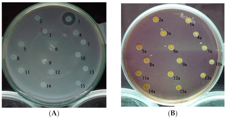
2.1. Isolation and Detection of Tn5 Insertion Mutants

When Pcc is exposed to rifampicin, it may undergo natural mutation and create progeny strains with rifampicin resistance genes. Even after mutations, these mutants continue to produce bacteriocins. We used rif-TO6, a rifampicin-resistant Pcc strain, as the recipient of bacterial conjugation and E. coli (1830) with the kanamycin resistance gene transposon Tn5 as the provider of conjugative reproduction in this study. Following mating, about 10,000 strains were tested on Drigalski’s and LB medium with antibiotics kanamycin and rifampicin. Following that, bacteriocin assays [21,22] were used to identify strains with successfully disrupted low-molecular-weight bacteriocin-related genes.
The size of the inhibitory zone around each isolate was compared to that of rif-TO6 in a bacteriocin assay. According to this evidence of mutation (Figure 1A), the absence of inhibitory zones confirmed that the mutant colonies’ transposon Tn5 had been inserted into LMW bacteriocin-related genes. The wild-type rif-TO6 and Tn5 insertional mutants that underwent bacteriocin testing verified that the strains are Pcc, according to the results from the Modified Drigalski medium (Figure 1B).

2.2. DNA Amplification at the Tn5 Insertion Junction

To investigate if Tn5 was incorporated into the genomic DNA of suspected isolates, we used a nested PCR using two primers, P3 and P4, to amplify the nptII resistance gene on Tn5 (about 500 bp in length). This PCR approach can be used to confirm whether the wild strain’s DNA has been inserted with transposon Tn5. All isolated putative mutants had a 500-bp band; however, because wild-type rif-TO6 lacked Tn5 insertion, there was no band (data not shown).
To identify Tn5-interrupted genes, genomic DNA from the various mutant strains was amplified with Thermal Asymmetric Interlaced PCR (TAIL-PCR) using an array of specific primers. The template DNA was extracted from Pcc’s insertional mutant’s genomic DNA, and the isolated chromosomal DNA was treated with EcoRI before being validated on a 0.7 percent agarose electrophoresis gel. Supplementary Figure S1 shows the results of the TAIL-PCR.
TAIL-PCR products with a length of 1070 bp were obtained and sequenced. DNASIS MAX 3.0 software was used to deduce the amino acid sequences from the putative mutants. Among the different insertion mutants, the TT6-6 DNA sequence revealed that this carries a diguanylate cyclase with a 30% homogeneity in Shigella boydii, according to results from the BLAST programs of the National Center for Biotechnology Information website (National Library of Medicine, Bethesda, MD, USA) Table 1.

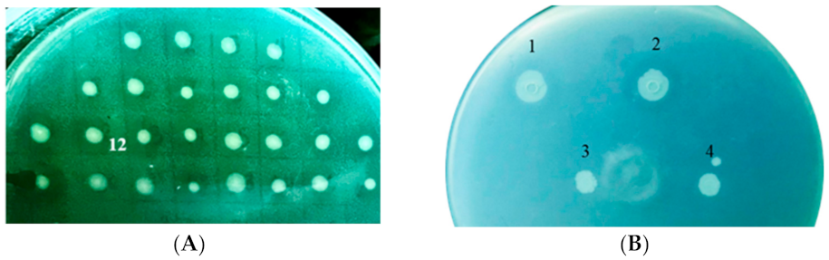
2.3. Role of DGC on Pcc’s Bacteriocin Secretion

We used rifampicin-resistant Pcc 89-H-12 bacteria as the gene knockout strain to better understand the link between DGC and bacteriocin production.
Because the 89-H-12 strain can synthesize carocin S2, S3, and S4, the low-molecular-weight bacteriocins (Figure 2A), using it as a gene selection strain will help the researcher better understand Pcc’s virulence mechanism.
The pG-dgc-Kan and pG-dgc-Tet constructs are utilized for double confirmation and undergo homologous recombination. Because the Pcc 89-H-12 strain is unable to produce kanamycin- and tetracycline-resistant bacteria, these antibiotic gene fragments were used as a selection marker for mutant screening.
Two dgc defective mutants, 89-H-rif-12/dgc-Kan, and 89-H-rif-12/dgc-Tet were compared to the wild-types Pcc 3F3 and Pcc 89H-rif-12 on their ability to secrete various bacteriocins, whereas sp33 was used as an indicator strain.
After transformations, in vivo tests revealed that the knockout strains had no LMWB secretions, whereas the wild types had normal LMWB secretion at the cellular level (Figure 2B). Carocin S1, S2, S3, and S4 cannot be transported extracellularly when the dgc gene is knocked out.

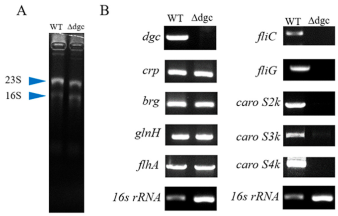
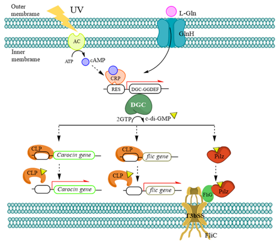
2.4. Regulation of DGC on Pcc’s Bacteriocin Secretion

A transcriptional study was also conducted to see how bacteria generated, regulated, and secreted bacteriocin. The wild-type and dgc knockout (89-H-rif-12/dgc-Kan and 89H-rif-12/dgc-Tet) strains were used in this experiment, which grew exponentially after induction. These induced strains were then reincubated to allow the cells to manufacture bacteriocin before being collected to extract the bacteriocin’s RNA for in vitro testing. The ability of the induced cells to create, regulate, and release bacteriocin was studied using a two-step endpoint reverse transcriptase polymerase chain reaction (endpoint RT-PCR) [23,24]. The 16 RNA expression, which is highly conserved among bacteria species, was used as a control for comparing gene expression in this technique.
The three bacteriocin genes, caroS2k, caroS3k, and caroS4k, all lose their ability to express on an agarose gel electropherogram (Figure 3), indicating that the dgc gene regulates the bacteriocin toxin gene to prevent the strain from secretion. We have also identified certain regulatory genes (crp, brg, and glnh) involved in the manufacture of bacteriocin based on earlier research. These genes can still be transcribed, indicating that the upstream end of the dgc gene is unaffected.
According to the findings of this study, further deletion of dgc genes inhibits the production of fliC and fliG. Because the T3SS genes fliC and fliG are involved in the synthesis of chaperones necessary for the delivery of LMWBs, knocking down dgc in Pcc limits bacteriocin secretion to the extracellular environment.

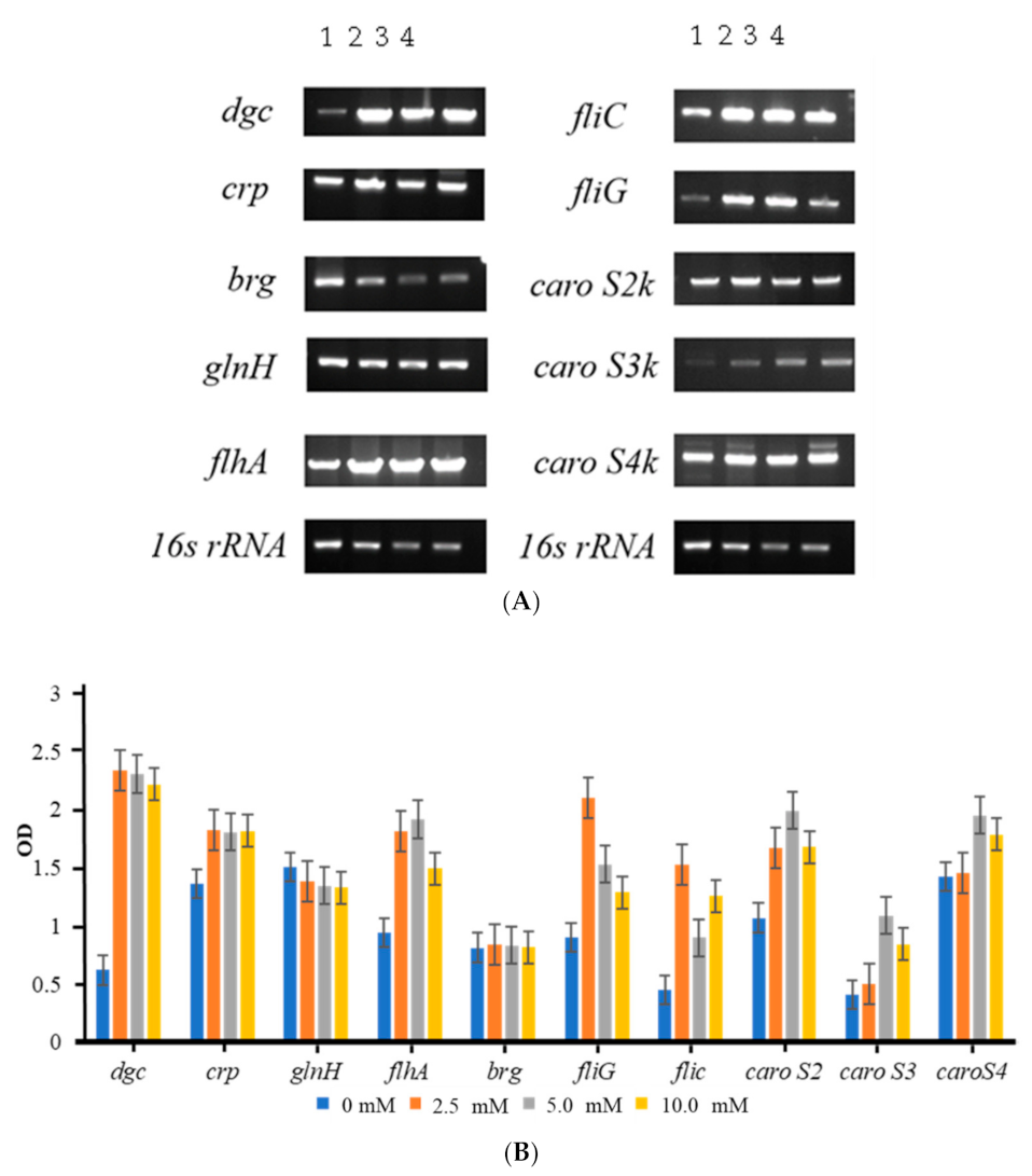
2.5. Extracellular L-Glutamine Concentrations Have Varied Effects on 89-H-rif-12

Bacteria’s tremendous adaptability to the environment is based on their ability to detect changes in the environment. To explore bacteriocin secretion in a nutrient-rich environment, we cultured 89-H-rif-12 in an environment containing L-glutamine to see if it affected bacteriocin secretion and regulatory gene expression. So, we cultured 89-H-rif-12 in 2.5 mM, 5.0 mM, and 10.0 mM L-glutamine LB medium and the control group without L-glutamine and incubated them overnight at 28 °C.
The RNA was extracted from exponentially grown cells induced at various L-glutamine concentrations for a two-step endpoint RT PCR analysis of genes associated with the carocin genes, regulatory genes, and type III secretory genes in both the wild-type and the dgc defective mutant.
The ability of the induced cells to create, regulate, and release bacteriocin was studied using a two-step endpoint reverse transcriptase polymerase chain reaction (endpoint RT-PCR). The gene segments 16s RNA, dgc, crp, brg, glnH, flhA, fliC, fliG, caroS2k, caroS3k, and caroS4k were successively amplified (Figure 4A).
Based on Figure 4B, there is no significant difference in the expression levels of brg and glnH in the L-glutamine environment. The expression levels of related protein genes dgc, crp, and the third secretion system-related protein genes flhA, fliC, and fliG were significantly increased compared to the control group, despite the fact that GlnH is an active transport protein that transports extracellular L-glutamine into the cell.
Increased production of these proteins associated with bacteriocin regulation also resulted in increased expression of the caroS2k, caroS3k, and caroS4k bacteriocin genes, proving that there is a causal relationship between bacteriocin expression and L-glutamine concentration.

3. Discussion

A number of transmission pathways are created by rapidly altering genes in cells in order to boost the ability of bacteria to compete for survival in the natural environment. The transmission pathway involves a large number of proteins and compounds. The secondary signal produced by DGC protein has been implicated in prior research, and c-di-GMP is required for the regulation of Pcc’s bacteriocin synthesis mechanism.
In addition to bacteriocin secretion, the regulatory mechanism for delivering bacteriocin to the extracellular environment comprises secreted proteins and immune proteins. In this study, blocking the dgc gene of 89-H-rif-12 resulted in no zone of inhibition around the mutant strains in the bacteriocin assay, indicating the strain’s inability to produce low-molecular-weight bacteriocin, and the related gene was turned off to prevent the production of bacteriocin. These findings clearly show how c-di-GMP, a secondary signal produced by DGC proteins, affects the production of bacteriocins.
DGC positively regulates the expression of fliC and fliG of the type III secretion system but cannot regulate flhA, despite the fact that blocking the T3SS prevents the secretion of bacteriocins into cells. Furthermore, the dgc defective mutant cannot transcribe bacteriocin toxin genes such as caros2k, caros3k, and caros4k. As a result, it is thought that DGC not only transports toxins in the Pcc regulatory system but also plays a role in the manufacture of bacteriocin.
Bacteria’s remarkable environmental adaptability is dependent on their ability to detect changes in their environment. The probable mechanism (Figure 5) of DGC regulating the bacteriocin gene is hypothesized based on the relevant literature.
DGC responds to UV irradiation and nutrient-rich environments, such as high glutamine concentrations. The signal created by cAMP and CRP changes the conformation of DGC when the sensor protein on bacteria is triggered by the outside world. DGC cyclizes c-di-GMP, and c-di-GMP generates a compound with CLP to remove CLP from the bacteriocin promoter-binding site; then, DGC activates the DNA-binding site and binds to the DGC promoter sequence to induce dgc transcription. It is released so that the bacteriocin promoter can be coupled with RNA polymerase to create bacteriocin toxin and immune proteins during transcription.
DGC was also observed to have an effect on the regulation of genes in the type III secretion system in this experiment. FliC encodes a flagellar filament-type structure and can export proteins more efficiently after the hook structure.
The c-di-GMP does not regulate fliC, according to earlier research. However, when the dgc gene was suppressed, the production of fliC was lost, and the FliC protein could not be transmitted out of the cell to construct a complete flagellar structure, resulting in the inability to send low molecular weight bacteriocin out of the cell.
Furthermore, prior research has revealed that fliC and flhA mutants are unable to manufacture bacteriocins, but the two genes do not control each other, and dgc mutants have no influence on flhA expression [25], although the exact mechanism is unknown at this time. To regulate the fliC gene, what receptor does c-di-GMP bind to? When clp is altered in Xcc, the transcriptional expression of fliC is decreased, and the clp mutant produces no flagella and cannot move under electron microscopy. As a result, c-di-GMP and CLP will alter fliC transcription in Xcc [26], and more research into how c-di-GMP regulates fliC transcription in Pcc is required.
FliG is a switch protein for flagellar movement on the T3bSS inner membrane group’s flagellar organelle, which dictates the composition and rotation direction of the flagella and mostly controls flagella torsion [27]. The bacteriocins secretion was found to have no effect on the fliG mutant generated in the previous research. The c-di-GMP is the signaling molecule that predominantly impacts flagellar motility in many studies, and variations in c-di-GMP concentration cause bacteria to go from a planktonic state to a surface-attached state; therefore, this protein has little to do with the transport of bacteriocins [19].
In 2021, the Pilz gene was identified in the NCBI database’s Pcc gene. Pilz is a polysaccharide synthesis, cell motility, and cell differentiation c-di-GMP receptor protein. The YcgR protein from Escherichia coli is the best example of the PilZ proteins in the current investigation. The YcgR-c-di-GMP complex interacts with the flagellar motor protein FliG, which regulates flagellar rotation and allows swimming speed, in addition to the PilZ domain [28,29,30].
We know from the findings of this experiment that the dgc mutant prevents fliG transcription, indicating that c-di-GMP is still vital in controlling flagella movement in the Pcc strain. When the speed of bacterial migration increases, the ability to infect increases as well.
Bacteria’s ability to notice changes in the environment is crucial to their high adaptability to the environment. Pcc penetrates plants via lenticels or wounds and remains in the intercellular space until environmental circumstances are favorable for bacterial growth. The growth of bacteria, as well as the performance of virulence, is influenced by pH, carbon source, and nitrogen source. Pcc must absorb nutrients through protein channels on the membrane in order to survive in nature.
GlnH is an ATP-binding cassette-binding active transporter protein that is necessary for L-glutamine transport. It may be deduced from the action of various exogenous quantities of L-glutamine on 89-H-rif-12 that when L-glutamine surpasses a specific concentration, crp interacts with it. The downstream gene dgc, the third secretion system-related protein genes flhA, fliC, fliG, and the bacteriocin poisoning protein genes caroS2k, caroS3k, and caroS4k are all significantly expressed, and L-glutamine has been shown to alter the expression of virulence in other bacteria as well.
Other studies have found that because ammonia makes up the majority of nitrogen in soil and water and L-glutamine is scarce, Listeria cells with low ammonia and high L-glutamine will activate the transcription of virulence genes [31]. However, mammalian cells with extremely little ammonia and very high L-glutamine will promote virulence gene transcription [32,33].
When the concentration of L-glutamine in Pcc cells exceeds a particular level, it is thought that some proteins can detect favorable environmental conditions for development and send out signals to enhance CRP expression, altering motility and toxicity. Our findings support previous evidence indicating that L-glutamine, an abundant nitrogen source, significantly increases the expression of all main virulence genes in pathogens [34,35].

4. Materials and Methods

Bacterial strains, plasmids, media, and growth conditions. The bacterial strains, plasmids, and primers utilized in this study are listed in Table 2 and Table 3. Pcc strains were cultured on a modified Luria-Bertani (LB) medium with 5 g sodium chloride per liter at 28 °C (half of the recommended quantity of NaCl). Strains of E. coli (cloning host) were grown in LB broth at 37 °C with rotary agitation at 125 rpm. The IFO-802 medium was supplemented with 1% polypeptin, 0.2% yeast extract, 0.1% MgSO4 (pH 7.0), and 1.5% agar. Antibiotic concentrations employed in the bacterial selection, 50 g/mL ampicillin, 50 g/mL kanamycin, and 50 g/mL rifampicin, were used to treat E. coli and Pcc strains. All bacterial growth densities were measured at 595 nm using a spectrophotometer (OD595).
Bacterial matting. The membrane-filter method was used for bacterial mating, as described by Gantotti et al. [37]. Pcc (receiver) H-rif-8-6 and E. coli cultures overnight E. coli (donor) 1830 were equally spread on 0.22 µm pore size membrane filters (Millipore, Inc. Bedford, MA, USA) and incubated overnight at 28 °C on LB agar media. After conjugation, progeny suspensions were properly diluted and cultured on modified Drigalski agar plates (with rifampicin and kanamycin, 100 g ml−1) at 28 °C for 24 to 48 h. For the bacteriocin production test, colonies were isolated.
Bacteriocin assays. The antibacterial activity of the bacteriocin-producing strain was assessed using the soft agar overlay method [39]. Hard IFO-802 (1.4% agar) and soft IFO-802 (0.65% agar) medium were used to grow the isolates. For colonies to develop, the cells were first cultured for 12 h. After that, the colonies were subjected to ultraviolet light before being incubated for another 12 h. The cells were then treated with chloroform before being covered with soft agar that contained the indicator cells. Bacteriocin production is indicated by an inhibitory zone of indicator-cell (SP33) development surrounding the colony.
Preparation of genomic DNA, plasmid DNA and RNA. Standard protocols for restriction endonuclease digestions, agarose gel electrophoresis, purification of DNA from agarose gels, DNA ligation, and other cloning-related techniques were followed [40,41]. E. coli DH5α cells that are exponentially growing (OD595 of about 1.0) were extracted for RNA extraction. RNA was extracted using Trizol reagent (Invitrogen, Waltham, MA, USA) and resuspended in DEPC-treated water according to the manufacturer’s instructions. The purity and concentration of total RNA were measured using a NanoVue PlusTM spectrophotometer (Biochrom, Holliston, MA, USA), followed by electrophoresis on a 1.5% formaldehyde-morpholinepropanesulfonic-agarose gel for evaluation.
TAIL-PCR and restriction DNA library screening. Detailed protocols have been utilized for the PCR, which used Promega’s Go-Taq DNA polymerase [42]. Thermal asymmetric interlaced PCR (TAIL-PCR) was utilized, as previously reported [41,43].
For TAIL-PCR, specific primers were developed at both ends of Tn5, namely PF1, PF2, PF3, PR1, PR2, and PR3. Two PCR products with unknown DNA sequences at both ends of Tn5 were produced using the above primers for amplification. However, after sequencing the two segments, it was discovered that they were the same PCR products. As a result, two sets of two PCR products were created utilizing this known 500-bp DNA sequence.
The TAIL-PCR products were sequenced using an ABI PRISM Dye Terminator Cycle Sequencing Ready Reaction kit (Applied Biosystems, Foster City, CA, USA). A GeneAmp System 9600 thermocycler was used to sequence the cycles (Applied Biosystems). According to the manufacturer’s procedure, the sequencing was conducted using an ABI 373S automated DNA sequencer 373S (Applied Biosystems).
Subcloning of dgc gene from 89-H-rif-12. Using the oligonucleotide primers Pcc DGC F upstream and Pcc DGC R downstream, the DNA fragment of dgc was amplified by PCR from 89-H-rif-12. TA cloning (Promega Inc., Madison, WI, USA) was used for subcloning the PCR product into the pGEM-T Easy vector, resulting in the plasmid pG-dgc. As previously described [35,38,39], SLIM excision of the tag element within the ribosome binding site and start codon of dgc in pG-dgc gave the construct pG-dgc-Kan. DGC Xho1 Fs, DGC Xho1 Ft, DGC Xho1 Rs, DGC Xho1 Rt, DGC Hpa1 Fs, DGC Hpa1 Ft, DGC Hpa1 Rs, and DGC Hpa1 Rt were used as primers. pG-dgc-Kan was then injected into E. coli. BL21 (DE3) cells were used.
Electroporation (1.25 kV/cm, 200, 25F) was used to insert plasmids into Pcc strains [44,45]. The competent E coli cells for heat-shock transformation were obtained using Hanahan’s method [33,37].

5. Conclusions

Under environmental stimulation, Pccs manufacture bacteriocin and defense protein molecules via the SOS mechanism. The c-di-GMP, which is formed from GTP by diguanylate cyclases, is a widely conserved bacterial signaling molecule that influences motility, biofilm formation, and virulence. This is the first study to show a link between dgc and the genes involved in Pcc LMWB extracellular export via the flagellar-type secretion system. The probable mechanism of dgc is hypothesized to synthesize the secondary signaling molecule c-di-GMP, which can bind to receptor proteins and positively influence bacteriocin transcription as well as toxin production, motility, and transport.

Supplementary Materials

The following supporting information can be downloaded at: https://www.mdpi.com/article/10.3390/ijms23105649/s1.

Author Contributions

R.B.J.S.L. led the investigation, wrote, and edited the manuscript. H.-L.C. and Y.-C.C. participated in the investigation and wrote the original draft of the manuscript. D.-Y.C. conceived the study, participated in its design, and corrected the manuscript. All authors have read and agreed to the published version of the manuscript.

Funding

This research received no external funding.

Institutional Review Board Statement

Not applicable.

Informed Consent Statement

Not applicable.

Data Availability Statement

Not applicable.

Conflicts of Interest

The authors declare that they have no conflict of interest.

References

  1. Tischler, A.D.; Camilli, A. Cyclic Diguanylate (C-Di-GMP) Regulates Vibrio Cholerae Biofilm Formation. Mol. Microbiol. 2004, 53, 857–869. [Google Scholar] [CrossRef] [PubMed] [Green Version]
  2. Zogaj, X.; Nimtz, M.; Rohde, M.; Bokranz, W.; Romling, U. The Multicellular Morphotypes of Salmonella Typhimurium and Escherichia coli Produce Cellulose As The Second Component of The Extracellular Matrix. Mol. Microbiol. 2001, 39, 1452–1463. [Google Scholar] [CrossRef] [PubMed]
  3. Aldridge, P.; Paul, R.; Goymer, P.; Rainey, P.; Jenal, U. Role of The GGDEF Regulator Pled In Polar Development of Caulobacter Crescentus. Mol. Microbiol. 2003, 47, 1695–1708. [Google Scholar] [CrossRef] [PubMed]
  4. Jenal, U.; Reinders, A.; Lori, C. Cyclic Di-GMP: Second Messenger Extraordinaire. Nat. Rev. Genet. 2017, 15, 271–284. [Google Scholar] [CrossRef] [Green Version]
  5. Chan, C.; Paul, R.; Samoray, D.; Amiot, N.C.; Giese, B.; Jenal, U.; Schirmer, T. Structural Basis of Activity and Allosteric Control Of Diguanylate Cyclase. Proc. Natl. Acad. Sci. USA 2004, 101, 17084–17089. [Google Scholar] [CrossRef] [Green Version]
  6. Teixeira, R.D.; Holzschuh, F.; Schirmer, T. Activation mechanism of a small prototypic Rec-GGDEF diguanylate cyclase. Nat. Commun. 2021, 12, 2162. [Google Scholar] [CrossRef]
  7. Römling, U.; Gomelsky, M.; Galperin, M. C-Di-GMP: The Dawning of A Novel Bacterial Signalling System. Mol. Microbiol. 2005, 57, 629–639. [Google Scholar] [CrossRef]
  8. Jenal, U. Cyclic Di-Guanosine-Monophosphate Comes of Age: A Novel Secondary Messenger Involved in Modulating Cell Surface Structures in Bacteria? Curr. Opin. Microbiol. 2004, 7, 185–191. [Google Scholar] [CrossRef]
  9. D’Argenio, D.A.; Miller, S.I. Cyclic Di-GMP as A Bacterial Second Messenger. Microbiology 2004, 150, 2497–2502. [Google Scholar] [CrossRef] [Green Version]
  10. Sudarsan, N.; Lee, E.R.; Weinberg, Z.; Moy, R.H.; Kim, J.N.; Link, K.H.; Breaker, R.R. Riboswitches in Eubacteria Sense The Second Messenger Cyclic Di-GMP. Science 2008, 321, 411–413. [Google Scholar] [CrossRef] [Green Version]
  11. Nelson, J.; Sudarsan, N.; Furukawa, K.; Weinberg, Z.; Wang, J.X.; Breaker, R.R. Riboswitches in Eubacteria Sense The Second Messenger C-Di-AMP. Nat. Chem. Biol. 2013, 9, 834–839. [Google Scholar] [CrossRef] [PubMed]
  12. Fazli, M.; O’Connell, A.; Nilsson, M.; Niehaus, K.; Dow, J.M.; Givskov, M.; Ryan, R.P.; Tolker-Nielsen, T. The CRP/FNR Family Protein Bcam1349 Is A C-Di-GMP Effector That Regulates Biofilm Formation in The Respiratory Pathogen Burkholderia Cenocepacia. Mol. Microbiol. 2011, 82, 327–341. [Google Scholar] [CrossRef] [PubMed]
  13. Chin, K.-H.; Lee, Y.-C.; Tu, Z.-L.; Chen, C.-H.; Tseng, Y.-H.; Yang, J.-M.; Ryan, R.P.; McCarthy, Y.; Dow, J.M.; Wang, A.H.-J.; et al. The Camp Receptor-Like Protein CLP Is A Novel C-Di-GMP Receptor Linking Cell–Cell Signaling to Virulence Gene Expression in Xanthomonas Campestris. J. Mol. Biol. 2010, 396, 646–662. [Google Scholar] [CrossRef] [PubMed]
  14. McCarthy, R.R.; Valentini, M.; Filloux, A. Contribution of Cyclic Di-GMP in The Control of Type III and Type VI Secretion in Pseudomonas aeruginosa. C-Di-GMP Signaling; Humana Press: New York, NY, USA, 2017; pp. 213–224. [Google Scholar] [CrossRef]
  15. Trampari, E.; Stevenson, C.E.M.; Little, R.H.; Wilhelm, T.; Lawson, D.M.; Malone, J.G. Bacterial Rotary Export Atpases Are Allosterically Regulated By The Nucleotide Second Messenger Cyclic-Di-GMP. J. Biol. Chem. 2015, 290, 24470–24483. [Google Scholar] [CrossRef] [Green Version]
  16. Haque, M.M.; Oliver, M.H.; Nahar, K.; Alam, M.Z.; Hirata, H.; Tsuyumu, S. Cytr Homolog of Pectobacterium Carotovorum Subsp. Carotovorum Controls Air-Liquid Biofilm Formation By Regulating Multiple Genes involved in Cellulose Production, C-Di-GMP Signaling, Motility, and Type III Secretion System in Response to Nutritional and Environmental Signals. Front. Microbiol. 2017, 8, 972. [Google Scholar] [CrossRef] [Green Version]
  17. Tseng, T.-T.; Tyler, B.M.; Setubal, J.C. Protein Secretion Systems in Bacterial-Host associations, and Their Description in The Gene Ontology. BMC Microbiol. 2009, 9, S2. [Google Scholar] [CrossRef] [Green Version]
  18. Baena, F.J.L.; Vinardell, J.-M.; Medina, C. Regulation of Protein Secretion Systems Mediated By Cyclic Diguanylate in Plant-interacting Bacteria. Front. Microbiol. 2019, 10, 1289. [Google Scholar] [CrossRef] [Green Version]
  19. Wu, H.-P.; Derilo, R.C.; Chen, H.-L.; Li, T.-R.; Lagitnay, R.B.J.S.; Chan, Y.-C.; Chuang, Y.; Chuang, D.-Y. Injectisome T3SS Subunits as Potential Chaperones in The Extracellular Export of Pectobacterium Carotovorum Subsp. Carotovorum Bacteriocins Carocin S1 and Carocin S3 Secreted Via Flagellar T3SS. BMC Microbiol. 2021, 21, 345. [Google Scholar] [CrossRef]
  20. Hengge, R. Principles of c-di-GMP signalling in bacteria. Nat. Rev. Microbiol. 2009, 7, 263–273. [Google Scholar] [CrossRef]
  21. Bolivar, F.; Rodriguez, R.L.; Greene, P.J.; Betlach, M.C.; Heyneker, H.L.; Boyer, H.W.; Crosa, J.H.; Falkow, S. Construction and Characterization of New Cloning Vehicle. II. A Multipurpose Cloning System. Gene 1977, 2, 95–113. [Google Scholar] [CrossRef]
  22. Hockett, K.L.; Baltrus, D.A. Use of the soft-agar overlay technique to screen for bacterially produced inhibitory compounds. J. Vis. Exp. 2017, 119, 55064. [Google Scholar] [CrossRef] [PubMed] [Green Version]
  23. Gause, W.C.; Adamovicz, J. The Use of The PCR to Quantitate Gene Expression. Genome Res. 1994, 3, S123–S135. [Google Scholar] [CrossRef] [Green Version]
  24. Tang, Y.W. Advanced Techniques in Diagnostic Microbiology; Springer: New York, NY, USA, 2012; ISBN 978-1461439691. [Google Scholar]
  25. Chan, Y.-C.; Wu, H.-P.; Chuang, D.-Y. Extracellular Secretion of Carocin S1 in Pectobacterium Carotovorum Subsp. Carotovorumoccurs Via The Type III Secretion System integral to The Bacterial Flagellum. BMC Microbiol. 2009, 9, 181. [Google Scholar] [CrossRef] [PubMed] [Green Version]
  26. Lee, M.-C.; Weng, S.-F.; Tseng, Y.-H. Flagellin Gene Flic of Xanthomonas Campestris Is Upregulated By Transcription Factor Clp. Biochem. Biophys. Res. Commun. 2003, 307, 647–652. [Google Scholar] [CrossRef]
  27. A Lloyd, S.; Tang, H.; Wang, X.; Billings, S.; Blair, D.F. Torque Generation in The Flagellar Motor of Escherichia coli: Evidence of a Direct Role for Flig But Not for Flim or Flin. J. Bacteriol. 1996, 178, 223–231. [Google Scholar] [CrossRef] [PubMed] [Green Version]
  28. Paul, K.; Nieto, V.; Carlquist, W.C.; Blair, D.F.; Harshey, R.M. The C-Di-GMP Binding Protein Ycgr Controls Flagellar Motor Direction and Speed to Affect Chemotaxis By A “Backstop Brake” Mechanism. Mol. Cell 2010, 38, 128–139. [Google Scholar] [CrossRef] [PubMed] [Green Version]
  29. Fang, X.; Gomelsky, M. A Post-Translational, C-Di-GMP-Dependent Mechanism Regulating Flagellar Motility. Mol. Microbiol. 2010, 76, 1295–1305. [Google Scholar] [CrossRef]
  30. Ryjenkov, D.A.; Simm, R.; Römling, U.; Gomelsky, M. The Pilz Domain Is A Receptor For The Second Messenger C-Di-GMP. J. Biol. Chem. 2006, 281, 30310–30314. [Google Scholar] [CrossRef] [Green Version]
  31. Planche, T. Plasma Glutamine and Glutamate Concentrations in Gabonese Children With Plasmodium Falciparum infection. QJM 2002, 95, 89–97. [Google Scholar] [CrossRef] [Green Version]
  32. Tullius, M.V.; Harth, G.N.; Horwitz, M.A. Glutamine synthetase GlnA1 is essential for growth of Mycobacterium tuberculosis in human THP-1 macrophages and guinea pigs. Infect. Immun. 2003, 71, 3927–3936. [Google Scholar] [CrossRef] [Green Version]
  33. Hanahan, D. Studies On Transformation of Escherichia coli with Plasmids. J. Mol. Biol. 1983, 166, 557–580. [Google Scholar] [CrossRef]
  34. Haber, A.; Friedman, S.; Lobel, L.; Burg-Golani, T.; Sigal, N.; Rose, J.; Livnat-Levanon, N.; Lewinson, O.; Herskovits, A.A. L-Glutamine induces Expression of Listeria Monocytogenes Virulence Genes. PLOS Pathog. 2017, 13, e1006161. [Google Scholar] [CrossRef] [PubMed]
  35. Minato, Y.; Fassio, S.R.; Wolfe, A.J.; Häse, C.C. Central Metabolism Controls Transcription of A Virulence Gene Regulator in Vibrio Cholerae. Microbiology 2013, 159, 792–802. [Google Scholar] [CrossRef] [PubMed] [Green Version]
  36. Reusch, R.N.; Hiske, T.W.; Sadoff, H.L. Poly-Beta-Hydroxybutyrate Membrane Structure and Its Relationship to Genetic Transformability in Escherichia coli. J. Bacteriol. 1986, 168, 553–562. [Google Scholar] [CrossRef] [Green Version]
  37. Gantotti, B.V.; Kindle, K.L.; Beer, S.V. Transfer of the drug-resistance transposon Tn5 to Erwinia herbicola and the induction of insertion mutations. Curr. Microbiol. 1981, 6, 377–381. [Google Scholar] [CrossRef]
  38. Hirsch, P.; Beringer, J. A Physical Map of Pph1ji and Pjb4ji. Plasmid 1984, 12, 139–141. [Google Scholar] [CrossRef]
  39. Sambrook, J.; Russel, D.W. Molecular Cloning: A laboratory Manual, 3rd ed.; Cold Spring Harbor Laboratory Press: New York, NY, USA, 2001. [Google Scholar]
  40. Liu, H.; Naismith, J.H. An Efficient One-Step Site-Directed Deletion, insertion, Single and Multiple-Site Plasmid Mutagenesis Protocol. BMC Biotechnol. 2008, 8, 91. [Google Scholar] [CrossRef] [Green Version]
  41. Addgene: Handling Plasmids from Addgene—Purifying Plasmid DNA. Addgene.org. Available online: https://www.addgene.org/protocols/purify-plasmid-dna/ (accessed on 15 March 2021).
  42. Liu, Y.-G.; Whittier, R.F. Thermal asymmetric interlaced PCR: Automatable Amplification and Sequencing of insert End Fragments From P1 and YAC Clones For Chromosome Walking. Genomics 1995, 25, 674–681. [Google Scholar] [CrossRef]
  43. Chiu, J. Site-Directed, Ligase-independent Mutagenesis (SLIM): A Single-Tube Methodology Approaching 100% Efficiency in 4 H. Nucleic Acids Res. 2004, 32, e174. [Google Scholar] [CrossRef]
  44. A Fyfe, J.; Harris, G.; Govan, J.R. Revised Pyocin Typing Method For Pseudomonas Aeruginosa. J. Clin. Microbiol. 1984, 20, 47–50. [Google Scholar] [CrossRef] [Green Version]
  45. Sambrook, J.; Fritsch, E.F.; Maniatis, T. Molecular Cloning: A Laboratory Manual; Cold Spring Harbor Laboratory Press: Cold Spring Harbor, NY, USA, 1989. [Google Scholar]
Figure 1. Bacteriocin of Pectobacterium carotovorum subsp. carotovorum. (A) The bacteriocin test was performed by soft agar overlay. (1) Wild-type rif-TO6 Pcc strain, (2–15) transposon Tn5 insertional mutants: 2—TT6-1, 3—TT6-3, 4—TT6-2, 5—TT6-4, 6—TT6-5, 7—TT6-6, 8—TT6-9, 9—TT6-12, 10—TT6-13, 11—TT6-14, 12—TT6-19, 13—TT6-21, 14—TT6-22, and 15—TT6-25. (B) Screening of the wild-type rif-TO6 and transposon Tn5 insertional mutants with Drigalski medium. If the strain is Pcc, the medium will be golden yellow, but if it is an Escherichia coli colony, the medium will be blue. (1) Wild Type rif-TO6 Pcc strain, (2–15) transposon Tn5 insertional mutants 2—TT6-1, 3—TT6-3, 4—TT6-2, 5—TT6-4, 6—TT6-5, 7—TT6-6, 8—TT6-9, 9—TT6-12, 10—TT6-13, 11—TT6-14, 12—TT6-19, 13—TT6-21, 14—TT6-22, and 15—TT6-25.
Ijms 23 05649 g001
Figure 2. Bacteriocin assay of the gene knockout strains. (A) Bacteriocin activity of the gene selection strain (89-H-rif-12). (B) Comparison of bacteriocin activity of wild types and dgc defective mutants: 1, Pcc 3F3; 2, 89-H-rif-89-12; 3, 89-H-rif-12/Δdgc-Tet; 4, 89-H-rif-12/Δdgc-Kan.
Ijms 23 05649 g002
Figure 3. Transcriptional analysis of bacteriocin genes and regulatory genes by two-step endpoint RT-PCR. (A) 16s RNA expression of the wild-type 89-H-rif-12 and the dgc defective mutant. (B) Shown are gel electropherogram of RT-PCR products exhibited by the wild-type and dgc defective mutants.
Ijms 23 05649 g003
Figure 4. Transcriptional analysis of bacteriocin genes and regulatory genes by two-step endpoint RT-PCR induced with different concentrations of L-glutamine. (A) Gel electropherogram of RT-PCR products exhibited by the wild-type and the dgc knockout strain: 1, 0 mM; 2, 2.5 mM; 3, 5.0 mM; 4, 10 mM. (B) Mean relative gene expression affected by the L-glutamine expression.
Ijms 23 05649 g004
Figure 5. DGC and bacteriocin regulation, synthesis, and secretion: a proposed molecular pathway.
Ijms 23 05649 g005
Table 1. List of genes identified by transposon mutagenesis.
StrainsProducts
TT6-1ATPase associated with various cellular activities, AAA_5, MoxR-like ATPase
TT6-2bacteriophage tail protein [Escherichia coli E22]
TT6-3peptidase S8 and S53 subtilisin kexin sedolisin; killer protein of pyocin S3
TT6-4bacteriophage tail protein [Escherichia coli E22]
TT6-5putative periplasmic ligand-binding sensor protein
TT6-6diguanylate cyclase AdrA [Shigella boydii Sb227]
TT6-9bacteriophage tail protein [Escherichia coli E22]
TT6-10putative phage tail fiber protein [Escherichia coli E24377A]
TT6-13killer protein of pyocin S3
TT6-14putative bacteriophage protein GP48
TT6-19putative phage tail fiber protein [Escherichia coli E24377A]
TT6-21killer protein of pyocin S3
TT6-22bacteriophage tail protein [Escherichia coli E22]
Table 2. Bacteria and plasmids used in this study.
Strain or PlasmidRelevant CharacteristicsSource of Reference
Escherichia coli
DH5αSupE44 ΔlacU169 (Φ80 lacZ ΔM15) hsdR17 recA1 endA1 gyrA96 thi-l relA1Hanahan; Reusch et al. [33,36]
1803prometKmrNmr, containing transposon Tn5 on the suicidal plasmid pBJ4JIGantotti et al. [37]
Pectobacterium carotovorum subsp. carotovorum
TO6Pcc, Ampr, wild type, putative biocontrol agentLaboratory stock
Rif-TO6Pcc, Ampr, Rifr, wild typeLaboratory stock
89-H-4Pcc, Ampr, wild-type, putative biocontrol agentLaboratory stock
89-H-rif-12Pcc, Ampr, Rifr, wild typeLaboratory stock
3F3Pcc, Ampr, wild typeLaboratory stock
SP33Pcc, wild-type indicator strainLaboratory stock
H-rif-89-12/∆dgc-KanPcc, Ampr, Rifr, Kanr, wild typeThis study
H-rif-89-12/∆dgc-TetPcc, Ampr, Rifr, Tetr, wild typeThis study
Plasmids
pJB4JIsuicide vector for Tn5, pJB4JI, contains pPH1JI, bacteriophage Mu, and Tn5P.R. Hirsch, J.E. Beringer, [38]
pGEM T-EasyAmpr, lacZ cloning vectorPromega
pBR322Ampr, TetrBolivar et al. [21]
pG-dgcpGEM T-Easy, Ampr, dgcThis study
pG-dgc-Xho1/Hpa1pGEM T-Easy, Ampr, dgcThis study
pG-TetpGEM T-Easy, Ampr, TetrLaboratory stock
pG-Δdgc-TpGEM T-Easy, Ampr, dgc, TetrThis study
pG-Δdgc-KpGEM T-Easy, Ampr, dgc, KanrThis study
Table 3. Primers used in this study.
PrimersSequence (5′-3′)
P3CTCGACGTTGTCACTGAAGCGGGAAG
P4AAAGCACGAGGA GCGGTCAGCCCAT
PF1AGAGA ACACAGATTTAGCCCAGTCGG
PF2CCGCACGATGAAGAGCAGAAGTTAT
PF3GATCCTGGAAAACGGGAAAGGTTC
PR1GCCGAAGAGAACACAGATTTAGCCCA
PR2CCGCACGATGAAGAGCAGAAGTT
Pcc_DGC_F_upstreamCTCACTGTTGCTGACATGC
Pcc_DGC_R_downstreamATTCAGGCAACTTCGGTTC
DGC_Xho1_FsCGACAATCCGTGGAATATAG
DGC_Xho1_FtCTCGAGCGACAATCCGTG
DGC_Xho1_RsGTACGATACTGTGCGCTC
DGC_Xho1_RtCTCGAGGTACGATACTGTG
DGC_Hpa1_FsGATAAGCTCTCCCGAATACG
DGC_Hpa1_FtGTTAACGATAAGCTCTCCCG
DGC_Hpa1_RsGCTGAAGTTTCTGAACCAG
DGC_Hpa1_RtGTTAACGCTGAAGTTTCTGAAC
F-16s- sense-ECCCTGGACAAAGACTGACGCTC
R-16s- sense-ECCTCGCTGGCAACAAAGGATAAG
caroS2K_RT_FGAGATACAATGACCGTGGATGG
caroS2K_RT_RGCAACTGGTGTTACCGTAACTG
caroS3K_RT_FATGATTAAGTACCGTTTATATGCTC
caroS3K_RT_RTCATTGCGACTCCCTCATAT
CaroS4KI_RT_RGGATCCATGATTAATTTTAAGG
CaroS4KI_RT_RGAGCTCTTAGAGACCGTAT
CRP_RT_FCTCTCGAATGGTTCCTTTCC
CRP_RT_RGAGATCAGGTTCTGGTCTTC
FlhA_RT_FTCACTCAAGCTTGCATCTAC
FlhA_RT_RAAGCTTTCACTTCTGAGCTTCC
GlnH_ RT_FACAGACCGGTGAATTACGCATCGG
GlnH_ RT_RGCCGCTACGCCTTCATCCATATTC
FliG senseATGACCCTGACAGGAACAG
FliG antisenseTTAGACATAAGCATCCTCGC
DY-F1GGTAGGATCCGTTGTTAGGTGCATAGGTTGG
DY-R1TTCAAGCTTGTGGTGAATTGACAATACGC
Publisher’s Note: MDPI stays neutral with regard to jurisdictional claims in published maps and institutional affiliations.

