Article

Phytochemical Profiling, Antioxidant, Anti-Inflammatory, Thrombolytic, Hemolytic Activity In Vitro and In Silico Potential of Portulacaria afra

1
Department of Pharmaceutical Chemistry, Faculty of Pharmacy, The Islamia University of Bahawalpur, Bahawalpur 63100, Pakistan
2
Department of Botany, University of Agriculture Faisalabad, Faisalabad 38000, Pakistan
3
Department of Environmental Sciences, The University of Lahore, Lahore 54590, Pakistan
4
Department of Botany and Microbiology, College of Science, King Saud University, P.O. Box 2455, Riyadh 11451, Saudi Arabia
5
The UWA Institute of Agriculture, School of Agriculture and Environment, The University of Western Australia, Perth, WA 6009, Australia
*
Authors to whom correspondence should be addressed.
Molecules 2022, 27(8), 2377; https://doi.org/10.3390/molecules27082377
Submission received: 6 March 2022 / Revised: 29 March 2022 / Accepted: 30 March 2022 / Published: 7 April 2022
(This article belongs to the Section Molecular Structure)

Abstract

:
The use of complementary herbal medicines has recently increased in an attempt to find effective alternative therapies that reduce the adverse effects of chemical drugs. Portulacaria afra is a rich source of phytochemicals with high antioxidant activity, and thus may possess health benefits. This study used the latest developments in GC-MS coupling with molecular docking techniques to identify and quantify the phytoconstituents in P. afra tissue extracts. The results revealed that n-butanol P. afra (BUT-PA) dry extracts contained total phenolic and flavonoids contents of 21.69 ± 0.28 mgGAE/g and 196.58 ± 6.29 mgGAE/g, respectively. The significant potential of antioxidants was observed through CUPRIC, FRAP, and ABTS methods while the DPPH method showed a moderate antioxidants potential for P. afra. Enzymatic antioxidants, superoxide dismutase, peroxidase and catalase also showed a better response in the BUT-PA dry extracts. The thrombolytic activity of the BUT-PA extracts ranged from 0.4 ± 0.32 to 11.2 ± 0.05%. Similarly, hemolytic activity ranged from 5.76 ± 0.15 to 9.26 ± 0.15% using the standard (triton x) method. The BUTPA and CHPA showed moderate acetylcholinesterase and butrylcholinesterase inhibition, ranging from 40.78 ± 0.52 to 58.97 ± 0.33, compared to galantamine. The carrageenan induced hind-paw edema assay, while BUT-PA extracts showed anti-inflammatory properties in a dose-dependent manner. Furthermore, 20 compounds were identified in the BUTPA extracts by GC-MS. Molecular docking was performed to explore the synergistic effect of the GC-MS-identified compounds on COX-1 and COX-2 inhibition. A high binding affinity was observed for Stigmastan-3, 5-diene, Phthalic acid, 3. Alpha-Hydroxy-5, 16-androstenol. The computed binding energies of the compounds revealed that all the compounds have a synergistic effect, preventing inflammation. It was concluded that active phytochemicals were present in P. afra, with the potential for multiple pharmacological applications as a latent source of pharmaceutically important compounds. This should be further explored to isolate secondary metabolites that can be employed in the treatment of different diseases.

1. Introduction

Medicinal plants are a rich source of novel lead compounds that can be used for a variety of therapeutic and pharmacological purposes [1]. The World Health Organization (WHO) estimates that up to 80% of the population in developing countries still depend on local medicinal plants to fulfill their primary healthcare needs. An estimated 25% of prescription drugs and 11% of drugs considered essential by the WHO are derived from plants, and a high number of synthetic drugs are obtained from precursor compounds originating from plants [2]. Their easy accessibility, efficacy in treatment and affordable cost are the main reasons to prefer traditional medicine to modern medication [3]. Thousands of polyphenolics compounds, such as phenolic and flavonoids, have been identified in these medicinal plants to date. The pharmacological and physiological potentials of phenolic compounds rely on their free radical scavenging and antioxidant activities to maintain the activity of the enzymes responsible for detoxification. Inflammation is a ubiquitous progression that occurs in a disturbed state of homeostasis, such as damage exposure to contaminating substances [4] and infection [5]. It is often triggered by innate immune system receptors to remove pathogens [6] when they are characterized by pain [7], redness, heat or warmth, and swelling [8]. Inflammation is classified into two types: acute and chronic. Acute inflammation could be the body’s early response to potentially damaging stimuli [9]. The symptoms of inflammation are severe pain, swelling, and loss of function of the affected area [10].
The inflammatory response is out of proportion in chronic inflammation, resulting in bodily harm [11]. Cyclooxygenase (COX) is a major enzyme in the production of prostacyclins, prostaglandins, and thromboxane, which play a role in inflammation, pain, and platelet aggregation [12,13]. Enzyme-glucuronidase has also been identified as a mediator in the beginning and during the progression of inflammation [14]. Free radicals include reactive species such as hydroxyl radicals, peroxyl radicals, superoxide radicals, hydrogen peroxide, superoxide anion, and other lipid peroxides [15].
Portulacaria afra is one of the dominant medicinally important plants, widely grown in the east of South Africa, belonging to the family Didiereaceae [16]. In this moist climate, it is relatively rare, and it tends to favor dryer rocky outcrops and slopes [17]. P. afra is an annual succulent herb with thick fleshy leaves for water storage [18]. This plant can grow up to 2 meters tall. It has red stems with round fleshy leaves, and produces pink flowers. It widely grows in warm climates in eastern South Africa [19]. The genus Portulaca contains approximately 100 species with a wide distribution, primarily in tropical and subtropical regions, and has a high degree of morphological variability [20].
Traditionally P. afra, was used to treat various skin disorders sores, ringworm, boils, burns, rash, wounds shingles, warts abscesses, and acne [21,22]. The identification of active phytochemicals from these natural sources, which can block COX-2 activity, may aid the production of anti-inflammatory pharmaceuticals. The present study aimed (1) to evaluate phytochemical components, total phenolic, total flavonoids, non-enzymatic antioxidants (DPPH, FRAP, ABTS, CUPRIC), enzymatic antioxidants (SOD, POD, CAT) and pharmacological activities in various fractions (n. hexane chloroform, n. butanol, methanol) using gas chromatography mass spectrometry (GC-MS), and (2) to perform in silico molecular docking analysis of bioactive compounds identified from P. afra.

2. Results

2.1. Phytochemical Screening, Total Phenolic and Flavonoids Contents

The presence of glycosides, saponins, tannins, flavonoids, phenols, fixed oils, and carbohydrates was discovered during phytochemical screening (Table 1). The total phenolic level identified in BUT-PA extracts of P. afra was 21.69 ± 0.28 mgGAE/g and flavonoid content was 196.58 ± 6.29 mgGAE/g in the dry extracts.

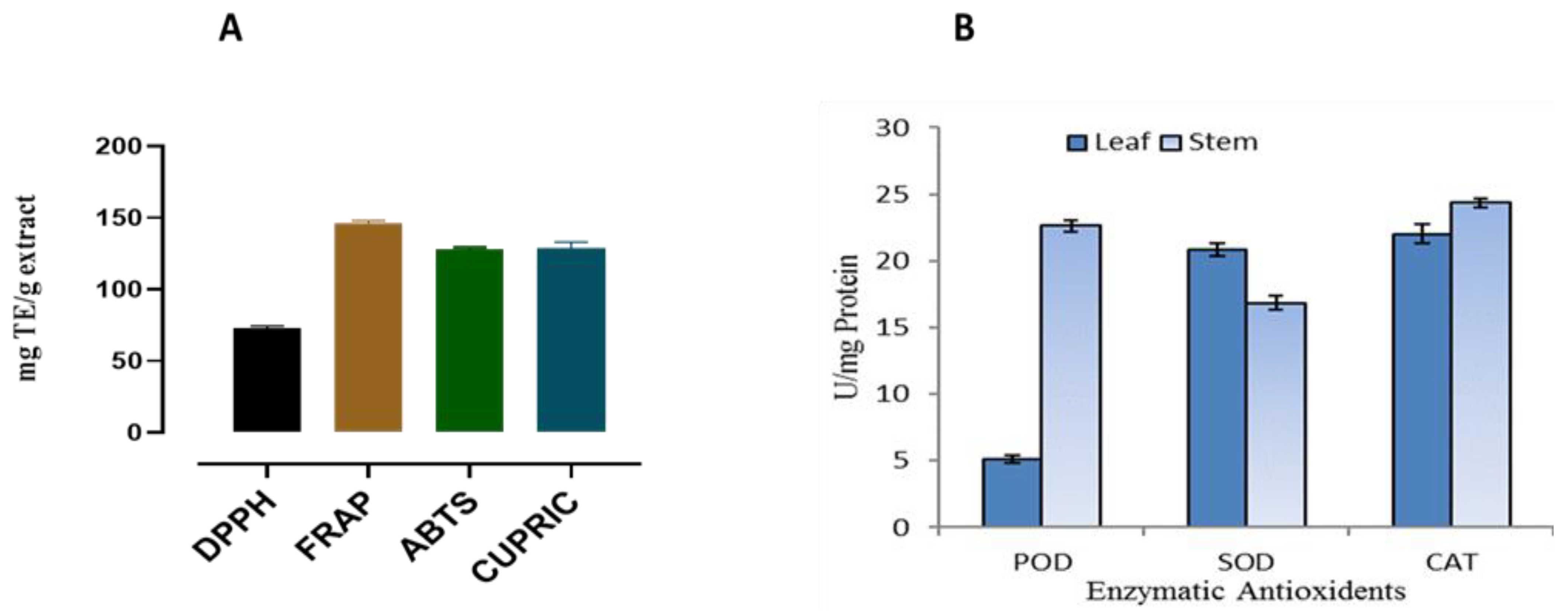
2.2. Antioxidant Potential

Analysis showed that the BUT-PA extracts of P. afra had significant antioxidant potential though CUPRIC, FRAP and ABTS methods, and moderate antioxidant potential through the DPPH method (Figure 1). The BUT-PA extracts showed the maximum antioxidant potential 144.74 ± 2.20 through the FRAP method, followed by a moderate potential via CUPRAC 124.19 ± 5.34 and ABTS 126.22 ± 0.64 methods, respectively. Enzymatic antioxidant in the BUT-PA extracts of P. afra had good potential for all tested enzymes: catalase (CAT), peroxidase (POD), and superoxide dismutase (SOD) (Figure 1).

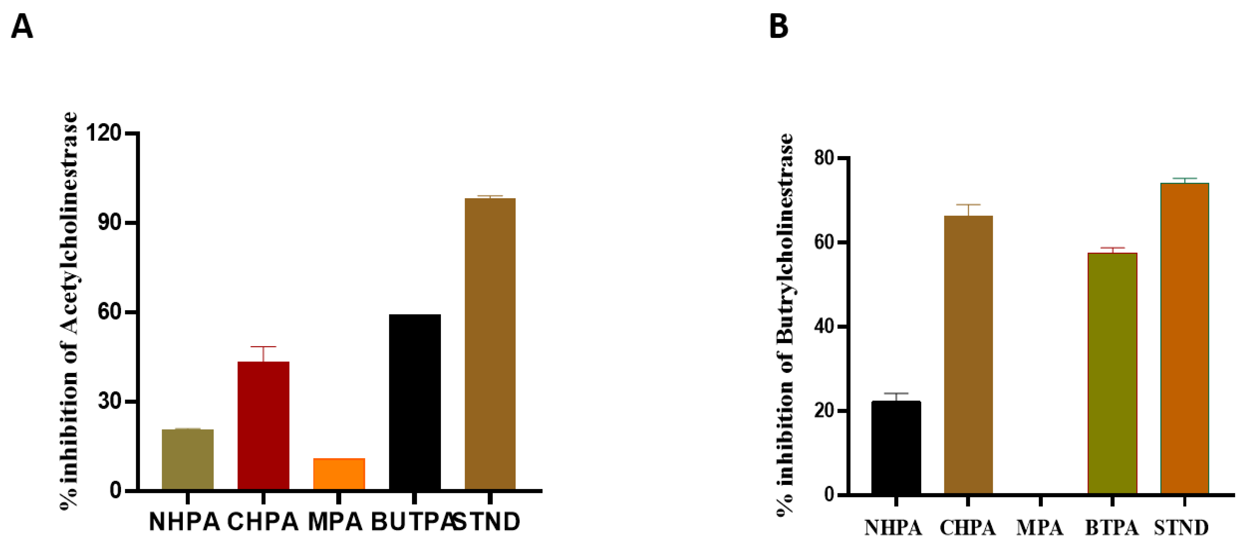
2.3. Acetylcholinesterase (AChE) and Butrylcholinesterase (BChE) Inhibition Activity

The decreasing order for AChE inhibition of the extract/fractions were obtained as: n-butanol fraction (BUTPA) > chloroform fraction (CHPA) > n-hexane fraction (NHPPA) > methanolic extract (MPA) (Figure 2). The % age acetylcholinesterase inhibition activity of the extract/fractions of P. afra was in the range from 20.55 ± 0.5 to 58.97 ± 0.33. The BChE inhibition results of different fractions were ordered as follows: chloroform fraction (CHPA) > n-butanol fraction (BUTPA) > n-hexane fraction (NHPA) > methanolic extract (MPA). The % inhibition of BChE of P. afra ranged from 22.29 ± 1.927 to 57.64 ± 1.192 (Figure 2).

2.4. Thrombolytic Activity

Thrombolytic activity was calculated for HPA, CHPA, MPA, and BUTPA. Thrombosis inhibition compared by Streptokinase (standard) showed significant clot lysis activity. While water was used as negative control, it showed minimum clot lysis. MPA showed minimum lysis 3.41 ± 0.32, followed by CHPA 7.32 ± 0.15 and HPA 11.21 ± 0.05. BUTPA showed a clot lysis of 5.51 ± 0.30, compared to the standard, 91.41 ± 0.06 (Table 2).

2.5. Hemolytic Activity

The data represented in Table 3 present the hemolytic activity of different extracts of P. afra. The MPA fraction has highest hemolytic percentage, 9.26 ± 0.15%, followed by BUTPA, 8.53 ± 0.25%, and CHPA, 7.46 ± 0.15%, which are statistically similar to each other. The lowest value, 5.76 ± 0.15%, was noticed in HPA extract. Overall, all four fractions have less than 30% hemolysis activity, so all fractions are nontoxic to humans and safe (Table 3).

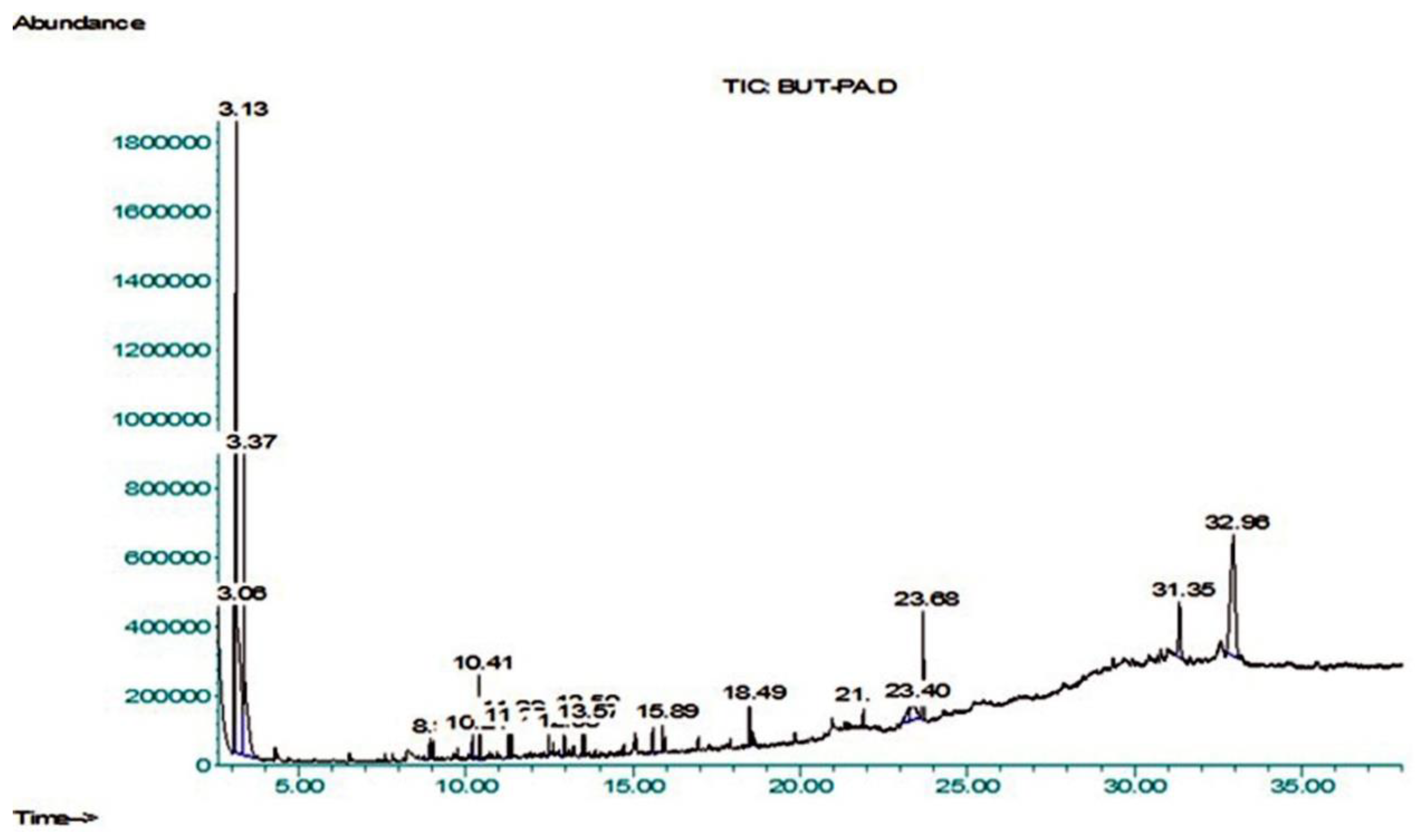
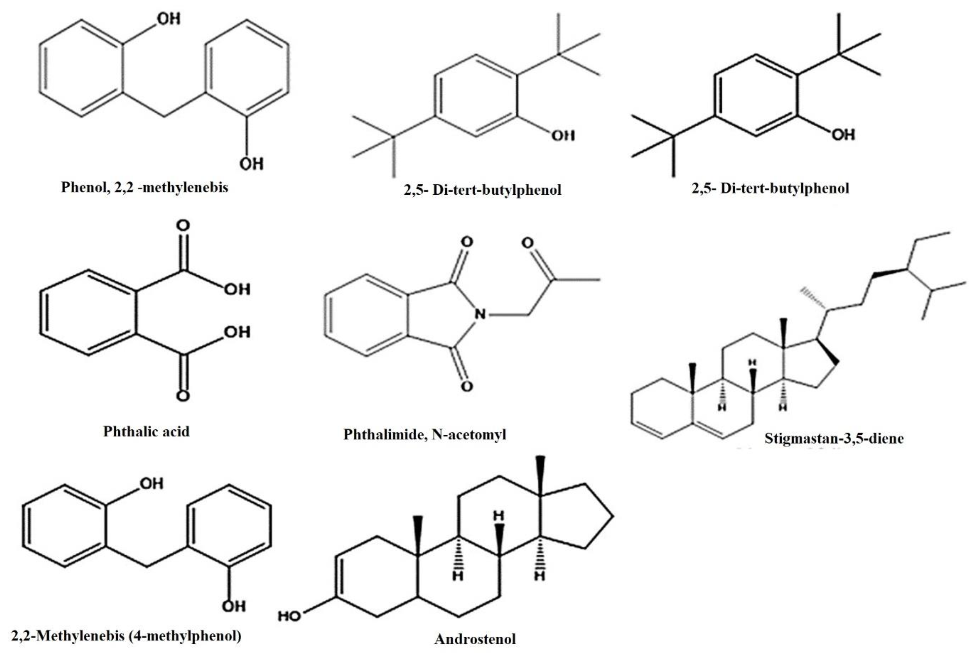
2.6. GC–MS Profiling

The existence of plant metabolites with documented pharmacological potential was revealed by GC–MS analysis (Figure 3 and Table 4). Twenty compounds were identified from the NIST library, i.e., 1-Tetradecene (8.93%), Pentadecane (10.2%), 2,5-Di-tert-butylphenol (10.414%), 7-Hexadecene (11.293%), Hexadecane (11.374%), Heptadecane (12.485%), Phthalimide-acetonyl (12.95%), E-15-Heptadecenal (13.50%), 1,2-Benzenedicarboxylic acid (15.60%), Dichloroacetic acid (15.89%), Hexadecanoic acid, butyl ester (18.49%), Phenol,2,2’-methylenebis (21.90%), 9-O-Pivaloyl-N-acetylcolchinol (23.43%), Phthalic acid (23.68%), Stigmastan-3,5-diene (31.35%), alpha.-Hydroxy-5, 16-androstad (32.96%). The size of the percent area was observed as follows: compounds 3. Alpha.-Hydroxy-5, 16-androstad, (19.95%), Phthalic acid (4.61%), Stigmastan-3, 5-diene (3.92%), 9-O-Pivaloyl-N-acetylcolchinol (3.07%), respectively. The structure of the major compounds detected is given in Figure 3 and Figure 4 and Table 4.

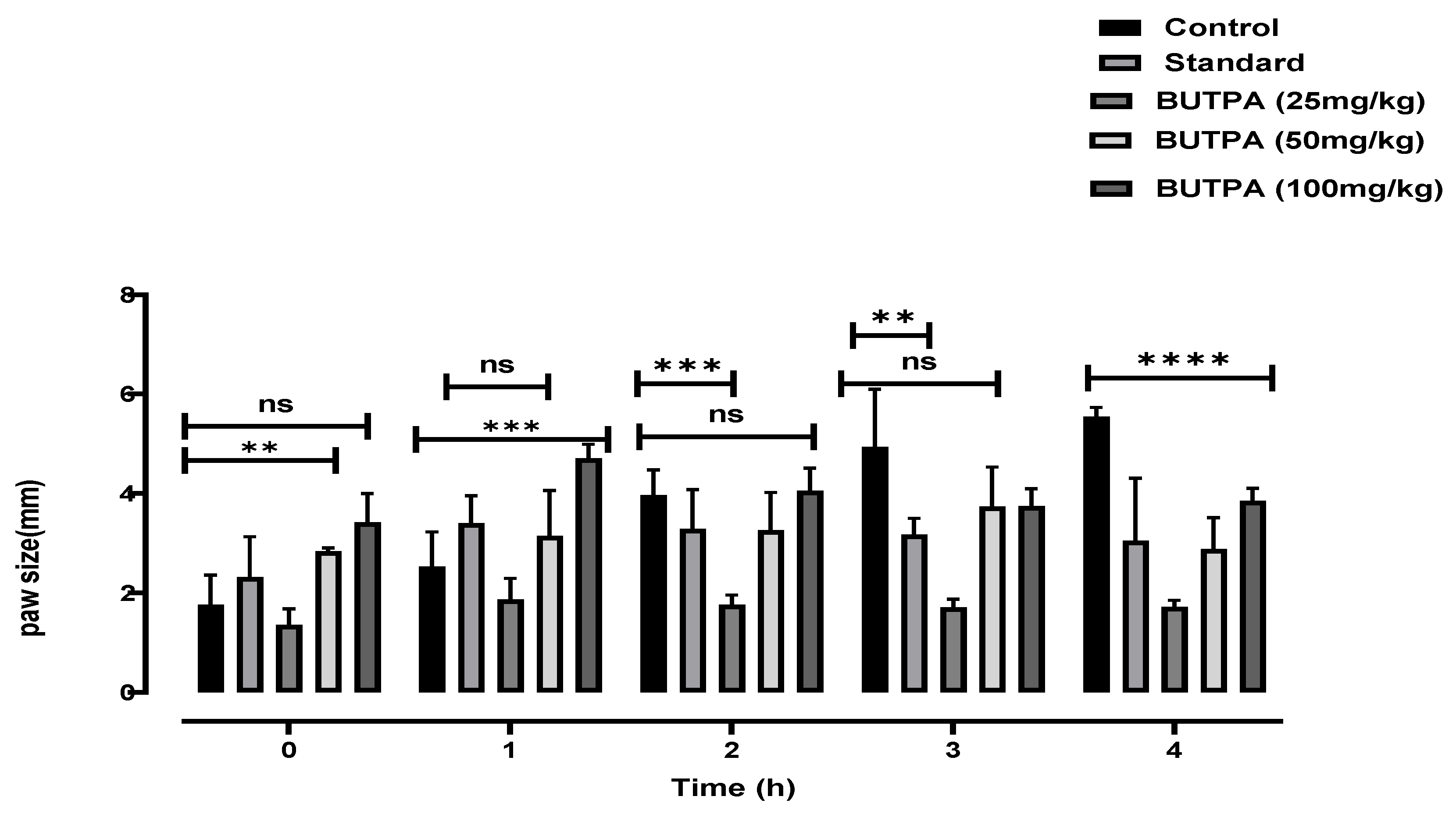
2.7. Carrageenan-Induced Hind Paw Edema

The BUTPA extract showed anti-inflammatory effect at various doses, i.e., 25, 50, 100 mg/kg. At 25 mg/kg, BUTPA extracts showed anti-inflammatory activity after the 2nd, 3rd and 4th hour, i.e., 3.44 ± 0.25, 3.37 ± 0.31 and 3.15 ± 0.42, respectively (Figure 5). At 50 mg/kg, BUTPA extracts showed anti-inflammatory activity after the 1st, 2nd, 3rd and 4th hour, i.e., 3.69 ± 0.15, 3.25 ± 0.08, 3.96 ± 0.86 and 2.81 ± 0.136, respectively. At 100 mg/kg, BUTPA extracts showed anti-inflammatory activity greater than the standard after the 1st, 2nd, 3rd and 4th hour, i.e., 4.14 ± 0.52, 4.07 ± 0.45, 3.09 ± 0.23 and 3.7 ± 0.74, respectively (Figure 5).

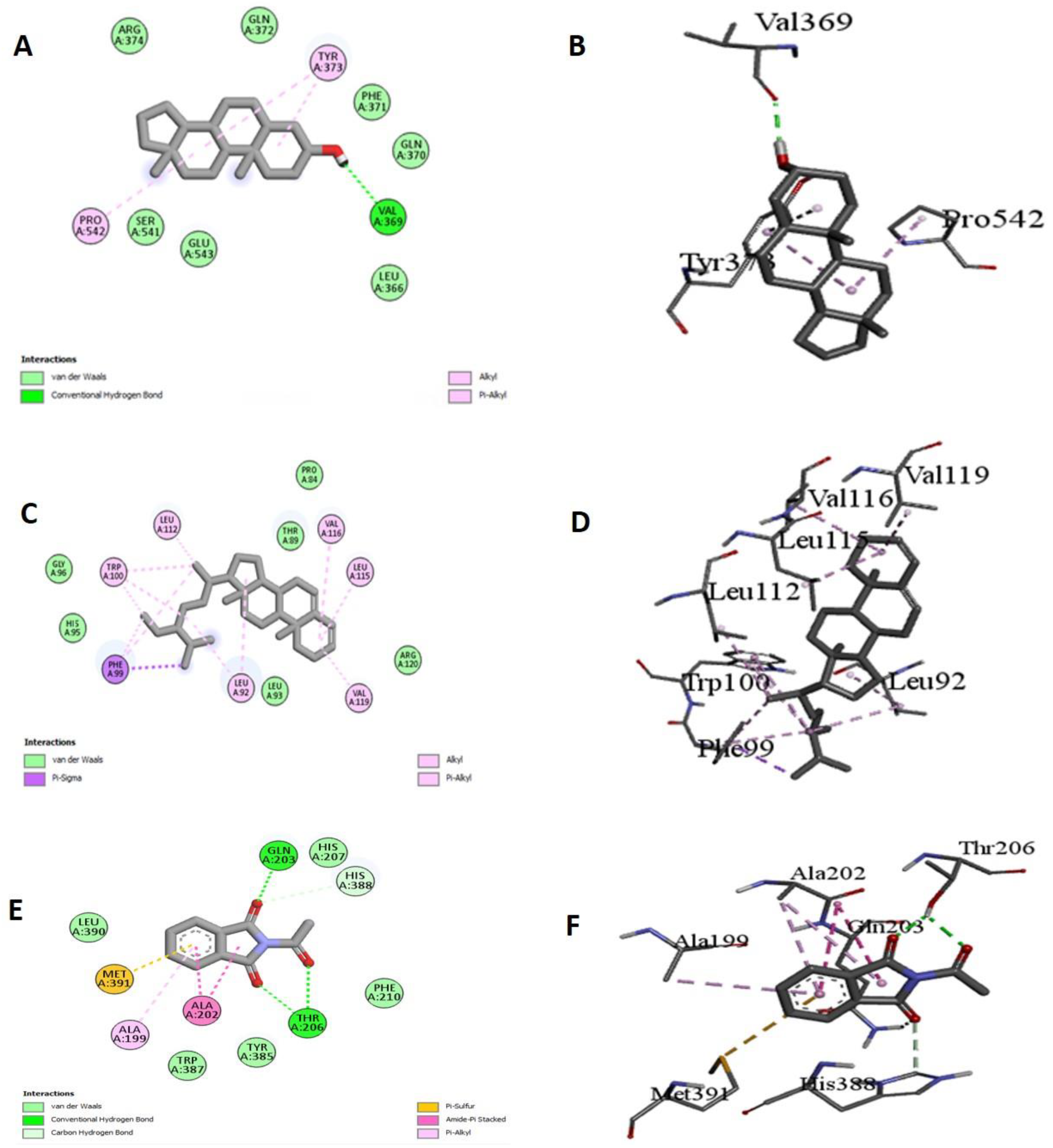
2.8. Molecular Docking

To obtain a better insight into the inhibition ability of the studied compounds and to correlate the experimental enzyme inhibition results, three compounds from the GC-MS profile of BUTPA extract were docked against COX-1 and COX-2. Stigmastan-3, 5-diene showed the maximum binding affinity, i.e., −8.2 against COX-1, while it showed a −7.32 binding affinity against COX-2. Androstenol showed a −8.1 binding affinity against COX-1, while the maximum binding was shown against COX-2, i.e., (−9.1). Phthalimide, N-acetonyl showed a binding affinity of −8.1 against COX-1 and a binding affinity −8.2 against COX-1, and showed Van der wall forces and hydrogen bonding with amino acid (Table 5 and Figure 6).

3. Discussion

Antioxidants are substances that prevent or delay the oxidation progressions caused by atmospheric oxygen. They are crucial in an organism’s defense mechanism against pathologies caused by free radicals [35]. Therefore, for the determination of phytochemicals and antioxidant potential, BUT-PA extract of P. afra was prepared in methanol by maceration. Initially, the phytochemical investigation was carried out using the guidelines mentioned in the phytochemistry manual [36]. We obtained positive results for the secondary metabolites of tannin, flavonoids, alkaloids, Phytosterol, saponins, cardiac glycoside quinolones, and triterpenoids (Table 1). Our results are consistent with studies on Purslane [37]. Major constituents, such as alkaloids, contribute to analgesic and antimicrobial activity; flavonoids and tannins act as antioxidant and antibacterial agents and saponins have antibacterial, anti-inflammatory, anticancer, and anti-diabetic activities [38]. The presence of these phytochemicals in the extracts/fractions of P. afra might contribute to its therapeutic potential. Flavonoid and phenolic are active metabolites that are produced in response to environmental stress and have received a lot of attention from researchers due to their potential benefits [39]. In the current situation, total phenolic levels were observed in BUT-PA extract of P. afra, (22.69 ± 0.28 mgGAE/g) and flavonoids contents (197.58 ± 6.29 mgGAE/g) in dry extract. These higher values may be attributed to the higher polyphenols content in roots compared to leaves.
In this work, for the first time, BUT-PA extracts of P. afra were investigated for their antioxidant potency in terms of the potential non-enzymatic and enzymatic antioxidants to support its traditional medicinal usage. The BUT-PA extracts of P. afra were identified to have significant antioxidant potential though CUPRIC, FRAP and ABTS methods, and moderate antioxidant potential through the DPPH method, as in Figure 1. The results for antioxidant enzymes, BUT-PA extracts of enzymatic antioxidants of P. afra namely, catalase (CAT), peroxidase (POD), superoxide dismutase (SOD), showed good enzyme activities for all the tested enzymes. Our results agree with the previous finding, in which antioxidant activity was determined using different methods [40].
The most common type of dementia is Alzheimer’s disease (AD). In this regard, numerous synthetic and plant-derived cholinesterase inhibitors are ordinarily used for the management and betterment of the disease [41]. For the first time NHPA, CHPA, MPA and BUTPA, of P. afra were evaluated for their acetylcholinesterase and butrylcholinesterase activity. The results were expressed as the % inhibition of enzymes ± standard deviation Figure 2. CHPA and BUTPA showed a higher % of inhibition of both enzymes then NHPA and MPA when compared to their respective standard, i.e., the inhibition of Acetylcholinesterase and butrylcholinesterase was assessed against galantamine (standard). The BUTPA showed an inhibition of acetylcholinesterase and butrylcholinesterase ranging from (57.64 ± 1.192) to (58.97 ± 0.33), compared to the standard, which ranged from (98.14 ± 1.00) to (74.14 ± 1.18). The lowest % of inhibition of acetylcholinesterase and butrylcholinesterase was shown by HPA, i.e., (20.55 ± 0.5) to (22.29 ± 1.927), when compared to standard Galanatmine. The present investigation revealed that BUTPA successfully contributed the maximum potential, indicating the positive relationship between phenolic content and cholinesterase’s enzymes inhibition assays [42].
To the best of our knowledge, the literature does not report on the %age thrombolytic Activity of the methanolic extract, n-hexane, chloroform, and n- butanol fractions of P. afra from Pakistan. In the present study, the thrombolytic percentages of four different plant extracts, HPA, CHPA, MPA, BUT PA, are presented in Figure 3, with a comparison to Streptokinase, the positive control. From the results, it is apparent that Streptokinase has (91.36 ± 0.06) clot lysis. Meanwhile, HPA exhibited the highest activity (11.2 ± 0.05%), followed by CHPA (7.3 ± 0.15%) and BUTPA (5.5 ± 0.30%), whereas MPA presented the lowest activity, which was negligible (3.4 ± 0.32%). Therefore, BUTPA has no significant thrombolytic activity. Staphylokinase and streptokinase are microbial plasminogen activators, which act as co-factoring molecules to promote development [43]. Our results are in line with the previous data [44].
Toxicology tests can identify several of the problems that may result from the use of medicinal plants/herbs, particularly in vulnerable people [45]. Hemolysis is the rupturing of red blood cells (erythrocytes), which indicates the cytotoxic effects on red blood cells [46]. If the degree of hemolysis is greater than 30%, the plant extracts are deemed hazardous towards erythrocytes [34]. Table 2 presents the hemolytic activity of different extracts of P. afra. The MPA fraction has highest hemolytic percentage (9.26 ± 0.15%), followed by BUTPA (8.53 ± 0.25%), AND CHPA has (7.46 ± 0.15%), and HPA has the lowest hemolytic activity (5.76 ±0.15%), as shown in Table 3. Overall, all four fractions have less than 30% hemolysis activity, so all fractions are nontoxic to humans and safe.
The genus Portulaca was shown to have anti-inflammatory properties by suppressing pro-inflammatory cytokine levels [47]. Certain natural fatty acid esters, such as methyl palmitate, have an inhibitory capacity against NF-B, which is accompanied by the downregulation of inflammatory channels [48]. The anti-inflammatory activity was measured in albino rats by measuring paw edema for four hours and inhibition was compared with standard indomethacin from 1- to 4-h intervals. The results exhibited that BUTPA extract at various doses (25, 50, and 100 mg/kg) had anti-inflammatory activity. The in vivo inhibition was assessed after 1 h, 2 h, 3 h, and 4 h. At 25 mg/kg, the BUTPA extract unveiled an anti-inflammatory effect. The BUTPA extract exhibited greater anti-inflammatory activity than the standard. At 400 mg/kg, the BUTPA extract showed an anti-inflammatory effect after the 2nd, 3rd, and 4th hour, as shown in Figure 5. Our results are consistent with the previous results for the same genus, which were reported by Reference [43].
The BUT-PA fraction of P. afra was subjected to gas chromatography-mass spectrometry (GCMS). The NIST library was used to identify the metabolites and 20 compounds were tentatively identified. The retention time in minutes (RT), area percentage (area %), compound name, molecular formula (M.F.), chemical class (Class), and molecular weight (M.W.) are shown in Table 3.The major identified compounds were Benzene, 1,3-dimethyl-, hexa- chloro derivative, p-Xylene, 2,5-Di-tert-butylphenol, Hexadecanoic acid, butyl ester, 9-O-Pivaloyl-N-acetylcolchinol, Phthalic acid, Stigmastan-3,5-diene, 3.alpha.-Hydroxy-5, Phthalimide, N-acetonyl, E-15-Heptadecenal, 1,2-Benzenedicarboxylic acid (Table 4 and Figure 3). 2,5-Di-tert-butylphenol showed antibacterial and antioxidant activity were as reported by Reference [49]. Aromatic compounds with methoxide group exhibit high antioxidant activity [50]. Such compounds have been identified in this study. Phenolic acids and their derivatives scavenge reactive oxygen species, showing antioxidant behavior. Compounds with one, two or three free hydroxyl groups on their aromatic ring exhibit low, medium and high antioxidant activity, respectively [51]. The highest antioxidant activity was observed in compounds with an -OH-group-oriented ortho or para position to the COOH group. The antioxidant activity decreases if the hydroxyl group is blocked, whereas no pronounced effect was served in the case of blocked carboxylic acid group regarding the compounds. Various environmental factors generate oxidative stress in plants. Consequently, the formation of phenolic metabolites with anti-oxidant properties is induced to overcome this stress.
The computational approach to molecular docking is now commonly employed in the drug development process. Docking has the advantage of determining the binding affinity of protein binding complexes, in this case, the chemical components from GCMS, as well as identifying the kind of interaction between the research molecules at the enzyme or receptor area through specific important interactions [52]. Molecular docking studies were performed for Cyclooxygenase-1 (COX-1) and cyclooxygenase 2 (COX-2). The PyRx virtual screening tool conducts flexible multiple ligand-docking studies within protein-binding sites [46]. A total of twenty compounds were docked against Cyclooxygenase 1 (PDB: 4O1Z) and Cyclooxygenase 2 (PDB: 5IKV). The hydrogen bond plays an essential role in proteinligand interactions and other hydrophobic interactions, such as alkyl and pi alkyl, as well as ensuring the stable binding of ligands with proteins [53]. The best RMSD values were found with meloxicam and flurbiprofen for the corresponding protein targets: COX-1 and COX-2. We identified three compounds with the best energy affinity against these two proteins. With Cox-1 Stigmastan-3, 5-diene has the lowest binding affinity (−8.2), and alkyl and pi sigma interact with the amino acids. It is worth noting that the Stigmastan-3, 5-diene also had a lot of interactions with amino acids near to the COX-1 active site’s amino acids. As a result, this molecule could change the local structure, which might have a biological effect. We observed a hydrophobic alkyl-type interaction with distance 4.99A for the amino acid LEU99, 4.21A for the amino acid LEU92, 4.75 and 5.41A for the amino acid VAL116, 4.60A for the amino acid VAL119, 4.28 and 4.78A for the amino acid ILE89, and 3.9A for the amino acid TRP100. There was no hydrogen bonding in this. For the other two compounds, Androstenol and Phthalimide, N-acetonyl- had 8.1 binding affinities, while androstenol has one hydrogen bond with GLN44, with a distance of 2.37A°, and Phthalimide, N-acetonyl- has hydrophobic interactions with different amino acids. The Androstenol molecule had hydrophobic alkyl-like interactions with the amino acids CYS36, CYS47, and PRO153, with varying distances in the COX-2 protein. At a distance of 1.64A, we observed an unfavorable donerdoner type interaction for the amino acids ARG44. Another compound, such as Stigmastan-3,5-diene, has alkyl type interactions with 7.3 binding affinities, and Phthalimide, N-acetonyl- has pi alkyl and pipi t-shaped interactions with −8.2 binding affinities (Table 5 and Figure 6).

4. Materials and Methods

4.1. Collection of Plant Materials

Plant materials of Portulacaria afra were amassed in March, 2019, from the Baghdad Campus, The Islamia University of Bahawalpur, Pakistan. The plant was identified, and a specified voucher number of 314 was allotted by Taxonomist from the Life Sciences Department, The Islamia University of Bahawalpur, Pakistan. Manually separated leaves and shoots were rinsed with tap water, chopped into small pieces with a razor blade, and dried in fresh air under shade. The dried samples were mechanically ground with a grinder and sieved through a fine sieve (400 mesh numbers) to obtain a uniform particle size sample, which was then stored in airtight, black-coated glass container for further analysis.

4.2. Extraction Method

Dried powder of the whole plant, of about 1 kg, was soaked in methanol for 72 h. This was routinely agitated through maceration. Afterward, the material was passed through muslin cloth to separate the material from solvent, and then the solvent was filtered with filter paper using a Buckner funnel. The obtained marc was then pre-soaked in n-butanol solvent for 72 h to collect all the remaining constituents from the plant material. This procedure was repeated for a third time to collect all the remaining constituents. The filtrate extracted with n-hexane, chloroform, and n-butanol solvent was concentrated using a rotary evaporator at 35 °C under lower pressure. The dried extract was weighed and stored in an airtight container for further analysis [54].

4.3. Chemicals and Drugs

Indomethacin was obtained from Shanxi Guangsheng pharmaceutical, carrageenan (Sigma–Aldrich, St. Louis, MI, USA). Indomethacin (15 mg/kg) was prepared for delivery in double-distilled water.

4.4. Phytochemical Screening

Preliminary phytochemical analysis of n. butanol extract (BUTPA), methanol fraction (MPA), chloroform fraction (CHPA), n-hexane fraction of P. afra was performed by using previously established protocols described by [47]

4.5. Estimation of Total Phenolic Contents (TPC)

The total phenolic contents (TPC) from each of the aforementioned extracts and fractions were determined using a colorimetric Folin–Ciocalteu reagent (FCR) technique with modifications. Then, they were incubated for 10 min with 10 µL of diluted FCR (10%), 100 µL of sample solution, and 90 µL of 15% w/v aqueous sodium carbonate solution. This mixture was incubated for another 90 min at 37 °C. At 750 nm, absorbance was measured. They were both positive and negative controls (gallic acid). TPC was computed using a calibration curve (range 0–100 g) and reported as milligram gallic acid equivalents per gram of dry material. The tests were performed in duplicate as described by [43].

4.6. Estimation of Total Flavonoid Contents (TFC)

An aluminum chloride colorimetric test was used to assess the total flavonoid content (TFC), and the number of total flavonoids was reported in milligram of rutin equivalents (mgRE/g extract) [55,56]. A calibration curve with a range of from 0 to 100 l (0–100 g) was created, using rutin as the standard, at 1 mg/mL in methanol. All the solutions were made with methanol. A total of 100 µL of test solution was mixed with 25 µL of 1% sodium nitrite solution and left to stand for 5 min, before adding 10 µL of 1% aluminum chloride solution and leaving it to react for another 5 min. Finally, 35 µL of 4% was added. At 510 nm, absorbance was read. The calibration curve equation was used to calculate TFC values, which were represented as milligram rutin equivalent per gram of dry extract (mg of RE/g extract).

4.7. Determination of Antioxidant Potential

The antioxidant activity of HMPA extract was assessed using two radical scavenging, i.e., DPPH, ABTS while reducing power was determined using, i.e., the protocol reported for FRAP and CUPRAC [57].

4.7.1. DPPH Assay

For the DPPH assay radical scavenging assay, 100 µL of extract/fractions solution and 400 µL of DPPH were mixed in a 96-microtiter plate. This mixture was incubated at ambient temperature for 30 min in darkness. Absorbance was measured at a wavelength of 517 nm, with the help of a BioTek Synergy HT (Winooski, VT, USA) microplate reader. Results were expressed as Trolox equivalents per gram of extract (mg TE/g extract).

4.7.2. ABTS Assay

For the ABTS reducing power assay, the formation of ABTS+ radical cation was due to the incubation of a mixture of 7 mM ABTS with 2.45 mM potassium persulfate in darkness at room temperature. The prepared stock solution of extract/fractions was diluted until its absorbance reached 0.700 ± 0.02 at 734 nm. A volume of 100 µL of extract/fraction solutions was combined with the previously prepared 200 µL ABTS+ solution in a 96-microtiter plate. This mixture was incubated at ambient temperature for 30 min. Absorbance was measured at 734 nm with the help of a BioTek Synergy HT (Winooski, VT, USA) microplate reader. Results were expressed as millimoles of Trolox equivalents per gram of dry extract. Results of the ABTS assay were expressed as Trolox equivalents per gram of extract (mg TE/g extract).

4.7.3. CUPRAC Assay

For the CUPRAC assay, 100 µL of extract/fractions solution were added to the reac- tion mixture [CuCl2 (200 µL, 10 mM), neocuproine (200 µL, 7.5 mM), NH4Ac buffer (200 µL, 1 M, pH 7.0)], and the absorbance was measured at 450 nm after 30 min of incubation at ambient temperature. Furthermore, a blank solution (without the extract/fractions) was prepared and analyzed according to this procedure.

4.7.4. FRAP Assay

For the FRAP assay, 0.05 mL of solution were added to 1 mL of reagent in acetate buffer (0.3 M, pH 3.6), 2,4,6-tris(2-pyridyl)-s-triazine (TPTZ) (10 mM) in 40-mM HCl and ferric chloride (20 mM) with a final concentration at the ratio of 10:1:1 (v/v/v). After incu- bation for 30 min at ambient temperature, the absorbance was measured at 593 nm. Simi- larly, a blank sample (prepared in the same manner but without the extract) was prepared. Furthermore, a blank solution (without the extract/fractions) was prepared and analyzed according to this procedure.

4.8. Enzymatic Antioxidants

4.8.1. Catalase Activity (CAT)

The method proposed in Reference [58] was performed to measure the catalase activity. In this method, the hydrogen peroxide (H2O2) was taken as a substrate and catalase enzymes were used for the decomposition of H2O2, detected using UV spectrophotometer (UV-1601, Shimadzu, Germany) by calculating the reduction in the absorbance for 5 min at 240 nm. The results of this activity were denoted as µM of consumed H2O2/min/mg of protein.

4.8.2. Peroxidase Activity (POD)

For POD activity, the guaiacol was used as a hydrogen. This was performed by measuring the variation at 470 nm for 1 min. The enzymatic activity was described as unit (one activity unit, defined as absorbance at 470 nm, with changes of 0.01 per min) per gram of freshly weighed tissue [59].

4.8.3. Superoxide Dismutase Activity (SOD)

Fresh leaves (1.0 g) were extracted in 50 mM phosphate buffer (pH~7.8) and the homogenate was centrifuged at 15,000× g for 10 min. The obtained supernatant was used to assay enzyme activity. The activity of superoxide dismutase was measured according to the method described by [60].

4.9. Enzyme Inhibition Assay

The potential of P. afra extract/fractions to inhibit the activity of acetylcholinesterase (AChE), and butrylcholinesterase (BChE), expressed as % inhibition, was determined according to procedures described in the literature, with minor modifications [61]. The detailed experimental methodology is explained below.

Acetylcholinesterase and Butrylcholinesterase Inhibition Assay

For the AChE and BChE inhibition assay, after 15 min of incubation at 25 °C, the reaction mixture, comprising 50 µL of the extract/fraction (1mg/mL) solution, 125 µL of DTNB (3 mM), and 25 µL of enzyme solution (0.265 U/mL AChE or 0.026 U/mL BChE) in Tris-HCl buffer (pH 8.0), was incubated at ambient temperature for 15 min. Then, 25 µL of substrate (15 mM acetylthiocholine iodide or butyrylthiocholine chloride) was added to the incubated mixture. The absorbance of the final solution was measured at 405 nm after 15 min. Galanatmine was used as the standard agent for both the acetylcholinesterase and butyryl cholinesterase inhibition assays. Likewise, a blank solution (without the extract/fractions) was prepared and analyzed according to the procedure described by [1].

4.10. Thrombolytic Activity

From each human volunteer who had not undergone seven days of anticoagulant and oral contraceptives therapy, venous blood (5 mL) was collected, which was placed in sterile and pre-weighed in six distinct centrifuge tubes. These tubes were incubated for 45 min at 37 °C. After the clot had formed, the fluid from each centrifuge tube was entirely discharged. The clot weight was computed by deducting the weight of empty tube from the tube containing the clot. Streptokinase (SK) was used as a standard to test the thrombolytic activity. Streptokinase (15, 00,000 I.U) was diluted with 5ml sterile water and shaken to mix properly. From this 100 μL, streptokinase (30000 I.U) and distilled water (100 μL) were used as positive and negative controls, respectively, and each sample fraction was separately added to the centrifuge tubes. After that, all tubes were incubated for 90 min at 37 °C and examined for clot lysis. The discharged fluid was discarded after incubation, and the tubes were weighed again to see the weight variation subsequent to clot lysis. Thereafter, the clot lysis percentage was calculated as [62].
Clot lysis percentage = (released clot weight/weight of clot) × 100

4.11. Hemolytic Activity

The hemolytic effect of different plant extract fractions was evaluated using the previously described method [58]. Ten milliliters of human blood was collected from human volunteers, added to a sterile, screw-top EDTA tube, and centrifuged at 850 g for 5 min. The upper layer was decanted, and the erythrocytes were rinsed several times with 10 mL cooled isotonic and sterile phosphate-buffered saline (PBS) with a pH of 7.4. The rinsed cells were resuspended in 20 mL sterile and cold PBS. The extracts (1000 µg/1 mL) were added to erythrocytes solution and incubated for 60 min at 37 °C. The absorbance of hemoglobin in the supernatant at 540 nm was used to calculate the hemolysis rate. A total of 0.1% Triton X-100 was used as a positive control and PBS as the negative control. Hemolysis percentage was calculated using the following formula.
Hemolysis percentage = (Ab of sample − Ab of negative control)/Ab of positive control × 100

4.12. Gas Chromatography Mass Spectroscopy (GC–MS) Analysis

The phytoconstituents composition of different P. afra extracts was determined using GC MS analysis for Agilent, 6890 series and Hewlett Packard, 5973 mass choosy detection systems, with HP-5MS column (250 m in diameter, 30 m in length 0.25 m in film thickness) was used for compound separation. (70 eV) was used for GC.MS-ionizing radiation of the sample. The temperature at the inlet was 220 °C. The oven temperature steadily increased from 60 °C to 280 °C at a rate of 3 °C/min. The carrier gas was pure helium gas (99.995 percent purity), flowing at a rate of 1 mL/min in constant flow mode. A total of 1.0 L of prepared extracts, diluted with respective solvents, were implanted in a split-less mode at 250 °C; 1.0 L of prepared extracts, diluted with individual solvents, were injected in split-less mode. The compound was discovered using an NIST library search (NIST 14).

4.13. Experimental Animals

Wistar albino rats weighing 150–230 g was kept in the animal house of the Pharmacology and Physiology research laboratory at the Islamia University of Bahawalpur’s Faculty of Pharmacy and Alternative Medicine in Pakistan. All the animals used in the study were housed in polycarbonate cages. The typical conditions of temperature (25 °C) and humidity (50–55 percent), as well as exposure to a 12:12-h light and dark cycle, were maintained throughout the study. The animals were fed conventional animal food and given free access to water. To reduce animal stress, animals were acclimatized to the test conditions for one week before the trial began. The institutional research committee approved the study protocols and procedures (PAEC, Pharmacy Animal Ethics Committee).

4.14. Anti-Inflammatory Activity

4.14.1. Carrageenan Induced Hind-Paw Edema

Except for the control group, rats received 0.1 mL of newly manufactured, one percent carrageenan, injected into the right hind paw [63]. The paw size of rats was measured using a digital Vernier caliper (Mitutoyo, Japan). The rats were given a vehicle (distilled water 5 mL/kg, i.p), various plant extract doses (25, 50, 100, mg/kg, i.p) and standard indomethacin 15 mg/kg, i.p, half an hour earlier. The thickness of the paws was experimentally validated before the carrageenan injection, at “0 h,” and subsequently at 1, 2, 3, and 4 h. The previously reported formula was used to compute the % inhibition of paw edema [23].
Percentage Inhibition = (Control means − Treated mean)/Control Means × 100

4.14.2. Molecular Docking Procedure

The crystal structures of the Cyclooxygenase 1 protein targets (PDB: 4O1Z, R: 2.40A) and Cyclooxygenase 2 (PDB: 5IKV, R: 2.51 A) were taken from the Protein Data Bank (PDB) (https://www.rcsb.org/, accessed on 25 March 2022) for the docking study. The polar hydrogens were added to the protein. Water, inhibitors, and extra chains were removed from the protein and then converted into the PDBQT format. The ligands were taken from the PubChem (https://pubchem.ncbi.nlm.nih.gov/, accessed on 25 March 2022) to evaluate their potential against the proteins. The ligands were entered into Open Babel and subjected to energy reductions using the PyRx program. After that, the chemicals were converted to PDBQT format for further investigation. Then, the grid box was formed in specific dimensions. Finally, interactions were visualized by the Discovery studio.

4.15. Statistical Analysis

The result values in the tables are represented as mean ± SD (n = 6). A two-way ANOVA, followed by Tukey test, was performed using Graph Pad Prism (San Diego, CA, USA) software. All values in the graphs are given in mean ± SE (n = 6). p < 0.001 (***) and p < 0.05 (*) compared to the control.

5. Conclusions

The present study revealed the in vitro antioxidant, thrombolytic, hemolytic activity, and % inhibition of acetylcholinesterase, as well as the butrylcholinesterase and in vivo anti-inflammatory potential of BUTPA extracts. Twenty compounds were identified through GC–MS analysis, which showed many pharmacological activities in in vivo and ex vivo experiments. A high binding affinity was observed for Stigmastan-3, 5-diene, Phthalic acid, 3. Alpha-Hydroxy-5, 16-androstenol. The computed binding energies of the compounds revealed that all the compounds had synergistic effects to prevent inflammation. Therefore, the findings of this study indicated that this plant is an excellent candidate for the treatment of inflammation and pain-related illnesses. The medicinal and pharmacological potential of P. afra revealed that it is quite auspicious as a versatile therapeutic plant and should be further investigated.

Author Contributions

Conceptualization, S.T., S.A. and K.u.R.K.; methodology, S.T.; software, Q.u.Z.; validation, F.T., S.T. and S.A.; formal analysis, A.K.; investigation, S.T.; resources, S.T.; data curation, S.T., F.T. and S.T.; writing—original draft preparation, S.T.; writing—review and editing, N.A.B., Y.C., A.A. and A.A.H. supervision, S.A.; project administration, S.A.; funding acquisition, N.A.B., A.A., A.A.H. All authors have read and agreed to the published version of the manuscript.

Funding

This research was funded by the Researchers supporting project number (RSP-2021/229) King Saud University, Riyadh, Saudi Arabia.

Institutional Review Board Statement

All the experiments were carried out in accordance with the NIH guidelines and were approved by the Department of Pharmaceutical Chemistry’s concerned committee (Pharmacy Animal Ethics Committee (PAEC/22/73).

Informed Consent Statement

Informed consent was obtained from all subjects involved in the study.

Data Availability Statement

Not applicable.

Acknowledgments

The authors extend their appreciation to the Researchers supporting project number (RSP-2021/229) King Saud University, Riyadh, Saudi Arabia.

Conflicts of Interest

The authors declare no conflict of interest.

References

  1. Ghalloo, B.A.; Khan, K.-u.-R.; Ahmad, S.; Aati, H.Y.; Al-Qahtani, J.H.; Ali, B.; Mukhtar, I.; Hussain, M.; Shahzad, M.N.; Ahmed, I. Phytochemical Profiling, In Vitro Biological Activities, and In Silico Molecular Docking Studies of Dracaena reflexa. Molecules 2022, 27, 913. [Google Scholar] [CrossRef] [PubMed]
  2. Akther, Y.; Nabi, J.; Tabassum, N. Comprehensive Overview of Some Edible Medicinal Plants from Kashmir Valley: Cultural, Economic, and Pharmacological Importance. Edi. Plants Health Dis. 2022, 137–159. [Google Scholar]
  3. Kabir, M.A.; Hussain, M.A.; Ahmad, Z. Candida albicans: A model organism for studying fungal pathogens. Int. Sch. Res. Not. 2012, 2012, 538694. [Google Scholar] [CrossRef] [PubMed] [Green Version]
  4. Ansari, M.Y.; Ahmad, N.; Haqqi, T.M. Oxidative stress and inflammation in osteoarthritis pathogenesis: Role of polyphenols. Biomed. Pharmacother. 2020, 129, 110452. [Google Scholar] [CrossRef]
  5. Kazemi, S.; Shirzad, H.; Rafieian-Kopaei, M. Recent findings in molecular basis of inflammation and anti-inflammatory plants. Current Pharm. Des. 2018, 24, 1551–1562. [Google Scholar] [CrossRef]
  6. Tasneem, S.; Liu, B.; Li, B.; Choudhary, M.I.; Wang, W. Molecular pharmacology of inflammation: Medicinal plants as anti-inflammatory agents. Pharma. Res. 2019, 139, 126–140. [Google Scholar] [CrossRef]
  7. Nakamura, J.S.; Kim, E.S.; Rentscher, K.E.; Bower, J.E.; Kuhlman, K.R. Early-life stress, depressive symptoms, and inflammation: The role of social factors. Aging Ment. Health 2021, 26, 843–851. [Google Scholar] [CrossRef]
  8. Carson, M.; Thurston, R. Physiologically-measured vasomotor symptoms and inflammation among midlife women. Psychoneuroendocrinology 2021, 131, 105567. [Google Scholar] [CrossRef]
  9. Schiattarella, G.G.; Sequeira, V.; Ameri, P. Distinctive patterns of inflammation across the heart failure syndrome. Heart Fail. Rev. 2021, 26, 1333–1344. [Google Scholar] [CrossRef]
  10. Tayab, M.A.; Islam, M.N.; Chowdhury, K.A.A.; Tasnim, F.M. Targeting neuroinflammation by polyphenols: A promising therapeutic approach against inflammation-associated depression. Biomed. Pharmacother. 2022, 147, 112668. [Google Scholar] [CrossRef]
  11. Negishi, Y.; Shima, Y.; Takeshita, T.; Morita, R. Harmful and beneficial effects of inflammatory response on reproduction: Sterile and pathogen-associated inflammation. Immunol. Med. 2021, 44, 98–115. [Google Scholar] [CrossRef] [PubMed]
  12. Ahmad, A.; Abuzinadah, M.F.; Alkreathy, H.M.; Banaganapalli, B.; Mujeeb, M. Ursolic acid rich ocimum sanctum L leaf extract loaded nanostructured lipid carriers ameliorate adjuvant induced arthritis in rats by inhibition of COX-1, COX-2, TNF-α and IL-1: Pharmacological and docking studies. PLoS ONE 2018, 13, e0193451. [Google Scholar] [CrossRef] [PubMed] [Green Version]
  13. Sami, A.; Usama, M.; Saeed, M.M.; Akram, M. Medicinal plants with non-steroidal anti-inflammatory-like activity. Mediterr. J. Pharm. Pharm. Sci. 2021, 1, 1–8. [Google Scholar]
  14. Yang, Q.; Ciebiera, M.; Victoria Bariani, M.; Ali, M.; Elkafas, H.; Boyer, T.G.; Al-Hendy, A. Comprehensive Review of Uterine Fibroids: Developmental Origin, Pathogenesis, and Treatment. Endocr. Rev. 2021, bnab039. [Google Scholar] [CrossRef]
  15. Zhang, Q.; Jia, S.; Bai, Y.; Zhou, X.; Ding, Y. Formation mechanisms of reactive carbonyl species from fatty acids in dry-cured fish during storage in the presence of free radicals. J. Future Foods 2021, 1, 203–210. [Google Scholar] [CrossRef]
  16. Ndhlovu, P.T.; Omotayo, A.O.; Otang-Mbeng, W.; Aremu, A.O. Ethnobotanical review of plants used for the management and treatment of childhood diseases and well-being in South Africa. S. Afr. J. Bot. 2021, 137, 197–215. [Google Scholar] [CrossRef]
  17. Bruyns, P.V.; Oliveira-Neto, M.; Melo-de-Pinna, G.F.; Klak, C. Phylogenetic relationships in the Didiereaceae with special reference to subfamily Portulacarioideae. Taxon 2014, 63, 1053–1064. [Google Scholar] [CrossRef]
  18. Iranshahy, M.; Javadi, B.; Iranshahi, M.; Jahanbakhsh, S.P.; Mahyari, S.; Hassani, F.V.; Karimi, G. A review of traditional uses, phytochemistry and pharmacology of Portulaca oleracea L. J. Ethnopharmacol. 2017, 205, 158–172. [Google Scholar] [CrossRef]
  19. De Vos, B. Investigating the Antidiabetic Potential of the Combination of Catharanthus roseus and Portulacaria afra Leaf Extracts. North-West University (South-Africa). 2021. Available online: https://repository.nwu.ac.za/bitstream/handle/10394/37735/De%20Vos%2C%20B.pdf?sequence=1 (accessed on 3 March 2021).
  20. Borsai, O.; Al Hassan, M.; Boscaiu, M.; Sestras, R.E.; Vicente, O. The genus as a suitable model to study the mechanisms of plant tolerance to drought and salinity. EuroBiotech. J. 2018, 2, 104–113. [Google Scholar] [CrossRef] [Green Version]
  21. Nciki, S.; Vuuren, S.; van Eyk, A.; de Wet, H. Plants used to treat skin diseases in northern Maputaland, South Africa: Antimicrobial activity and in vitro permeability studies. Pharm. Biol. 2016, 54, 2420–2436. [Google Scholar] [CrossRef] [Green Version]
  22. De Wet, H.; Nciki, S.; van Vuuren, S.F. Medicinal plants used for the treatment of various skin disorders by a rural community in northern Maputaland, South Africa. J. Ethnobiol. Ethnomed. 2013, 9, 51. [Google Scholar] [CrossRef] [PubMed] [Green Version]
  23. Sudjarwo, S.A. The potency of piperine as antiinflammatory and analgesic in rats and mice. Folia Med. Indones. 2005, 41, 190–194. [Google Scholar]
  24. Rahbar, N.; Shafaghat, A.; Salimi, F. Antimicrobial activity and constituents of the hexane extracts from leaf and stem of Origanum vulgare L. ssp. Viride (Boiss.) Hayek. growing wild in Northwest Iran. J. Med. Plant Res. 2012, 6, 2681–2685. [Google Scholar]
  25. Ugbogu, E.A.; Akubugwo, I.E.; Ude, V.C.; Gilbert, J.; Ekeanyanwu, B. Toxicological evaluation of phytochemical characterized aqueous extract of wild dried Lentinus squarrosulus (Mont.) mushroom in rats. Toxicol. Res. 2019, 35, 181–190. [Google Scholar] [CrossRef] [Green Version]
  26. Ren, J.; Wang, J.; Karthikeyan, S.; Liu, H.; Cai, J. Natural anti-phytopathogenic fungi compound phenol, 2,4-bis (1,1-dimethylethyl) from Pseudomonas fluorescens TL-1. Indian J. Biochem. Biophys. 2019, 56, 162–168. [Google Scholar]
  27. Mou, Y.; Meng, J.; Fu, X.; Wang, X.; Tian, J.; Wang, M.; Peng, Y.; Zhou, L. Antimicrobial and antioxidant activities and effect of 1-hexadecene addition on palmarumycin C2 and C3 yields in liquid culture of endophytic fungus Berkleasmium sp. Dzf12. Molecules 2013, 18, 15587–15599. [Google Scholar] [CrossRef] [Green Version]
  28. Bruno, F.; Castelli, G.; Migliazzo, A.; Piazza, M.; Galante, A.; Verde, V.L.; Calderone, S.; Nucatolo, G.; Vitale, F. Cytotoxic screening and in vitro evaluation of pentadecane against Leishmania infantum promastigotes and amastigotes. J. Parasitol. 2015, 101, 701–705. [Google Scholar] [CrossRef] [Green Version]
  29. Sharma, U.; Kumar, P.; Kumar, N.; Singh, B. Recent advances in the chemistry of phthalimide analogues and their therapeutic potential. Mini. Rev. Med. Chem. 2010, 10, 678–704. [Google Scholar] [CrossRef]
  30. Zhao, S.; Wu, Q.; Zhang, J.; Wu, K.; Guo, W.; Wu, J. Identification of actinomycete strain PY-C-21 and structural analysis of its active products. Xiandai Shipin Keji 2016, 32, 36–41. [Google Scholar]
  31. Tan, Y.N.; Zhang, J.H.; Chen, W.N. GC-MS-Based Metabolomics Analysis of Prawn Shell Waste Co-Fermentation by Lactobacillus plantarum and Bacillus subtilis. Polysaccharides 2020, 1, 31–50. [Google Scholar] [CrossRef]
  32. Huang, L.; Zhu, X.; Zhou, S.; Cheng, Z.; Shi, K.; Zhang, C.; Shao, H. Phthalic acid esters: Natural sources and biological activities. Toxins 2021, 13, 495. [Google Scholar] [CrossRef] [PubMed]
  33. Mallu, M.R.; Vemula, S.; Kante, R.K. Molecular docking studies of bioactive compounds from Stevia rebaudiana for its anti-cancer activity. J. Pharma. Sci. Res. 2019, 11, 2016–2018. [Google Scholar]
  34. Rasmussen, M.; Zamaratskaia, G.; Ekstrand, B. Gender-related differences in cytochrome P450 in porcine liver–implication for activity, expression and inhibition by testicular steroids. Reprod. Domest. Anim. 2011, 46, 616–623. [Google Scholar] [CrossRef] [PubMed]
  35. Aziz, M.; Ahmad, S.; Iqbal, M.N.; Khurshid, U.; Saleem, H.; Alamri, A.; Anwar, S.; Alamri, A.S.; Chohan, T.A. Phytochemical, pharmacological, and In-silico molecular docking studies of Strobilanthes glutinosus Nees: An unexplored source of bioactive compounds. S. Afr. J. Bot. 2022, 147, 618–627. [Google Scholar] [CrossRef]
  36. Zheljazkov, V.D.; Semerdjieva, I.B.; Stevens, J.F.; Wu, W.; Cantrell, C.L.; Yankova-Tsvetkova, E.; Koleva-Valkova, L.H.; Stoyanova, A.; Astatkie, T. Phytochemical Investigation and Reproductive Capacity of the Bulgarian Endemic Plant Species Marrubium friwaldskyanum Boiss. (Lamiaceae). Plants 2021, 11, 114. [Google Scholar] [CrossRef]
  37. D’Imperio, M.; Durante, M.; Gonnella, M.; Renna, M.; Montesano, F.F.; Parente, A.; Mita, G.; Serio, F. Enhancing the Nutritional Value of Portulaca oleracea L. by using Soilless Agronomic Biofortification with Zinc. Food Res. Int. 2022, 155, 111057. [Google Scholar] [CrossRef]
  38. Urzúa, A.; Rezende, M.C.; Mascayano, C.; Vásquez, L. A structure-activity study of antibacterial diterpenoids. Molecules 2008, 13, 882–891. [Google Scholar] [CrossRef] [Green Version]
  39. Fukalova Fukalova, T.; García-Martínez, M.D.; Raigón, M.D. Nutritional Composition, Bioactive Compounds, and Volatiles Profile Characterization of Two Edible Undervalued Plants: Portulaca oleracea L. and Porophyllum ruderale (Jacq.) Cass. Plants 2022, 11, 377. [Google Scholar] [CrossRef]
  40. Ghauch, A.; Tuqan, A. Reductive destruction and decontamination of aqueous solutions of chlorinated antimicrobial agent using bimetallic systems. J. Hazard. Mater. 2009, 164, 665–674. [Google Scholar] [CrossRef]
  41. Seyrekoglu, F.; Temiz, H.; Eser, F.; Yildirim, C. Comparison of the antioxidant activities and major constituents of three Hypericum species (H. perforatum, H. scabrum and H. origanifolium) from Turkey. S. Afr. J. Bot. 2022, 146, 723–727. [Google Scholar] [CrossRef]
  42. Khurshid, U.; Ahmad, S.; Saleem, H.; Nawaz, H.A.; Zengin, G.; Locatelli, M.; Mahomoodally, M.F.; Abidin, S.A.Z.; Tousif, M.I.; Ahemad, N. Phytochemical composition and in vitro pharmacological investigations of Neurada procumbens L. (Neuradaceae): A multidirectional approach for industrial products. Ind. Crops Prod. 2019, 142, 111861. [Google Scholar] [CrossRef]
  43. Alu’datt, M.H.; Rababah, T.; Alhamad, M.N.; Al-Tawaha, A.; Al-Tawaha, A.R.; Gammoh, S.; Ereifej, K.I.; Al-Karaki, G.; Hamasha, H.R.; Tranchant, C.C. Herbal yield, nutritive composition, phenolic contents and antioxidant activity of purslane (Portulaca oleracea L.) grown in different soilless media in a closed system. Ind. Crops Prod. 2019, 141, 111746. [Google Scholar] [CrossRef]
  44. Prasad, S.; Kashyap, R.S.; Deopujari, J.Y.; Purohit, H.J.; Taori, G.M.; Daginawala, H.F. Development of an in vitro model to study clot lysis activity of thrombolytic drugs. Thromb. J. 2006, 4, 14. [Google Scholar] [CrossRef] [PubMed] [Green Version]
  45. Kauser, A.; Shah, S.M.A.; Iqbal, N.; Murtaza, M.A.; Hussain, I.; Irshad, A.; Nasir, S.; Akram, M.; Munir, N.; Riaz, M. In vitro antioxidant and cytotoxic potential of methanolic extracts of selected indigenous medicinal plants. Prog. Nutr. 2018, 20, 706–712. [Google Scholar]
  46. Velika, B.; Kron, I. Antioxidant properties of benzoic acid derivatives against superoxide radical. Free Radic. Antioxid. 2012, 2, 62–67. [Google Scholar] [CrossRef] [Green Version]
  47. Baradaran Rahimi, V.; Rakhshandeh, H.; Raucci, F.; Buono, B.; Shirazinia, R.; Samzadeh Kermani, A.; Maione, F.; Mascolo, N.; Askari, V.R. Anti-inflammatory and anti-oxidant activity of Portulaca oleracea extract on LPS-induced rat lung injury. Molecules 2019, 24, 139. [Google Scholar] [CrossRef] [Green Version]
  48. El-Demerdash, E. Anti-inflammatory and antifibrotic effects of methyl palmitate. Toxicol. Appl. Pharmacol. 2011, 254, 238–244. [Google Scholar] [CrossRef]
  49. Hegde, S.; Jayaraj, M.; Bhandarkar, A. Pharmacognostic studies and preliminary phytochemical pnalysis of cold and hot Extracts of leaf of Tinospora malabarica Miers-An Important Medicinal Plant. Int. J. Pharm. Sci. Rev. Res. 2015, 34, 19–25. [Google Scholar]
  50. Khan, K.; Firdous, S.; Ahmad, A.; Fayyaz, N.; Nadir, M.; Rasheed, M.; Faizi, S. GC-MS profile of antimicrobial and antioxidant fractions from Cordia rothii roots. Pharm. Biol. 2016, 54, 2597–2605. [Google Scholar] [CrossRef] [Green Version]
  51. Oliveira Calil, N.; Senra Goncalves de Carvalho, G.; Farah da Silva, A.; David da Silva, A.; Rezende Barbosa Raposo, N. Antioxidant activity of synthetic resveratrol analogs: A structure-activity insight. Lett. Drug Des. Discov. 2012, 9, 676–679. [Google Scholar] [CrossRef]
  52. Chandak, N.; Kumar, P.; Kaushik, P.; Varshney, P.; Sharma, C.; Kaushik, D.; Jain, S.; Aneja, K.R.; Sharma, P.K. Dual evaluation of some novel 2-amino-substituted coumarinylthiazoles as anti-inflammatory–antimicrobial agents and their docking studies with COX-1/COX-2 active sites. J. Enzyme Inhib. Med. Chem. 2014, 29, 476–484. [Google Scholar] [CrossRef] [PubMed]
  53. Borges, R.S.; Lima, E.S.; Keita, H.; Ferreira, I.M.; Fernandes, C.P.; Cruz, R.A.S.; Duarte, J.L.; Velázquez-Moyado, J.; Ortiz, B.L.S.; Castro, A.N. Anti-inflammatory and antialgic actions of a nanoemulsion of Rosmarinus officinalis L. essential oil and a molecular docking study of its major chemical constituents. Inflammopharmacology 2018, 26, 183–195. [Google Scholar] [CrossRef] [PubMed]
  54. Gilani, A.H.; Jabeen, Q.; Khan, A.U.; Shah, A.J. Gut modulatory, blood pressure lowering, diuretic and sedative activities of cardamom. J. Ethnopharmacol. 2008, 115, 463–472. [Google Scholar] [CrossRef] [PubMed]
  55. Slinkard, K.; Singleton, V.L. Total phenol analysis: Automation and comparison with manual methods. Am. J. Enol. Vitic. 1977, 28, 49–55. [Google Scholar]
  56. Zengin, G.; Nithiyanantham, S.; Locatelli, M.; Ceylan, R.; Uysal, S.; Aktumsek, A.; Selvi, P.K.; Maskovic, P. Screening of in vitro antioxidant and enzyme inhibitory activities of different extracts from two uninvestigated wild plants: Centranthus longiflorus subsp. longiflorus and Cerinthe minor subsp. auriculata. Eur. J. Integ. Med. 2016, 8, 286–292. [Google Scholar] [CrossRef]
  57. Asif, M.; Yehya, A.H.S.; Al-Mansoub, M.A.; Revadigar, V.; Ezzat, M.O.; Ahamed, M.B.K.; Oon, C.E.; Murugaiyah, V.; Majid, A.S.A.; Majid, A.M.S.A. Anticancer attributes of Illicium verum essential oils against colon cancer. S. Afr. J. Bot. 2016, 103, 156–161. [Google Scholar] [CrossRef]
  58. Diaconu, M.; Pavel, L.V.; Hlihor, R.-M.; Rosca, M.; Fertu, D.I.; Lenz, M.; Corvini, P.X.; Gavrilescu, M. Characterization of heavy metal toxicity in some plants and microorganisms—A preliminary approach for environmental bioremediation. New Biotechnol. 2020, 56, 130–139. [Google Scholar] [CrossRef]
  59. Güneş, A.; Kordali, Ş.; Turan, M.; Bozhüyük, A.U. Determination of antioxidant enzyme activity and phenolic contents of some species of the Asteraceae family from medicanal plants. Ind. Crops Prod. 2019, 137, 208–213. [Google Scholar] [CrossRef]
  60. Ramadan, K.M.; Alharbi, M.M.; Alenzi, A.M.; El-Beltagi, H.S.; Darwish, D.B.E.; Aldaej, M.I.; Shalaby, T.A.; Mansour, A.T.; El-Gabry, Y.A.E.-G.; Ibrahim, M.F. Alpha Lipoic Acid as a Protective Mediator for Regulating the Defensive Responses of Wheat Plants against Sodic Alkaline Stress: Physiological, Biochemical and Molecular Aspects. Plants 2022, 11, 787. [Google Scholar] [CrossRef]
  61. Grochowski, D.M.; Uysal, S.; Zengin, G.; Tomczyk, M. In vitro antioxidant and enzyme inhibitory properties of Rubus caesius L. Int. J. Environ. Health Res. 2019, 29, 237–245. [Google Scholar] [CrossRef]
  62. Saleem, A.; Nasir, S.; Rasool, N.; Bokhari, T.H.; Rizwan, K.; Shahid, M.; Abbas, M.; Zubair, M. In vitro antimicrobial and haemolytic studies of Kalanchoe pinnata and Callistemon viminalis. Int. J. Chem. Biochem. Sci 2015, 7, 29–34. [Google Scholar]
  63. Saleem, M.; Khalid, I.; Akhtar, M.F.; Saleem, A.; Rasul, A.; Hamid, I.; Shah, M.A.; Basheer, E.; Javaid, Z.; Sohail, K. HPLC analysis and pharmacological study of Quercus dilatata Lindle. ex Royle aqueous methanolic extract in Sprague Dawley rats. Pak. J. Pharm. Sci. 2020, 33, 1327–1332. [Google Scholar]
Figure 1. Non-enzymatic (A), and enzymatic anti-oxidant (B) potential of BUT-PA extracts. For each parameter, vertical bars represent mean data ± SE (n = 3).
Molecules 27 02377 g001
Figure 2. (A) Acetylcholinesterase inhibition of galantamine (standard) and extract/fractions, and (B) butrylcholinesterase inhibition of galantamine (standard) and extract/fractions of P. afra. NHPA, n-hexane fraction; CHPA, chloroform fraction; BUTPA, n-butanol fraction; STND, galantamine (Standard); MPA, methanolic extract. For each parameter, vertical bars represent mean data ± SE (n = 3).
Molecules 27 02377 g002
Figure 3. GC-MS chromatogram of BUT-PA extract.
Molecules 27 02377 g003
Figure 4. Structures of some phytoconstituents identified in GC-MS of BUT-PA extracts.
Molecules 27 02377 g004
Figure 5. Carrageenan-induced inflammation assay, paw diameter in carrageenan-induced edema was significantly reduced at different doses of plant extract. All result values are represented as mean ± SE (n = 6). p < 0.001 (*** and ****) and p < 0.05 (**) when compared to the control group (two-way ANOVA followed by Tukey test).
Molecules 27 02377 g005
Figure 6. Docking of androstenol, Phthalimide, Stigmastan-3,5-diene performing interaction with Cyclooxygenase-1 (A) Androstenol 2d, (B) 3d, (C) Phthalimide 2d (D) (3d) Cyclooxygenase-2 compounds, (3d) Phthalimide2d (E), (3d) Cyclooxygenase-2 (F) compounds.
Molecules 27 02377 g006
Table 1. Qualitative phytochemical analysis of different fractions in tissue extracts of P. afra.
No.MetabolitesTestExtracts
MPAHPACHPABUTPA
Primary Metabolites
1CarbohydratesMoloch’s+++
Fehling’s+++
2Amino acidsNinhydrin
3ProteinsBurette++++
4LipidsSaponification++++
Secondary Metabolites
5AlkaloidsHager’s+++++++
Wagner’s+++++++
Mayer’s+++++++
6TanninsLead Acetate+++++++
7PhenolsFerric chloride++++++++++
8FlavonoidsReaction with NaOH++++++++++
9SaponinsFroth++++++++++++
10GlycosidesErdmann’s+++++++
11ResinsAcetic Anhydride++++++++
MPA, methanolic extract; HPA, n-hexane fraction; CHPA, chloroform fraction; BUTPA, n-butanol fraction; +++, strongly present; ++, moderately present; +, present; , not present, or not detected.
Table 2. Thrombolytic activity of the extract/fractions of BUT-PA extracts and streptokinase blood samples.
No.FractionThrombolytic Activity (%)
1HPA11.21 ± 0.05 B
2CHPA7.32 ± 0.15 C
3MPA3.41 ± 0.32 E
4BUTPA5.51 ± 0.30 D
5Streptokinase (standard)91.41 ± 0.06 A
All the procedures were carried out thrice. Data are mean ± standard deviation (n = 3). Streptokinase was used as the standard. Significantly different results were exhibited by different letters when compared to the standard (p < 0.05).
Table 3. Results of Hemolytic Activity of P. afra.
No.FractionHemolytic Activity (%)
1HPA5.76 ± 0.15 C
2CHPA7.46 ± 0.15 B
3MPA9.26 ± 0.15 B
4BUTPA8.53 ± 0.25 B
5Triton × 10094.53 ± 0.35 A
All the procedures were carried out thrice. Data are mean ± standard deviation (n = 3). Triton was utilized as the standard. Significantly different results were exhibited by different letters when compared to the standard (p < 0.05).
Table 4. GC-MS spectra of GC-MS analysis of BUT-PA extracts (compound identification was based on the NIST library).
Peak NumberRT (min)Area %Identified CompoundsMolecular FormulaMolecular WeightClassPharm. Activity
13.063.93EthylbenzeneC8H10106.16Aromaticn/f
23.1333.60Benzene,1,3-dimethyl-, hexachloro derivC8H4Cl6312.8Aromatic hydrocarbonsn/f
33.3716.28p-XyleneC8H10106.16Benzene DerivativesCNS depression [23]
48.930.501-TetradeceneC14H28196.37AlkenesAntimicrobial [24]
510.210.68PentadecaneC15H32212.41alkaneAntibacterial
Antifungal [25]
610.412.302,5-Di-tert-butylphenol 5875-45-6C14H22O206.32Aromatic PhenolsAntifungal [26]
711.290.927-Hexadecene, (Z)-C16H32224.42AlkenesAntimicrobial, antioxidant [27]
811.370.77HexadecaneC16H34226.44AlkanesAntimicrobial cytotoxic [28]
912.480.60HeptadecaneC17H36240.5AlkanesAntibacterial [1]
1012.950.88Phthalimide, N-acetonylC10H7NO3189.17Cyclic imideAntiinflammatory
Analgesic,anti-convulsant [29]
1113.501.09E-15-HeptadecenalC17H32O252.4AldehydesAntimicrobial [1]
1315.601.091,2-Benzenedicarboxylic acid,C14H22O4Si2310.49Aromatic Dicarboxylic acid EstersAntimicrobial [1]
1415.890.89Dichloroacetic acidC19H36Cl2O2367.4Fatty Acid Es ters -Antimicrobial [1]
1518.491.48Hexadecanoic acid, butyl esterC20H40O2312.5Fatty acid methyl esterAntioxidant, hypocholesterolemic nematicide [1]
1621.900.93Phenol,2,2’-methylenebisC15H16O2228.29aromatic organic compoundantimicrobial activity [30]
1723.393.079-O-Pivaloyl-N-acetylcolchinolC25H31NO6441.5Aromatic
Steroid
Antibacterial
Antifungal [31]
1823.684.61Phthalic acidC14H16O4248.27aromatic dicarboxylic acid,Antibacterial activity [32]
1931.353.92Stigmastan-3,5-dieneC29H48396.7tetracyclic triterpeneAnticancer activity [33]
2032.9619.953.alpha.-Hydroxy-5, 16-androstadC19H30O274.4endogenous steroidinhibits CYP2E1 dependent activity [34]
R.T., retention time (minutes); % Area, percent peak area; M.F., molecular formula; M.W., molecular weight; Pharm. Activity, pharmacological activity; Class, chemical class.
Table 5. The binding scores and interactions of the examined three compounds, isolated from P. afra against COX-1 and COX-2.
Compounds NameCOX-1COX-2
Binding AffinityAmino AcidsTypes of InteractionBinding AffinityAmino AcidsTypes of Interaction
Stigmastan-3,5-diene−8.2LEU A:99
LEU A:92
VAL A:116
VAL A:119
ILE A:89
TRP A:100
Alkyl
Alkyl
Alkyl
Alkyl
Alkyl
Pi Sigma
−7.3VAL A:89
ALA A:111
ILE A:112
ILE A:92
LEU A:93
Alkyl
Alkyl
Alkyl
Alkyl
Alkyl
Androstenol−8.1PRO A:153
CYS A:47
CYS A:41
GLN A:44
ARG A:469
LYS A:468
LEU A:152
Alkyl
Alkyl
Alkyl
H bond
Alkyl
Alkyl
Alkyl
−9.1CYS A:36
CYS A:47
PRO A:153
ARG A:44
Alkyl
Alkyl
Alkyl
Doner-doner
Phthalimide, N-acetonyl-−8.1HIS A: 388
GLN A: 203
ALA A: 202
THR A: 206
Amide pi stacked
Van der walls
Pi alkyl
Pi pi T shaped
CH bond
−8.2HIS A:388
ALA A:202
Pi Pi T shaped
Pi alkyl
Publisher’s Note: MDPI stays neutral with regard to jurisdictional claims in published maps and institutional affiliations.

