pco.edge 5.5 CLHS

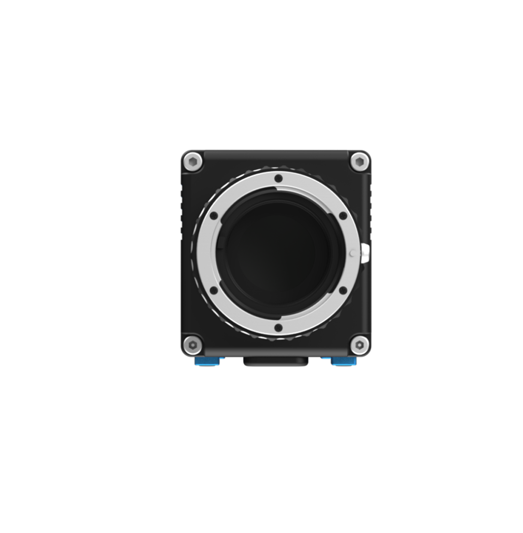
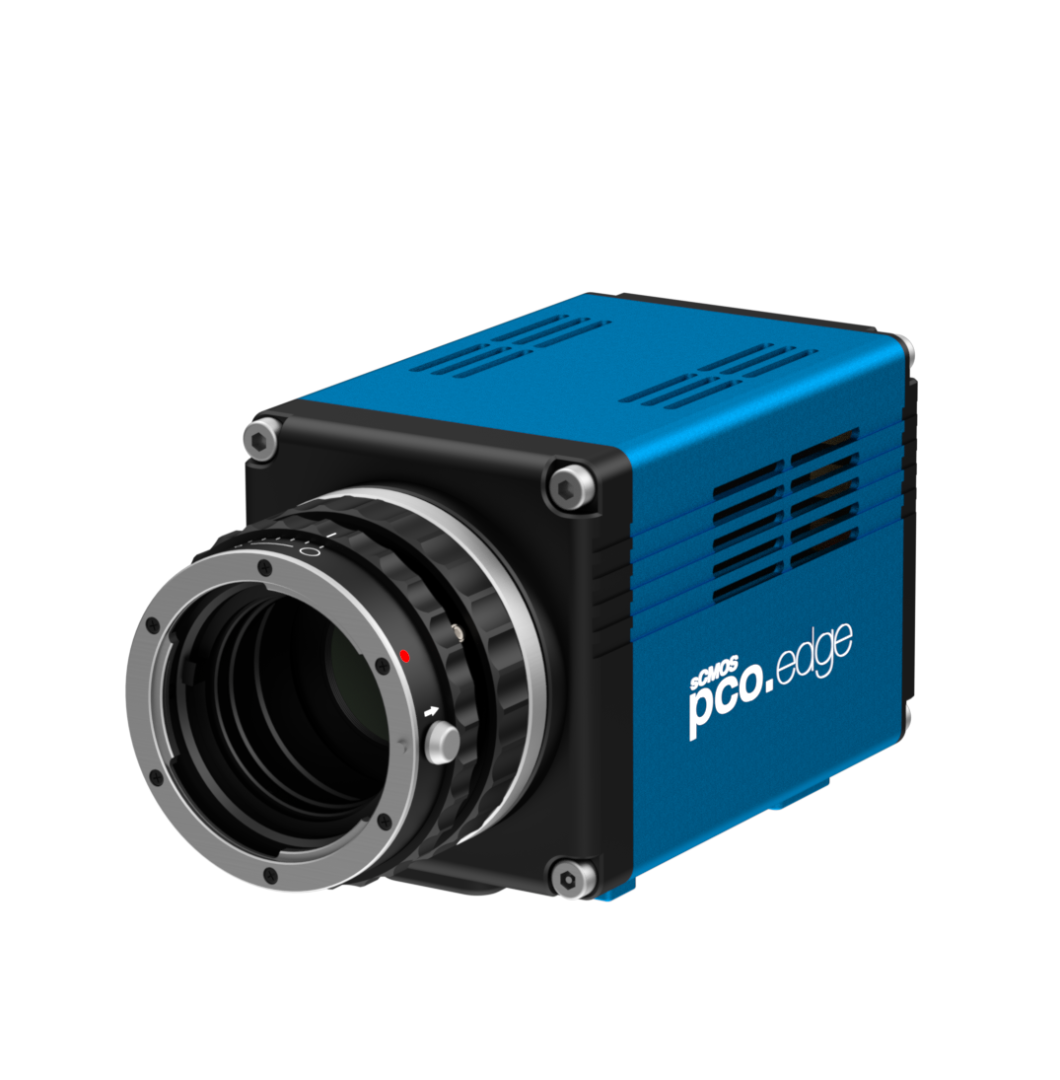
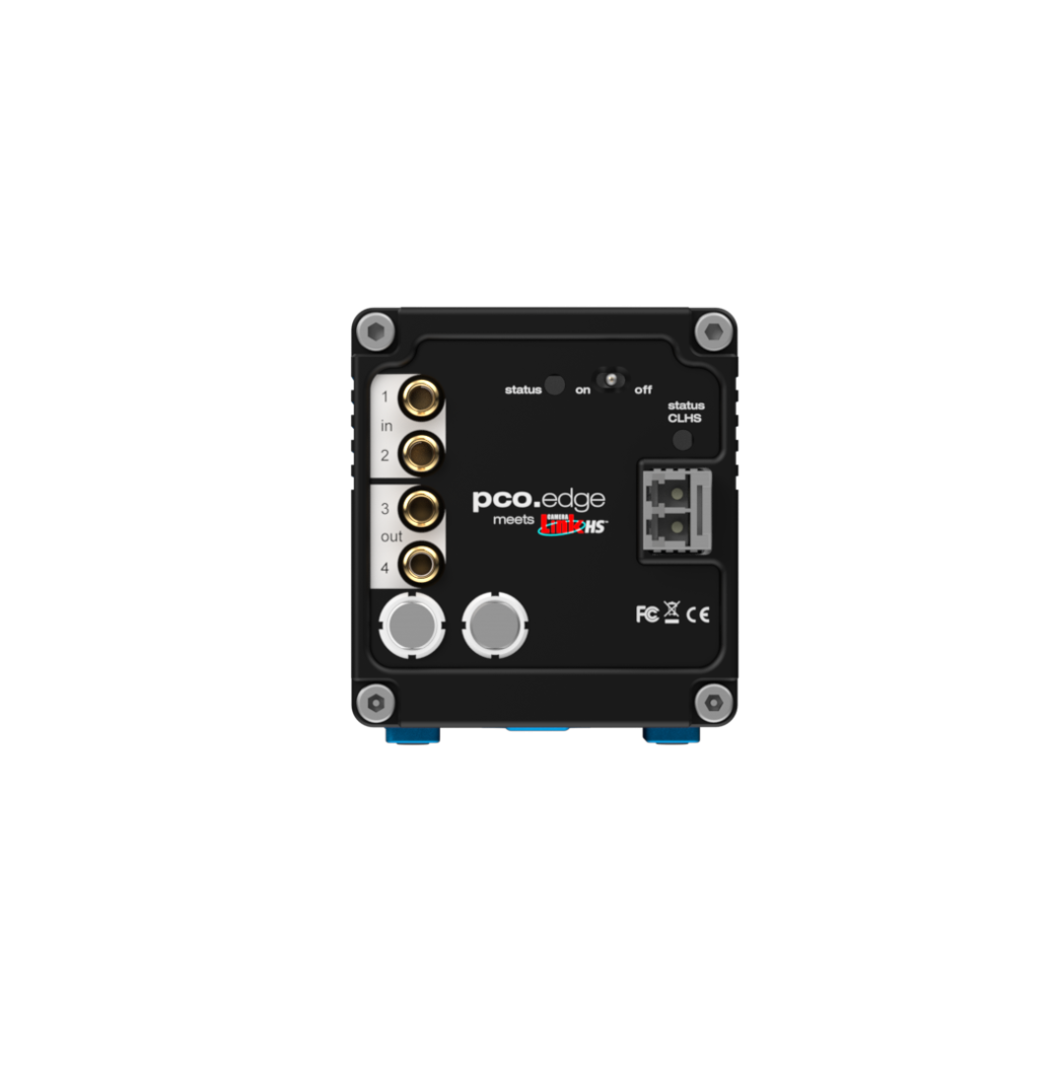
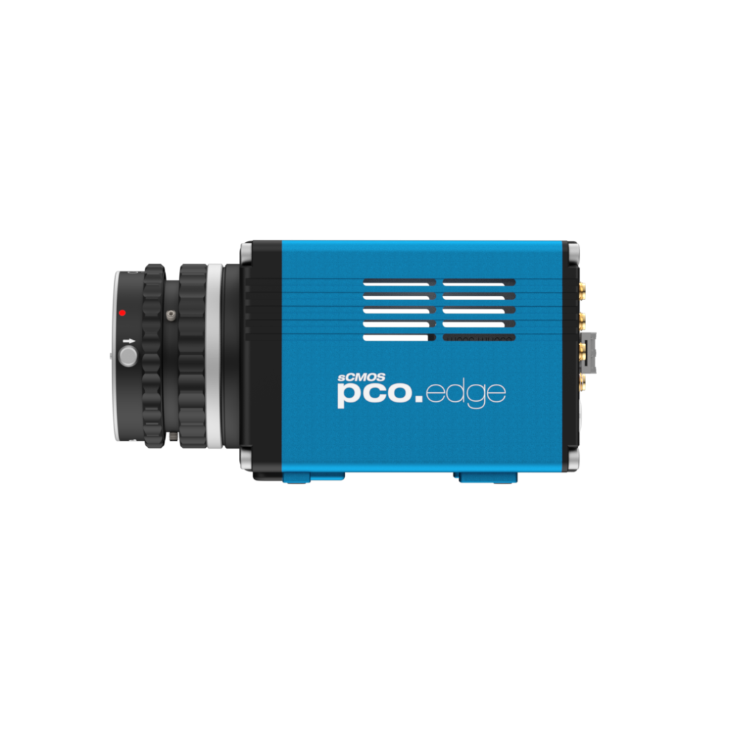
pco.edge 5.5 CLHS sCMOS Camera

The pco.edge 5.5 is a high resolution cooled sCMOS camera utilizing a CLHS interface and 10G FOL (Camera Link HS) to allow direct processing of large data transfers via a computer making it easy to securely bridge distances of up to 10 km. This camera is equipped with a 16-bit scientific CMOS sensor for capturing crisp images and precise measurements. The pco.edge 5.5 camera also comes with an option to upgrade with a water cooling system.

Also, try our MachVis Lens Selector tool to help identify the perfect lens solution and PCO camera for your imaging and machine vision requirements.


Interface

CLHS FOL

Sensor technology

sCMOS

Color type

monochrome or color

Resolution 

2560 x 2160 pixels

Sensor diagonal

21.8 mm

Pixel size

6.5 x 6.5 μm

Maximum frame rate @ full resolution 

100 fps

Maximum pixel rate

572 MPixel/s

Peak QE

60 % @ 600 nm1

Typ. read noise2 

1.0 e-

Dark current @ sensor temperature

< 0.6 RS/GR e-/pixel/s

< 0.9 GS @ 7 °C e-/pixel/s


Maximum dynamic range

30,000 : 1

Shutter type

Rolling Shutter, Global Shutter, Global Reset

Sensor cooling3

air, optional: water

Additional options

double shutter, lens control

Dimensions (H x W x L)

76 x 70 x 122 mm

 
1 Monochrome version
2 The readout noise values are given as median (med). All values are raw data without any filtering.
3 air = air forced with fan | water = external water connection
 
pco.edge 5.5 CLHS QE Curve - Monochrome
pco.edge 5.5 CLHS QE Curve - Color
   
  • Bright-field microscopy
  • Fluorescence microscopy
  • Digital pathology
  • Single molecule localization microscopy (SMLM) – PALM, STORM, dSTORM, GSDIM
  • Lightsheet fluorescence microscopy (LSFM)
  • Structured illumination microscopy (SIM)
  • Calcium imaging
  • Förster resonance energy transfer (FRET)
  • Fluorescence recovery after photobleaching (FRAP)
  • High-speed bright-field ratio imaging
  • High throughput screening
  • High content screening
  • Biochip reading
  • Total internal reflection microscopy (TITF)
  • Spinning disk confocal microscopy
  • 3D metrology
  • Ophthalmology
  • Photovoltaic inspection
  • Industrial quality inspection
  • Wafer inspection
  • Image intensifier imaging
  • Lucky astronomy
  • Disaster recovery
  • Tunnel inspection
  • Particle tracking velocimetry (PTV)


Download asset:
To download this item, please complete the form below.
NOTE: If you have disabled our marketing cookies, you will not be able to access our videos or downloads. To update your cookie preferences, please click here to review and enable cookies.
CAPTCHA
This question is for testing whether or not you are a human visitor and to prevent automated spam submissions.
Close