2AMV

THE STRUCTURE OF GLYCOGEN PHOSPHORYLASE B WITH AN ALKYL-DIHYDROPYRIDINE-DICARBOXYLIC ACID


Experimental Data Snapshot

  • Method: X-RAY DIFFRACTION
  • Resolution: 2.30 Å
  • R-Value Free: 0.282 
  • R-Value Work: 0.201 
  • R-Value Observed: 0.201 

wwPDB Validation   3D Report Full Report


This is version 1.2 of the entry. See complete history


Literature

The structure of glycogen phosphorylase b with an alkyldihydropyridine-dicarboxylic acid compound, a novel and potent inhibitor.

Zographos, S.E.Oikonomakos, N.G.Tsitsanou, K.E.Leonidas, D.D.Chrysina, E.D.Skamnaki, V.T.Bischoff, H.Goldmann, S.Watson, K.A.Johnson, L.N.

(1997) Structure 5: 1413-1425

  • DOI: https://doi.org/10.1016/s0969-2126(97)00292-x
  • Primary Citation of Related Structures:  
    2AMV

  • PubMed Abstract: 

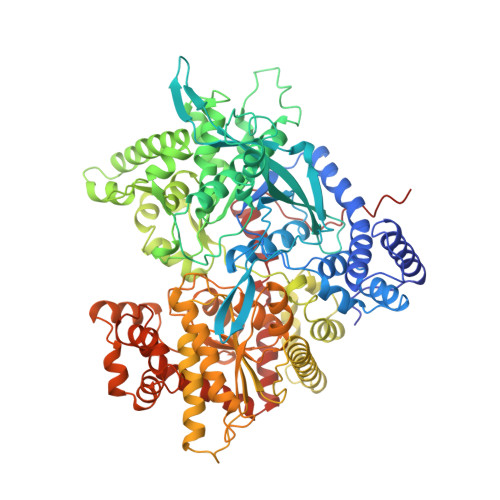
    In muscle and liver, glycogen concentrations are regulated by the reciprocal activities of glycogen phosphorylase (GP) and glycogen synthase. An alkyl-dihydropyridine-dicarboxylic acid has been found to be a potent inhibitor of GP, and as such has potential to contribute to the regulation of glycogen metabolism in the non-insulin-dependent diabetes diseased state. The inhibitor has no structural similarity to the natural regulators of GP. We have carried out structural studies in order to elucidate the mechanism of inhibition.


  • Organizational Affiliation

    Institute of Biological Research and Biotechnology, The National Hellenic Research Foundation 48, vas Constantinou Avenue, Athens, 11635, Greece.


Macromolecules
Find similar proteins by:  (by identity cutoff)  |  3D Structure
Entity ID: 1
MoleculeChains Sequence LengthOrganismDetailsImage
PROTEIN (GLYCOGEN PHOSPHORYLASE)842Oryctolagus cuniculusMutation(s): 0 
EC: 2.4.1.1
UniProt
Find proteins for P00489 (Oryctolagus cuniculus)
Explore P00489 
Go to UniProtKB:  P00489
Entity Groups  
Sequence Clusters30% Identity50% Identity70% Identity90% Identity95% Identity100% Identity
UniProt GroupP00489
Sequence Annotations
Expand
  • Reference Sequence
Small Molecules
Ligands 3 Unique
IDChains Name / Formula / InChI Key2D Diagram3D Interactions
BIN
Query on BIN

Download Ideal Coordinates CCD File 
C [auth A]2,3-DICARBOXY-4-(2-CHLORO-PHENYL)-1-ETHYL-5-ISOPROPOXYCARBONYL-6-METHYL-PYRIDINIUM
C20 H21 Cl N O6
HMSIYRVIPQHZBI-UHFFFAOYSA-O
PLP
Query on PLP

Download Ideal Coordinates CCD File 
B [auth A]PYRIDOXAL-5'-PHOSPHATE
C8 H10 N O6 P
NGVDGCNFYWLIFO-UHFFFAOYSA-N
GOL
Query on GOL

Download Ideal Coordinates CCD File 
D [auth A]GLYCEROL
C3 H8 O3
PEDCQBHIVMGVHV-UHFFFAOYSA-N
Binding Affinity Annotations 
IDSourceBinding Affinity
BIN PDBBind:  2AMV Ki: 1.6 (nM) from 1 assay(s)
Binding MOAD:  2AMV Ki: 1.6 (nM) from 1 assay(s)
Experimental Data & Validation

Experimental Data

  • Method: X-RAY DIFFRACTION
  • Resolution: 2.30 Å
  • R-Value Free: 0.282 
  • R-Value Work: 0.201 
  • R-Value Observed: 0.201 
  • Space Group: P 43 21 2
Unit Cell:
Length ( Å )Angle ( ˚ )
a = 127.11α = 90
b = 127.11β = 90
c = 115.46γ = 90
Software Package:
Software NamePurpose
X-PLORmodel building
X-PLORrefinement
DENZOdata reduction
SCALEPACKdata scaling
X-PLORphasing

Structure Validation

View Full Validation Report


Entry History 

Deposition Data

Revision History  (Full details and data files)

  • Version 1.0: 1998-10-21
    Type: Initial release
  • Version 1.1: 2008-04-27
    Changes: Version format compliance
  • Version 1.2: 2011-07-13
    Changes: Derived calculations, Version format compliance