Article

Optimized Method for Mapping Inorganic Pigments by Means of Multispectral Imaging Combined with Hyperspectral Spectroscopy for the Study of Vincenzo Pasqualoni’s Wall Painting at the Basilica of S. Nicola in Carcere in Rome

by
Lucilla Pronti
1,
Giuseppe Capobianco
2,*,
Margherita Vendittelli
3,
Anna Candida Felici
3,*,
Silvia Serranti
2 and
Giuseppe Bonifazi
2
1
INFN-Laboratori Nazionali di Frascati, via Enrico Fermi 54, 00044 Frascati, Italy
2
Department of Chemical Engineering, Materials and Environment, Sapienza University of Rome, Via Eudossiana 18, 00184 Rome, Italy
3
Department of Basic and Applied Science for Engineering, Sapienza University of Rome, Via A. Scarpa 16, 00161 Rome, Italy
*
Authors to whom correspondence should be addressed.
Minerals 2021, 11(8), 839; https://doi.org/10.3390/min11080839
Submission received: 30 June 2021 / Revised: 24 July 2021 / Accepted: 29 July 2021 / Published: 2 August 2021
(This article belongs to the Special Issue Spectral Behavior of Mineral Pigments)

Abstract

:
Multispectral imaging is a preliminary screening technique for the study of paintings. Although it permits the identification of several mineral pigments by their spectral behavior, it is considered less performing concerning hyperspectral imaging, since a limited number of wavelengths are selected. In this work, we propose an optimized method to map the distribution of the mineral pigments used by Vincenzo Pasqualoni for his wall painting placed at the Basilica of S. Nicola in Carcere in Rome, combining UV/VIS/NIR reflectance spectroscopy and multispectral imaging. The first method (UV/VIS/NIR reflectance spectroscopy) allowed us to characterize pigment layers with a high spectral resolution; the second method (UV/VIS/NIR multispectral imaging) permitted the evaluation of the pigment distribution by utilizing a restricted number of wavelengths. Combining the results obtained from both devices was possible to obtain a distribution map of a pictorial layer with a high accuracy level of pigment recognition. The method involved the joint use of point-by-point hyperspectral spectroscopy and Principal Component Analysis (PCA) to identify the pigments in the color palette and evaluate the possibility to discriminate all the pigments recognized, using a minor number of wavelengths acquired through the multispectral imaging system. Finally, the distribution and the spectral difference of the different pigments recognized in the multispectral images, (in this case: red ochre, yellow ochre, orpiment, cobalt blue-based pigments, ultramarine and chrome green) were shown through PCA false-color images.

1. Introduction

Pigments used as pictorial materials in Cultural Heritage are inorganic and organic compounds. Among the inorganic compounds, natural and synthetic minerals have been largely employed since ancient times [1].
The identification of pigments involves many research fields in Cultural Heritage concerning authentication purposes, restoration treatments, technical methods of production and development of proper analytical instruments and approaches.
Mineral pigments have been used in paintings since the Paleolithic age (iron oxide, such as natural hematite or heated goethite and charcoal or manganese oxides) [2]. At first, the choice of the pictorial materials was correlated with the availability in nature of opaque and colored materials, but over time, new synthetic minerals have been developed to obtain specific artistic effects and the durability of artworks.
During the creation of mural paintings, it is important considering that some pigments are not alkali-resistant and therefore cannot be used in fresco, such as organic pigments, white and red lead, malachite, or azurite [3]. However, even if these materials are mixed with organic binders [4], they suffer from chemical alterations caused by the exposition to some environmental conditions, such as high moisture, capillary rise, light direct exposition, etc. This means that the analyses of the used pigments are extremely important both for the knowledge of the execution techniques of artists and for restoration purposes.
Many analytical techniques could be applied directly in situ; however, since mural paintings are generally large-size works, the analyses are performed by selecting few points and extending the results to the other areas with similar colors.
In the last few decades, the research trend is to address the development of advanced methods for non-destructive in situ analyses that return imaging output, allowing for us to achieve the distribution of the chemical composition of artworks [5].
In this scenario, hyperspectral imaging (HSI) is a powerful diagnostic tool, due to the possibility of recognizing several pigments and returning chemical imaging or mapping. The capability to identify specific materials from hyperspectral images is correlated to the use of an advanced algorithm (chemometric approach) coupled to the creation of huge spectral databases acquired with the same technique [6].
Hyperspectral imaging, combined with chemometrics, is considered an upgrade of multispectral imaging [7,8]; however, although it remains a good method for the identification of the pigments, it requires long-time acquisitions and scan support. On the other hand, the limited wavelengths selection of the multispectral systems entails being less advisable for the classification of pictorial materials and complex mixtures.
In the last decade, snapshot cameras have been developed [9,10]; they capture hyperspectral images during a single integration time through a detector array, and the data cube, namely the multi-dimensional array of values (X, Y, and λ), is taken at once, without any time offset.
All the imaging techniques that involve scanning or automatic process require surfaces parallel to the acquisition camera and stability of the support on which they are placed; all of these aspects are often disregarded when they are applied on mural paintings of architectural surfaces, such as apses or domes of churches, or in those situations where the analyses have to be performed on scaffolds, which are generally less stable than a floor. Moreover, although the snapshot cameras could be performed at a certain distance from the object, they have low spatial resolution concerning multispectral imaging with IR-converted reflex cameras or monochromatic CCD cameras.
To simplify multispectral systems, reducing the number of filters employed to the most significant ones, some methods have been proposed, trying to obtain similar performances for hyperspectral data [11,12]. Based on these methods, Principal Component Analysis (PCA) seems to be effective to select the relevant wavebands that allow the best discrimination among pigments [13,14].
In this paper, we propose an optimized method for mapping inorganic pigments by using multispectral imaging combined with hyperspectral spectroscopy for the study of Vincenzo Pasqualoni’s wall painting at the Basilica of S. Nicola in Carcere in Rome by performing the following:
(1)
The identification of the main pigments used through point hyperspectral spectroscopy, confirmed by other diagnostic analyses: X-ray fluorescence (XRF) and Raman Spectroscopy [15].
(2)
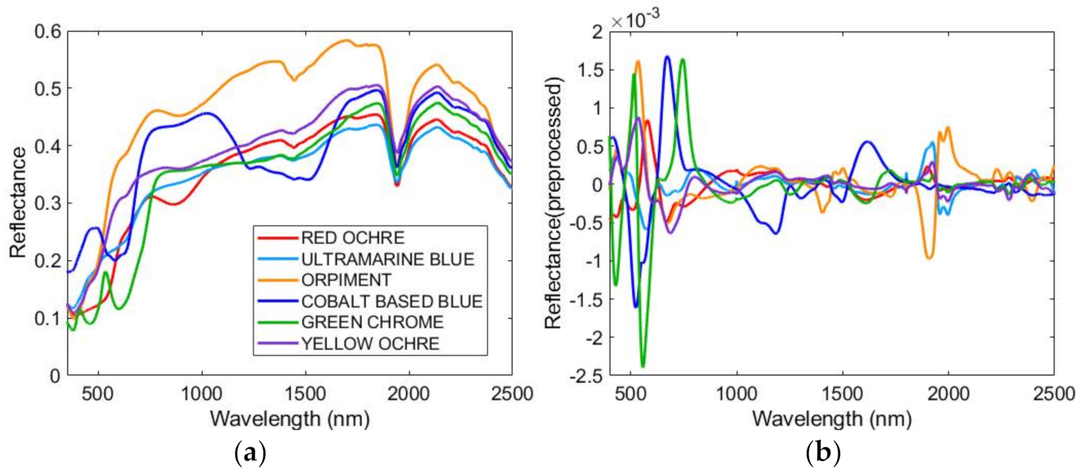
A PCA on the hyperspectral data to evaluate the effective discrimination among the identified pigments;
(3)
A PCA on a wavelength reduction of the hyperspectral data to evaluate if a limited spectral range, corresponding to the filters set of the multispectral system, was sufficient to discriminate the pigments;
(4)
Acquisition of the multispectral images coupled with PCA to individuate all the areas in which the identified pigments are located.

2. Materials and Methods

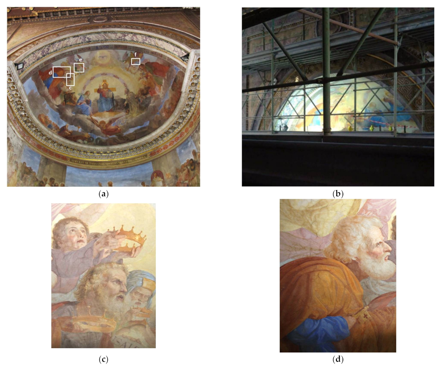
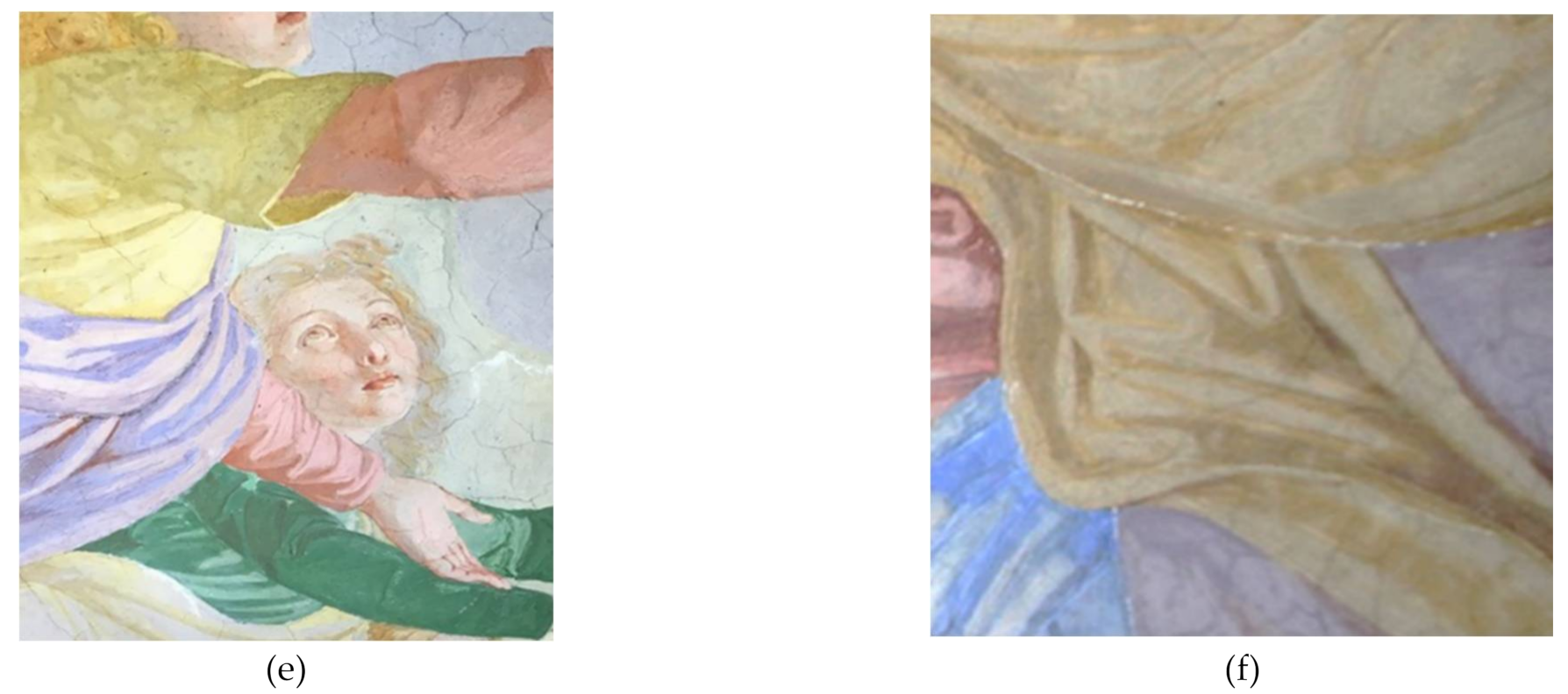
2.1. Vincenzo Pasqualoni’s Wall Painting

Vincenzo Pasqualoni’s wall painting is a fresco decoration of the apse of the basilica of S. Nicola in Carcere (Rome), dated 1865–1866. The analyzed portion depicts Christ in Glory between the Virgin and S. Nicholas. Since the wall painting was painted in the XIX century, the painter might have used both industrial products (i.e. cobalt blue, zinc white, etc.) and natural pigments (ochres, lapis, green earth, etc.).
The analyses were performed during the restoration intervention, and the results were used for the choice of proper conservative treatments. For this painting, a multi-analytical approach was performed, as published in a previous paper [15], but in this work, we focus on four areas examined with UV/VIS/NIR hyperspectral and multispectral techniques, as shown in Figure 1. The investigated areas were suggested by the restorer during the restoration of the basilica (Figure 1b).

2.2. Multispectral Imaging System

The multispectral images were acquired with a Peltier cooled monochromatic camera (QSI 583-w) equipped with a Kodak KAF8300 CCD sensor (3348 × 2574 pixel, pixel area 5.4 µm2). The selection of the spectral ranges was performed with 15 narrow band interferential filters (10 nm FWHM) respectively centered at 380, 390, 400, 410, 420, 450, 470, 500, 550, 600, 650, 680, 700, 750, and 800 nm and a high pass filter at 1000 nm. Dark frames were employed to reduce the electronic noise of the images.
The multispectral images were transformed into reflectance images by applying a normalization of the images to the average values of a diffuse reflectance standard (Spectralon SRM-99, Labsphere, North Sutton, NH, USA) inserted in the scene.
Multispectral reflectance datasets were aligned by using a routine written in the MatlabTM environment, where the parameters of a generic affine transformation for the alignment of two adjacent datasets were calculated by selecting suitable control points.
Finally, the multispectral images were concatenated into a multispectral data cube composed of 16 channels (one channel for each filter).

2.3. Hyperspectral Spectrometers

The ASD FieldSpec® 4 Standard-Res (ASD Inc., Boulder, CO, U.S.A.) is a portable spectroradiometer that works in the spectral range of 350–2500 nm and has a spectral resolution of 3 nm at 700 nm, and 10 nm at 1400/2100 nm. This instrument essentially consists of a detector case and a fiber optic cable with a contact probe that is connected to a laptop computer. Inside the detector case, the system has different separate holographic diffraction gratings with three separate detectors. Order separation filters cover each detector to eliminate second- and higher-order light. The detector system is made up of a VNIR detector (350–1000 nm; 512 element silicon array), the SWIR1 detector (1001–1800 nm; Graded Index InGaAs. Photodiode, Two-Stage TE Cooled), and the SWIR2 detector (1801–2500 nm; Graded Index InGaAs. Photodiode, Two-Stage TE Cooled) 61. The ASD Contact Probe consists of a halogen bulb light source with a color temperature equal to 2901 ± 10% K [16]. It has a length of 25.4 cm (including the probe grip) and weights 0.7 kg. The light source is placed at 12° from the normal axis to the contact probe spot plane (light-source angle). The fiber optic head is placed at 35° from the normal axis to the contact probe spot plane (measurement angle). Since the spot size of the contact probe is 10 mm, the Field of View (FOV) is about 1 cm2. The native software of the ASD instrument, called RS3, was used for data acquisition [17].

2.4. Principal Component Analysis (PCA)

PCA is a chemometric method enabling an overview of multivariate data, and it is widely adopted to analyze hyperspectral data [18]. This method can be used to uncover relations between variables (i.e., wavelengths) and samples (i.e., measurement areas), generating hypothetical data clusters. PCA consists of the transformation of the spectral data into several principal components (PCs), which are linear combinations of the original spectral data containing the spectral variations of each collected spectral dataset [19]. One of the results is the reduction of the data to those that provide an accurate description of the entire dataset. The first few PCs, resulting from PCA, are generally used to analyze the maximum variance among samples and those characterized by similar spectral signatures tend to aggregate in the plots of the first two or three components (namely “score plots”). To evaluate how strongly each variable (i.e., wavelength) influences a principal component, loading plots might be displayed.
In this work, PCA was performed on both the hyperspectral spectra and the multispectral data cube. To emphasize the differences between the spectral signatures of the pigment classes, preprocessing treatments were performed:
-
The hyperspectral data were preprocessed with the sequential application of detrend, derivative, and mean center (MC) algorithms;
-
The multispectral data cube was preprocessed with the sequential application of Probabilistic Quotient Normalization (PQN) and autoscale algorithms.
In detail, detrend removes a linear or quadratic trend from an image. PQN is a robust normalization method similar to MSC, but it uses the median as the target and a robust fitting of each row to the target [20]. Spectral differences among the different polymers were emphasized, and the baseline background was removed by applying a Savitzky–Golay (SG) polynomial derivative [21]. PQN is a robust normalization method, similar to multiple scatter correction, but it uses the median as the target and a robust fitting of each row to the target [22].
Centering and scaling are the two most common types of preprocessing normally applied before PCA. MC has the effect to include an adjustable intercept in multivariate models [23]. Autoscale is a common preprocessing method that uses mean-centering, followed by division of each column (variable) by the standard deviation of that column [24].

3. Results and Discussion

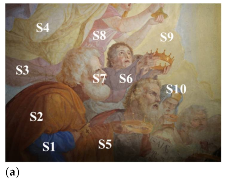
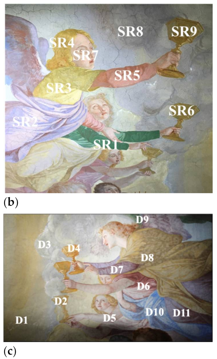
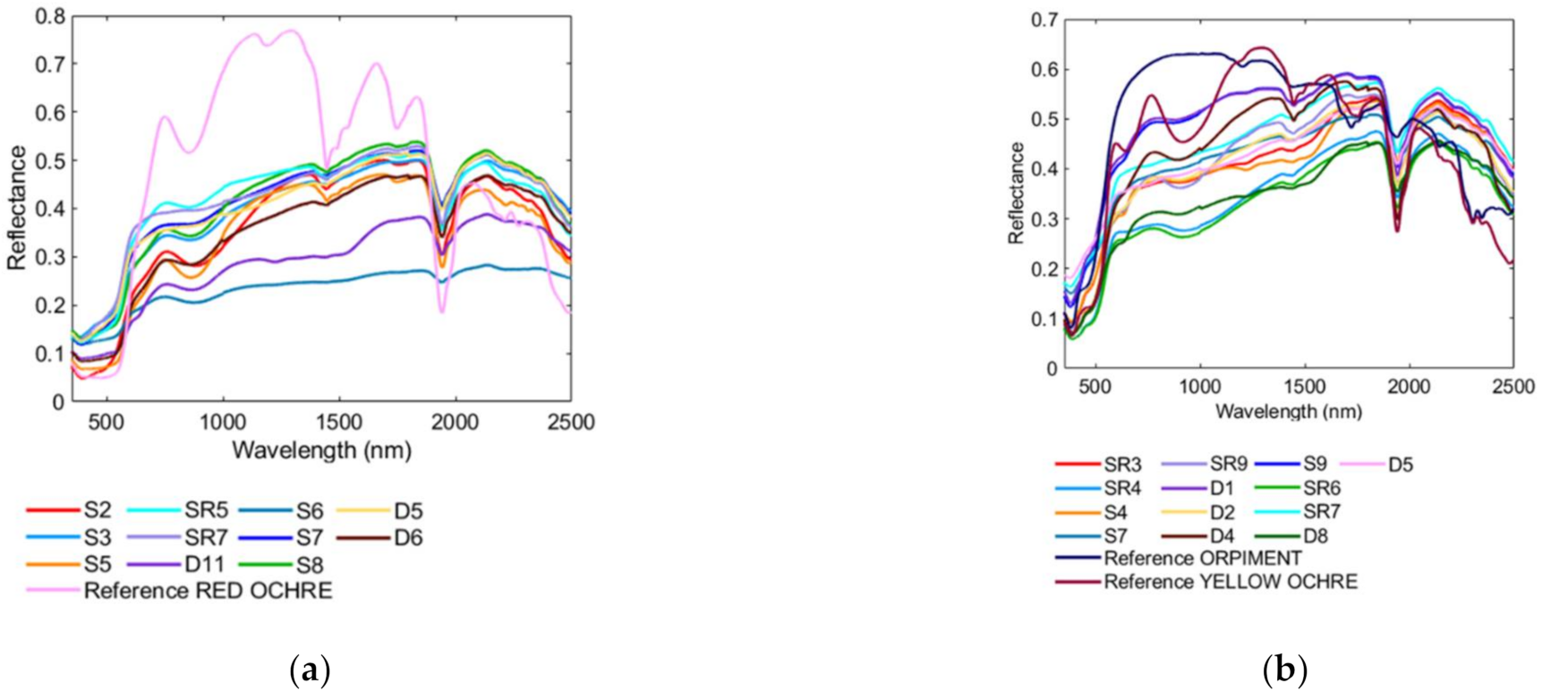
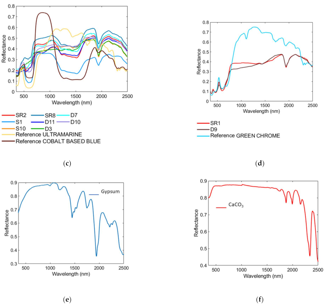
3.1. Pigment Identification by Full Range VIS/NIR/SWIR Spectroscopy

Full Range VIS/NIR/SWIR spectroscopy was performed point-by-point, selecting the main colors used in the wall painting.
Since the painting is dated 1865–1866, modern and ancient pigments might have been used. In Figure 2, we report the areas analyzed. Figure 3 summarizes the average reflectance spectra obtained compared with the reference pigments assigned to each area shown in Figure 2.
In detail, the red and pink areas (Figure 2: S3, S5, S8, SR5, D5, and D6) are characterized by an inflection point at 580 nm and absorption at 869 nm; these spectral features could be attributed to Fe3+ cations of hematite (Fe2O3) (Figure 3a) [10]. Hematite is one of the minerals constituting red ochres that is largely used in frescoes paintings due to their high durability, but also due to their abundance and low cost compared to other red pigments [25,26,27,28,29].
For the yellow areas (Figure 2: S4, S9, SR3, SR4, SR6, SR9, D1, D2, D4, and D8), we identify two different spectral behaviors: (1) an inflection point at 540 nm and absorption at 650 nm and (2) an inflection point at 480 nm. The first spectral features are related to Fe3+ cations of goethite (FeO(OH)) (Figure 3b) [10], the main component of yellow ochre. Similar to red ochre, yellow ochre has been largely used in frescoes paintings since ancient times [30,31,32]. The inflection point at 480 nm is attributed to orpiment (As2S3) (Figure 3b). Orpiment exists as minerals, as well as synthetic pigments; it was even used in Roman and Egyptian cultures [33,34]. The different ratios between yellow ochre and orpiment allow us to obtain the various shades of color in the yellow pictorial layers being analyzed. The colors found in these areas are obtained by combining red ochre, yellow ochre and orpiment (Figure 2: S7 and SR7). All the As-based pigments display some degradation effects in frescoes [35]; this suggests the use of a “secco” technique to spread orpiment. Orange hues (i.e., Figure 2: S2) are created by the combination between yellow ochre and red ochre, since both their spectral features are detected.
Most of the gray, blue, and purple areas (Figure 2: S1, S6, S10, SR8, SR2, SR8, D3, D7, and D11) are characterized by a continuous absorption from 538 to 663 nm and absorption regions in the SWIR range from 1223 to 1600 nm; these features are related to cobalt oxide, such as cobalt blue (CoAl2O4) or smalt (SiO2 + K2O + Al2O3 + CoO) [36,37] (Figure 3c). Purple hues are created by combining cobalt-based blue and red ochre, since both of their spectral features are detected. In some light blue areas (Figure 2: S10, SR8, D10, and D3), we also found ultramarine blue (Al6Na8O24S3Si6) characterized by a continuous absorption from 530 to 678 nm (Figure 3c). Ultramarine blue was probably used to improve the chromatic variability of the blue-colored paint layer, increasing the perspective effect.
Finally, the green areas (Figure 2: SR1 and D9) are identified as chrome green (Cr2O3), defined also as viridian, by a double absorption at 606 and 696 nm [38] (Figure 3d).
Almost all the spectra present the absorption regions in the SWIR range, at 1451, 1491, and 1545 nm, due to the presence of gypsum (reference spectrum was showed in Figure 3e) [39]. Moreover, an absorption band at 2140 nm was observed, attributed to impurities such as materials containing anion groups (Al-OH, Mg-OH, Fe-OH, and Si-OH). Finally, an absorption band at 2200 nm was detected due to calcium carbonate [39]. These two compounds are the constituents of the mural support [40,41,42] (reference spectrum was shown in Figure 3f).
This pigment identification was confirmed also with other diagnostic techniques, reported in a previous work [15] and summarized in Table 1.

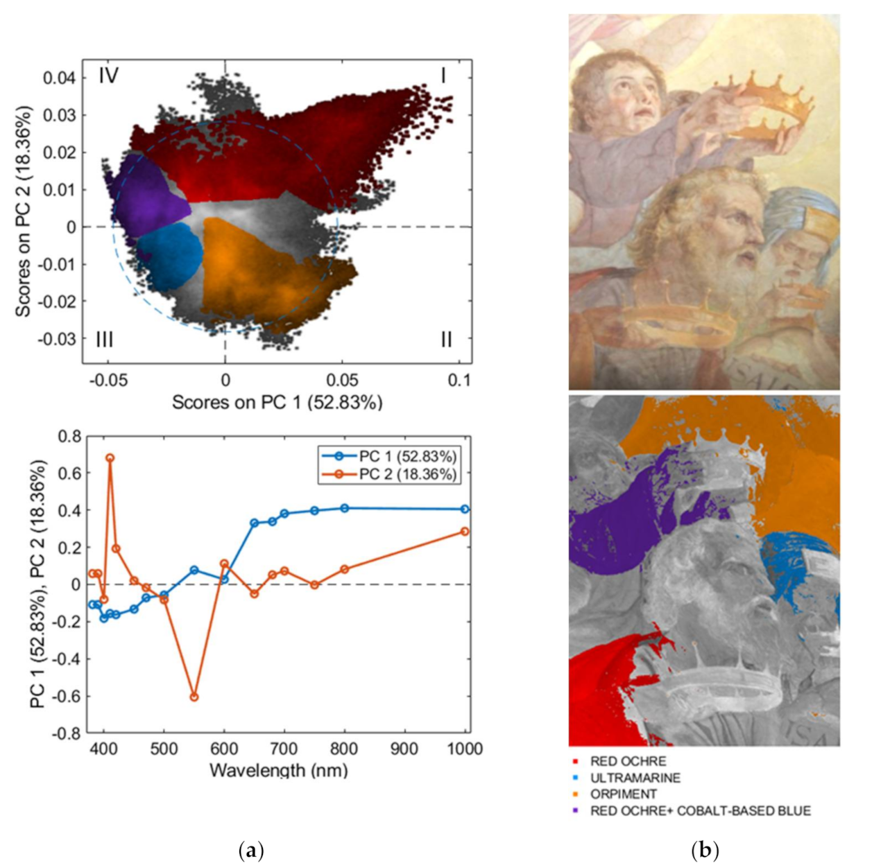
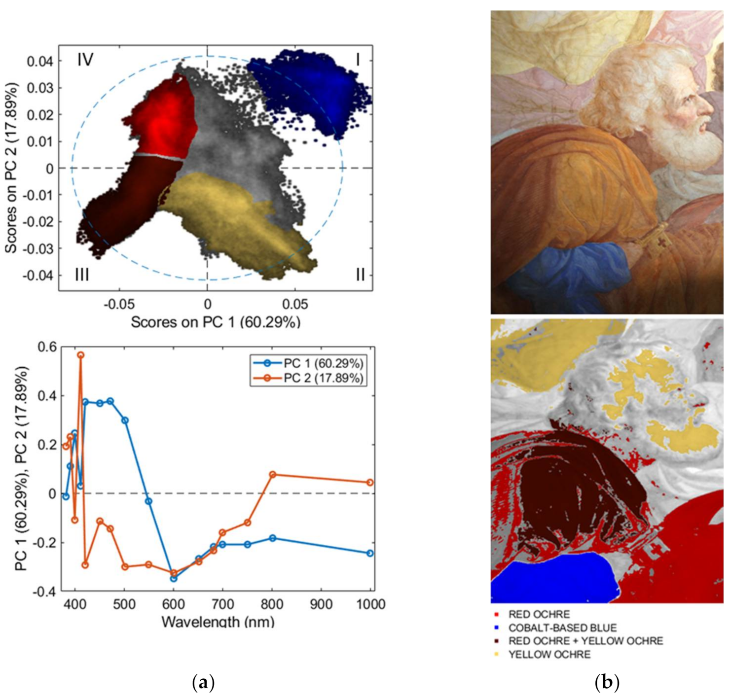
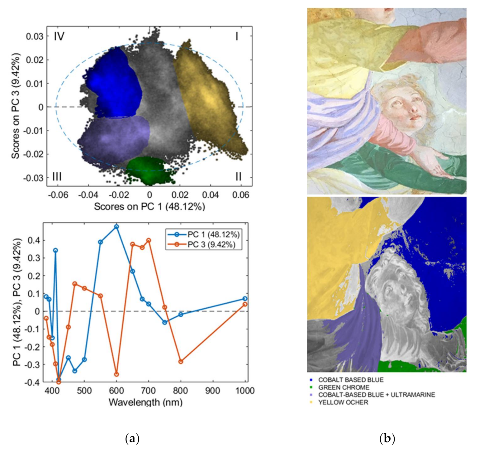
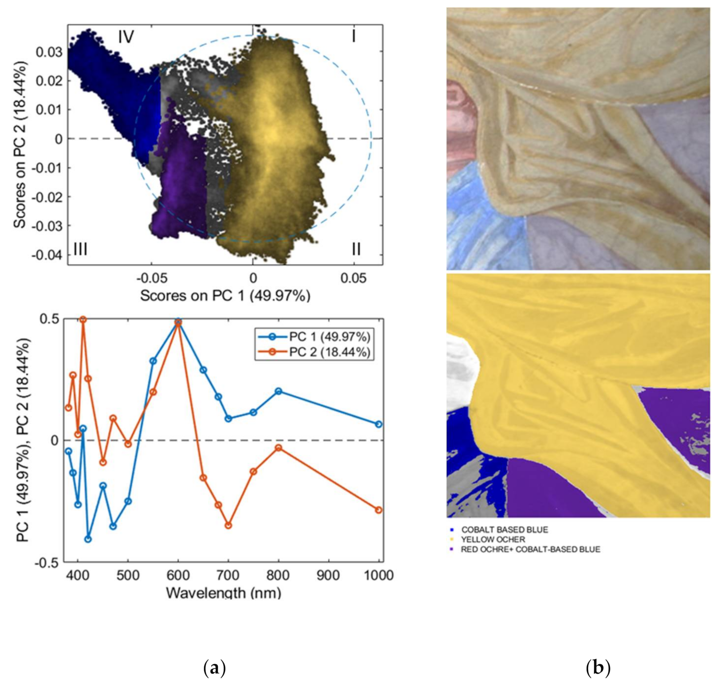
3.2. UV/VIS/NIR Multispectral Imaging

The entire multispectral data cube was analyzed by PCA to evaluate the spectral features and variability of the six color layers. The raw and preprocessed spectra are shown in Figure 4.
PCA model requires 5 PCs to express a total captured variance of 92.98%. In more detail, in the PC1–PC3 score plot (Figure 5a) it is possible to note that pixels belonging to the “cobalt-based blue” class can be easily distinguished from spectra of the other classes, being clustered in the first and fourth quadrants, whereas pixels belonging to the “red ochre”, “yellow ochre”, and “orpiment” classes occur in different regions of the plot, mainly in the second and third quadrants.
The variability inside each class is given using pigments with a different ratio to obtain the nuances and volumetric effects on the wall painting. In addition, in some areas, there is the co-presence of pigments (i.e., yellow ochre with orpiment or cobalt-based blue with ultramarine). All of these features contribute to the high variance found on some pictorial layers, such as cobalt-based blue, green chrome, or the proximity of the red ochre and yellow ochre clusters or between yellow ochre and orpiment, for which the pigments are combined to obtain shades of clothes or the volumetric effects of the cups or the sky.
Green chrome spectra are mainly clustered in the positive values of PC1 and PC3. Finally, the “ultramarine blue” class is mainly clustered in the center part of the PCA score plot. The loading plots of PC1 and PC3 (Figure 5b) show how the variance detected in the PCA score plots mainly depends on the spectral regions around 395, 514, 580, 673, and 744 nm (Figure 3b).
Starting from the spectral features detected by mentioned PCA, a variable selection approach was adopted, taking into account the wavelengths acquired by the multispectral imaging device (i.e., 380, 390, 400, 410, 420, 450, 470, 500, 550, 600, 650, 680, 700, 750, 800, and 1000 nm). Subsequently, a new PCA on hyperspectral data with the selected variables was performed. The PCA model of mentioned wavelength requires five PCs to express a total captured variance of 99.91%. In more detail, in the PC1–PC3 score plot (Figure 5c), it is possible to note that pixels belonging to the “cobalt-based blue” class can be easily distinguished from spectra of the other classes, being clustered in the second and third quadrants, whereas pixels belonging to “red ochre”, “yellow ochre”, and “orpiment” classes occur in different regions of the plot, mainly in the first and fourth quadrants. Green chrome spectra are mainly clustered by positive values of PC3 and negative values of PC1. Finally, the “ultramarine blue” class is mainly clustered in the third and fourth quadrants.
The loading plot of PC1 vs. PC3 (Figure 5d) displays how the score plot is influenced by the wavelengths 400, 500, 550, 680, 800, and 1000 nm. By comparing the result of full-spectrum PCA with variable selection and the “selected variable” PCA, it is possible to notice how the clouds regarding yellow ochre, orpiment, and red ochre in the “selected variable” PCA are more close than full-spectrum PCA (Figure 5c). However, despite the low number of wavelengths, the cloud overlapping is low, allowing the differentiation of the six paint layers considered. In general, the result obtained by PCA confirms that we can use the wavelengths selected in this case study, without losing sensitivity in identifying the colorimetric classes.
Starting from the results obtained by hyperspectral VIS/NIR/SWIR spectroscopy, we performed PCA on the multispectral imaging set to map the identified pigments on four big areas of the wall painting. The results of the four areas analyzed are as follows:
AREA 1: The PCA model requires 5 PCs to express a total captured variance of 91.43%. In detail, in the PC1–PC2 score plot (Figure 6a), it is possible to note that pixels belonging to the “red ochre” class can be easily distinguished from pixels of the other classes, being clustered in the first and fourth quadrants. The purple areas are clustered near the “red ochre” one, meaning that the contribution of this pigment is more marked concerning the blue pigment one. “Orpiment” clouds are mainly clustered in the second quadrant (negative values of PC2 and positive values of PC1), whereas pixels belonging to the “ultramarine” class mainly occur in the third quadrant, corresponding to negative values of PC1 and PC2. The loading plots of PC1 and PC2 show how the variance detected in the PCA score plot mainly depends on the wavelengths at 410 and 550 nm and the spectra region over 700 nm (Figure 6a). Figure 6b shows the multispectral image highlighting the classes clustered in the corresponding PCA score plots. In detail, the gray areas, and in particular, the area of the faces, correspond to areas in which there is a greater combination of pigments or pure pigments mixed with white ones.
AREA 2: The PCA model requires five PCs to express a total captured variance of 93.14% (Figure 7). In detail, in the PC1–PC2 score plot (Figure 7a), it is possible to note that pixels belonging to the “cobalt-based blue” class can be easily distinguished from pixels of the other classes, being clustered in the first quadrant. Red ochre clouds are mainly clustered in the fourth quadrant (positive values of PC2 and negative values of PC1), whereas pixels belonging to a mixture of “red-ochre and yellow ochre” mainly occur in the third quadrant. “Yellow ochre” clouds are mainly clustered in the second quadrant (negative values of PC2 and positive values of PC1) and relative to the yellow clothes and on San Pietro’s face. The loading plots of PC1 and PC2 show how the variance detected in the PCA score plot mainly depends on the spectral region from 400 to 500 nm (Figure 6). Figure 6b shows the multispectral image highlighting the classes clustered in the corresponding PCA score plots. In detail, the gray areas correspond to areas in which there is a greater combination of pigments or pure pigments mixed with white ones.
AREA 3: The PCA model requires six PCs to express a total captured variance of 94.98% (Figure 8). In detail, the PC1–PC3 score plot (Figure 8a) shows a cluster separation between the “cobalt-based blue”, “green chrome”, “yellow ochre”, and “cobalt-based blue + ultramarine” classes. Pixels belonging to the “yellow ochre” class mainly occur in the first and second quadrants (positive values of PC1). The spectra of ‘’green chrome” can be identified by the negative value of PC3 in the second and third quadrants. The spectra belonging to “cobalt-based blue” classes occur mainly in the fourth quadrant, whereas, the “cobalt-based blue + ultramarine” class is in the third quadrant. The loading plots of PC1 and PC3 show how the variance detected in the PCA score plot mainly depends on the spectral region from 400 to 800 nm (Figure 8a). Figure 8b shows the multispectral image highlighting the classes clustered in the corresponding PCA score plots. In detail, the gray areas—and, in particular, the area of the faces—correspond to areas in which there is a greater combination of pigments or pure pigments mixed with white ones.
AREA 4: The PCA model requires five PCs to express a total captured variance of 91.11% (Figure 9). In detail, in the PC1–PC2 score plot (Figure 9a), it is possible to note that pixels belonging to the “cobalt-based blue” class can be easily distinguished from pixels of the other classes, being clustered in the fourth quadrant. “Yellow ochre” clouds are mainly clustered in the first and second quadrants, whereas pixels belonging to the “red ochre + cobalt-based blue” class mainly occur in the third quadrant, corresponding to negative values of PC1 and PC2. The loading plots of PC1 and PC2 show how the variance detected in the PCA score plot mainly depends on the wavelengths at 420, 550, 650, 680, 700, and 800 nm (Figure 9a). Figure 9b shows the multispectral image highlighting the classes clustered in the corresponding PCA score plots. The greater variability of the yellow ochre is given by the different yellow shades required to create the prospective effects of the clothes.
The PCA model obtained from the 16 wavelengths allows us, through the loadings, to highlight the significant wavelengths for each pigment investigated. In detail, the separation of cobalt-based blue is given in all PCAs by the wavelengths at 420 and 470 nm. Red ochre discrimination is due to the wavelengths at 600, 800, and 1000 nm. The ultramarine separation is most influenced by the region from 400 to 420 nm. Orpiment discrimination is caused by the region around 550 nm. Yellow ochre is mainly due to the region from 550 to 650 nm. Finally, chrome green separation is mainly to the wavelength at 420, 600, and 800 nm.

4. Conclusions

In this work, the application of a combined hyperspectral and multispectral approach for the identification of mineral pigments and their distribution, respectively, was proposed and applied on the wall painting of Vincenzo Pasqualoni in the apse of S. Nicola in Carcere in Rome.
The analytical procedure adopted has foreseen a first analysis of the UV/VIS/SWIR reflectance spectra, coupled with a chemometric approach, that allowed us to compare the spectral signatures obtained and their variability. Starting from this first exploration, a variable selection approach was adopted to evaluate the possibility to detect the same color variability with a minor number or wavelength. Finally, the same wavelengths were acquired and concatenated to obtained a multispectral data cube. The result allows the possibility to show the distribution and variability of the paint layer detected by hyperspectral imaging.
UV/VIS/NIR reflectance spectroscopy permits to characterize pigment layers with a high spectral resolution, while the multispectral imaging allows us to display the pigment distribution by utilizing a restricted number of wavelengths. The combination of the results obtained from both devices, coupled with PCA, allows us to explain the pigments variance and to obtain their distribution maps for future monitoring actions with the same methodological approaches.
In this case, the selection of the filters used to acquire the multispectral images was effective to evaluate the pigment distribution on Vincenzo Pasqualoni’s wall painting, but, in general, the proposed method could be useful to test several wavelengths combinations to achieve the best pigment identification with a multispectral system for any polychromatic artwork.

Author Contributions

Conceptualization, G.C. and L.P; methodology, G.C., G.B., S.S., A.C.F. and M.V.; software, G.C., G.B., S.S., A.C.F. and L.P.; formal analysis, G.C., A.C.F. and M.V.; data curation, G.C., A.C.F. and L.P.; writing—original draft preparation, G.C. and L.P.; writing—review and editing, G.B., S.S., A.C.F. and M.V.; supervision, A.C.F., S.S. and G.B. All authors have read and agreed to the published version of the manuscript.

Funding

This research was funded by The Lazio Region (Italy) with the ADAMO project (Analysis, Diagnostic and Monitoring Technologies for the Conservation and Restoration of Cultural Heritage) (CUP: B86C18001220002, Grant No. G06970, 30 May 2018), within DTC Lazio (Cultural and Heritage Activities Technology District, Center of Excellence).

Data Availability Statement

The data presented in this study are available from the corresponding authors upon request.

Acknowledgments

The authors want to thank M. Mangano (EURESARTE s.r.l.) and A. Acconci (Soprintendenza Speciale Archeologia Belle Arti e Paesaggio di Roma) for their precious notes relating to the restoration and the historical–artistic framework of the wall painting. Moreover, the authors want to thank “Soprintendenza Speciale Archeologia Belle Arti e Paesaggio di Roma” and the whole team of restorers who supported us during the in situ analyses. Finally, the authors want to sincerely thank “l’Ufficio diocesano per l’edilizia di culto della Diocesi di Roma”, don Pier Luigi Stolfi, and Stefano di Stefano. Mauro Missori is warmly thanked for his help during the measurements.

Conflicts of Interest

The authors declare that they have no known competing financial interests or personal relationships that could have appeared to influence the work reported in this paper.

References

  1. Natalia, B.; Leonardo, B.; Adrover Gracia, I. I Pigmenti Nell’arte Dalla Preistoria Alla Rivoluzione Industriale; Prato, I., Ed.; Saonara: Padova, Italy, 2010; ISBN 8863360901. [Google Scholar]
  2. Chalmin, E.; Menu, M.; Vignaud, C. Analysis of rock art painting and technology of Palaeolithic painters. Meas. Sci. Technol. 2003, 14, 1590–1597. [Google Scholar] [CrossRef]
  3. Cuní, J. What do we know of Roman wall painting technique? Potential confounding factors in ancient paint media analysis. Herit. Sci. 2016, 4, 1–13. [Google Scholar] [CrossRef] [Green Version]
  4. A Casoli, S.S.S. Organic materials in the wall paintings in Pompei: A case study of Insula del Centenario. Chem. Cent. J. 2012, 6, 107. [Google Scholar] [CrossRef] [Green Version]
  5. Fischer, C.; Kakoulli, I. Multispectral and hyperspectral imaging technologies in conservation: Current research and potential applications. Stud. Conserv. 2006, 51, 3–16. [Google Scholar] [CrossRef]
  6. Capobianco, G.; Bracciale, M.P.; Sali, D.; Sbardella, F.; Belloni, P.; Bonifazi, G.; Serranti, S.; Santarelli, M.L.; Cestelli Guidi, M. Chemometrics approach to FT-IR hyperspectral imaging analysis of degradation products in artwork cross-section. Microchem. J. 2017, 132, 69–76. [Google Scholar] [CrossRef]
  7. Pelagotti, A.; Mastio, A.D.; De Rosa, A.; Piva, A. Multispectral imaging of paintings: A way to material identification. IEEE Signal Process. Mag. 2008, 25, 27–36. [Google Scholar] [CrossRef]
  8. Cosentino, A. Identification of pigments by multispectral imaging; a flowchart method. Herit. Sci. 2014, 2, 8. [Google Scholar] [CrossRef] [Green Version]
  9. Bodkin, A.; Sheinis, A.; Norton, A.; Daly, J.; Beaven, S.; Weinheimer, J. Snapshot hyperspectral imaging: The hyperpixel array camera. In Proceedings of the Algorithms and Technologies for Multispectral, Hyperspectral, and Ultraspectral Imagery XV, Orlando, FL, USA, 13–16 April 2009; SPIE: Orlando, FL, USA; Volume 7334, p. 73340H. [Google Scholar]
  10. Geelen, B.; Blanch, C.; Gonzalez, P.; Tack, N.; Lambrechts, A. A tiny VIS-NIR snapshot multispectral camera. In Proceedings of the Advanced Fabrication Technologies for Micro/Nano Optics and Photonics VIII, San Francisco, CA, USA, 8–11 February 2015; SPIE: San Francisco, CA, USA; Volume 9374, p. 937414. [Google Scholar]
  11. Pronti, L.; Felici, A.C.; Ménager, M.; Vieillescazes, C.; Piacentini, M. Spectral Behavior of White Pigment Mixtures Using Reflectance, Ultraviolet—Fluorescence Spectroscopy, and Multispectral Imaging. Appl. Spectrosc. 2017, 71. [Google Scholar] [CrossRef] [Green Version]
  12. Legnaioli, S.; Lorenzetti, G.; Cavalcanti, G.H.; Grifoni, E.; Marras, L.; Tonazzini, A.; Salerno, E.; Pallecchi, P.; Giachi, G.; Palleschi, V. Recovery of archaeological wall paintings using novel multispectral imaging approaches. Herit. Sci. 2013, 1, 1–9. [Google Scholar] [CrossRef] [Green Version]
  13. Pronti, L.; Ferrara, P.; Uccheddu, F.; Pelagotti, A.; Piva, A. Identification of pictorial materials by means of optimized multispectral reflectance image processing. In Proceedings of the 2015 IEEE International Workshop on Information Forensics and Security (WIFS), Rome, Italy, 16–19 November 2015. [Google Scholar] [CrossRef]
  14. Baronti, S.; Casini, A.; Lotti, F.; Porcinai, S. Multispectral imaging system for the mapping of pigments in works of art by use of principal-component analysis. Appl. Opt. 1998, 37, 1299. [Google Scholar] [CrossRef]
  15. Romani, M.; Capobianco, G.; Pronti, L.; Colao, F.; Seccaroni, C.; Puiu, A.; Felici, A.C.; Verona-Rinati, G.; Cestelli-Guidi, M.; Tognacci, A.; et al. Analytical chemistry approach in cultural heritage: The case of Vincenzo Pasqualoni’s wall paintings in S. Nicola in Carcere (Rome). Microchem. J. 2020, 156, 104920. [Google Scholar] [CrossRef]
  16. Jamaludin, M.I.; Matori, A.N.; Myint, K.C. Application of NIR to determine effects of hydrocarbon microseepage in oil palm vegetation stress. In Proceedings of the International Conference on Space Science and Communication, IconSpace, Langkawi, Malaysia, 10–12 August 2015; IEEE Computer Society: Washington, DC, USA, 2015; Volume 2015, pp. 215–220. [Google Scholar]
  17. ASD Full Range, Portable Spectrometers & Spectroradiometers | Malvern Panalytical. Available online: https://www.malvernpanalytical.com/en/products/product-range/asd-range (accessed on 15 June 2021).
  18. Amigo, J.M.; Martí, I.; Gowen, A. Hyperspectral Imaging and Chemometrics. A Perfect Combination for the Analysis of Food Structure, Composition and Quality. In Data Handling in Science and Technology; Elsevier Ltd.: Amsterdam, The Netherlands, 2013; Volume 28, pp. 343–370. [Google Scholar]
  19. Bro, R.; Smilde, A.K. Principal component analysis. Anal. Methods 2014, 6, 2812–2831. [Google Scholar] [CrossRef] [Green Version]
  20. Otto, M. Chemometrics: Statistics and Computer Application in Analytical Chemistry, 3rd ed.; Wiley-VCH: Weinheim, Germany, 2016; ISBN 978-3-527-34097-2. [Google Scholar]
  21. Rinnan, Å.; van den Berg, F.; Engelsen, S.B. Review of the most common pre-processing techniques for near-infrared spectra. TrAC-Trends Anal. Chem. 2009, 28, 1201–1222. [Google Scholar] [CrossRef]
  22. Dieterle, F.; Ross, A.; Schlotterbeck, G.; Senn, H. Probabilistic quotient normalization as robust method to account for dilution of complex biological mixtures. Application in1H NMR metabonomics. Anal. Chem. 2006, 78, 4281–4290. [Google Scholar] [CrossRef]
  23. Sun, D.-W. Infrared Spectroscopy for Food Quality Analysis and Control; Elsevier: London, UK, 2009; ISBN 978-0-12-374136-3. [Google Scholar]
  24. Varmuza, K.; Filzmoser, P. Introduction to Multivariate Statistical Analysis in Chemometrics; CRC Press: Boca Raton, FL, USA, 2016. [Google Scholar]
  25. Bikiaris, D.; Daniilia, S.; Sotiropoulou, S.; Katsimbiri, O.; Pavlidou, E.; Moutsatsou, A.P.; Chryssoulakis, Y. Ochre-differentiation through micro-Raman and micro-FTIR spectroscopies: Application on wall paintings at Meteora and Mount Athos, Greece. Spectrochim. Acta-Part A Mol. Biomol. Spectrosc. 2000, 56, 3–18. [Google Scholar] [CrossRef]
  26. Erdogu, B.; Ulubey, A. colour symbolism in the prehistoric architecture of central anatolia and raman spectroscopic investigation of red ochre in chalcolithic çatalhöyük. Oxford J. Archaeol. 2011, 30, 1–11. [Google Scholar] [CrossRef]
  27. Clementi, C.; Ciocan, V.; Vagnini, M.; Doherty, B.; Tabasso, M.L.; Conti, C.; Brunetti, B.G.; Miliani, C. Non-invasive and micro-destructive investigation of the Domus Aurea wall painting decorations. Anal. Bioanal. Chem. 2011, 401, 1815–1826. [Google Scholar] [CrossRef]
  28. Mazzocchin, G.A.; Agnoli, F.; Salvadori, M. Analysis of Roman age wall paintings found in Pordenone, Trieste and Montegrotto. Talanta 2004, 64, 732–741. [Google Scholar] [CrossRef]
  29. Sbroscia, M.; Cestelli-Guidi, M.; Colao, F.; Falzone, S.; Gioia, C.; Gioia, P.; Marconi, C.; Mirabile Gattia, D.; Loreti, E.M.; Marinelli, M.; et al. Multi-analytical non-destructive investigation of pictorial apparatuses of “Villa della Piscina” in Rome. Microchem. J. 2020, 153, 104450. [Google Scholar] [CrossRef]
  30. Marcaida, I.; Maguregui, M.; Morillas, H.; Perez-Diez, S.; Madariaga, J.M. Raman imaging to quantify the thermal transformation degree of Pompeian yellow ochre caused by the 79 AD Mount Vesuvius eruption. Anal. Bioanal. Chem. 2019, 411, 7585–7593. [Google Scholar] [CrossRef]
  31. Mazzocchin, G.A.; Agnoli, F.; Mazzocchin, S.; Colpo, I. Analysis of pigments from Roman wall paintings found in Vicenza. Talanta 2003, 61, 565–572. [Google Scholar] [CrossRef]
  32. Daniilia, S.; Minopoulou, E.; Andrikopoulos, K.S.; Tsakalof, A.; Bairachtari, K. From Byzantine to post-Byzantine art: The painting technique of St Stephen’s wall paintings at Meteora, Greece. J. Archaeol. Sci. 2008, 35, 2474–2485. [Google Scholar] [CrossRef]
  33. Uda, M. In situ characterization of ancient plaster and pigments on tomb walls in Egypt using energy dispersive X-ray diffraction and fluorescence. Nucl. Instrum. Methods Phys. Res. Sect. B: Beam Interact. Mater. At. 2004, 226, 75–82. [Google Scholar] [CrossRef]
  34. Siddall, R. Mineral Pigments in Archaeology: Their Analysis and the Range of Available Materials. Minerals 2018, 8, 201. [Google Scholar] [CrossRef] [Green Version]
  35. Baldini, F.; Bacci, M.; Carlà, R.; Linari, R. A Color Analysis of the Brancacci Chapel Frescoes. Appl. Spectrosc. 1991, 45, 26–31. [Google Scholar]
  36. Agresti, G.; Bonifazi, G.; Capobianco, G.; Lanteri, L.; Pelosi, C.; Serranti, S.; Veneri, A. Hyperspectral imaging as powerful technique for evaluating the stability of Tattoo Wall®. Microchem. J. 2020, 157, 104866. [Google Scholar] [CrossRef]
  37. Yivlialin; Galli; Raimondo; Martini; Sassella Detecting the NIR Fingerprint of Colors: The Characteristic Response of Modern Blue Pigments. Heritage 2019, 2, 2255–2261. [CrossRef] [Green Version]
  38. Cosentino, A. FORS Spectral Database of Historical Pigments in Different Binders. e-Conserv. J. 2014, 54–65. [Google Scholar] [CrossRef]
  39. Kokaly, R.F.; Clark, R.N.; Swayze, G.A.; Livo, K.E.; Hoefen, T.M.; Pearson, N.C.; Wise, R.A.; Benzel, W.M.; Lowers, H.A.; Driscoll, R.L.; et al. USGS Spectral Library Version 7; Data Series 1035; U.S. Geological Survey: Reston, VA, USA, 2017. [CrossRef]
  40. Capobianco, G.; Prestileo, F.; Serranti, S.; Bonifazi, G. Hyperspectral imaging-based approach for the in-situ characterization of ancient Roman wall paintings. Period. Mineral. 2015, 84, 407–418. [Google Scholar]
  41. Capobianco, G.; Bonifazi, G.; Prestileo, F.; Serranti, S. Pigment identification in pictorial layers by Hyper-Spectral Imaging. In Proceedings of the Advanced Environmental, Chemical, and Biological Sensing Technologies XI, Baltimore, MD, USA, 5–9 May 2014; p. 91060B. [Google Scholar]
  42. Bonifazi, G.; Capobianco, G.; Pelosi, C.; Serranti, S. Hyperspectral imaging as powerful technique for investigating the stability of painting samples. J. Imaging 2019, 5, 8. [Google Scholar] [CrossRef] [Green Version]
Figure 1. (a) Vincenzo Pasqualoni’s wall painting (Basilica of S. Nicola in Carcere in Rome). (b) Photo during the restoration. (cf) Detail of the four areas analyzed through multispectral imaging.
Minerals 11 00839 g001aMinerals 11 00839 g001b
Figure 2. Areas measured by means of Full Range VIS/NIR/SWIR spectroscopy (ac).
Minerals 11 00839 g002aMinerals 11 00839 g002b
Figure 3. Mean spectra and reference class pigments of the main color group identify: red (a), yellow (b), blue and purple (c), green (d). Reference spectra of gypsum (e) and calcium carbonate, CaCO3 (f).
Minerals 11 00839 g003aMinerals 11 00839 g003b
Figure 4. Raw reflectance spectra (a) preprocessed spectra (b) obtained by detrend (2nd order), 1st derivative (order, 2; window, 55 pt; tails, weighted), and mean center.
Minerals 11 00839 g004
Figure 5. Score plot of PC1 and PC3 of the hyperspectral data (a) and relative loading plot (b). Score plot of PC1 and PC3 of the multispectral data (c) and relative loading plot (d).
Minerals 11 00839 g005
Figure 6. PCA score and loading plot of PC1 and PC2 (a) and relative false color image of AREA 1 (b).
Minerals 11 00839 g006
Figure 7. PCA score and loading plot of PC1 and PC2 (a) and relative false color image of AREA 1 (b).
Minerals 11 00839 g007
Figure 8. PCA score and loading plot of PC1 and PC3 (a) and relative false color image of AREA 1 (b).
Minerals 11 00839 g008
Figure 9. PCA score and loading plot of PC1 and PC2 (a) and relative false color image of AREA 1 (b).
Minerals 11 00839 g009
Table 1. Summary of the identified pigments by means of XRF and Raman Spectroscopy.
ColorsXRFRamanPigment Identification
bluesilicon, aluminum, sulfur550 cm−1ultramarine
cobalt, nickel, arsenic, bismuthshoulder at 500 cm−1smalt and/or cobalt blue
yellowiron, calcium391 cm−1 (goethite)yellow ochre
arsenic, sulfur345 cm−1orpiment
greenchrome299, 351, 553, 613 cm−1chrome green
iron, copper-green earth + malachite
rediron, calcium224, 290, 411 cm−1red ochre
lead, chromium106, 144, 340, 380, 826 cm−1phoenicochroite
Publisher’s Note: MDPI stays neutral with regard to jurisdictional claims in published maps and institutional affiliations.

