
The Physiological Roles of the Exon Junction Complex in Development and Diseases
by
 , , , ,
Shravan Asthana
 ,
,
Hannah Martin
 ,
,
Julian Rupkey
 ,
,
Shray Patel
, 
Joy Yoon
,
Abiageal Keegan
and
Yingwei Mao
* Department of Biology, Pennsylvania State University, University Park, PA 16802, USA
*
Author to whom correspondence should be addressed.
Cells 2022, 11(7), 1192; https://doi.org/10.3390/cells11071192
Submission received: 28 January 2022
/
Revised: 19 March 2022
/
Accepted: 24 March 2022
/
Published: 1 April 2022
(This article belongs to the Special Issue Molecular Genetics of Neuropsychiatric Diseases)
Abstract
:The exon junction complex (EJC) becomes an increasingly important regulator of early gene expression in the central nervous system (CNS) and other tissues. The EJC is comprised of three core proteins: RNA-binding motif 8A (RBM8A), Mago homolog (MAGOH), eukaryotic initiation factor 4A3 (EIF4A3), and a peripheral EJC factor, metastatic lymph node 51 (MLN51), together with various auxiliary factors. The EJC is assembled specifically at exon-exon junctions on mRNAs, hence the name of the complex. The EJC regulates multiple levels of gene expression, from splicing to translation and mRNA degradation. The functional roles of the EJC have been established as crucial to the normal progress of embryonic and neurological development, with wide ranging implications on molecular, cellular, and organism level function. Dysfunction of the EJC has been implicated in multiple developmental and neurological diseases. In this review, we discuss recent progress on the EJC’s physiological roles.
1. Introduction
Since the 1960s, the complex nature of the spliceosome in mRNA metabolism has been a focus of intense molecular and biochemical research [1,2]. At the turn of the century, exon-specific splicing associated factors were investigated as enhancers of endogenous alternative splicing, resulting in growing mechanistic insight into the differential processing of pre-mRNA transcripts [3,4,5,6,7]. In 2001, seminal publications implicated both RBM8A (also named Y14) and MAGOH as core components of the EJC [8,9]. Soon after, the core components EIF4A3 and MLN51 (also called CASC3) were defined and corroborated as significant components of the core EJC [10,11,12,13,14]. However, later studies demonstrate that MLN51 is a peripheral EJC factor with distinct function from the other 3 core factors (RBM8A, MAGOH and EIF4A3) [15,16,17]. Other auxiliary factors, such as CWC22, ALYREF, Acinus, RNPS1, SAP18, UPF3, PYM, join with EJCs and play critical roles in subsequent postsplicing functions [18,19,20,21,22,23,24,25,26,27,28,29,30,31,32]. Here, the EJC emerges as a crucial regulator of early gene expression, acting on a multifarious set of mRNA pathways to directly control mRNA splicing, export, translation, and degradation. Cumulatively, the EJC is composed of this trimeric core together with peripheral components whose structural integration is activity dependent [17,33,34,35,36]. EJC components have frequently been discussed using different names and these aliases are listed in Table 1.
1.1. Formation of the EJC
It is known that the EJC displays a high affinity for mRNA transcripts requiring translation to render EJC and mRNA mutual dissociation [44]. This stable association is described as a lock-on function delivered by EIF4A3 DEAD-box domain and ATPase activity, further stabilized by a heterodimer of RBM8A and MAGOH (Figure 1) [11,45]. In the early stages of the splicing process, a trimeric immature pre-EJC is therefore composed, and upon exon ligation MLN51 completes the complex forming the mature and tetrameric EJC. Furthermore, MLN51 stimulates both ATPase and RNA helicase activity, although only the former is inhibited by the RBM8A/MAGOH heterodimer, functionally locking EIF4A3 onto the mRNA transcript by preventing the release of hydrolyzed ATP [11,46]. Assembly of the EJC takes place on the mRNA about 20–24 nucleotides upstream of the exon-exon junction, and occurs in a regimented splicing-dependent manner, not requiring a specific sequence to attach [47,48]. Recruitment of each of the EJC components into a mature complex is feasible in in vitro conditions amenable to RNA and ATP, however, physiological assembly is highly splicing-dependent [49]. In fact, a direct link between EJC deposition and splicing activity is provided by the spliceosome protein CWC22, an essential splicing factor and binding partner of EIF4A3. CWC22 protein contains a MIF4G domain that upon binding maintains EIF4A3 in an open binding conformation [45,50]. In addition to its abundance in the spliceosome, CWC22 knockout significantly decreases spliceosome efficiency demonstrating its necessity in splicing.
1.2. The EJC at the Cellular Level
Within the nucleus, the EJC therefore influences splicing events. Studies of core component orthologues in C. elegans and Drosophila demonstrate that dysregulation due to variable depletion or knockdown of either RBM8A, EIF4A3, or MAGOH results in nuclear RNA leakage and alters transcriptome wide alternative splicing products [51,52]. Studies further identify additional deregulation of neural microexon networks in mammalian cells [53,54,55]. Hence, it has been hypothesized that the EJC plays a critical role in the temporal order of splicing events [54,55]. Early structural insight into the inner and outer shell composition of the EJC demonstrated that the different mutations of outer shell, activity-dependent peripheral factors could be responsible for the location, mobility, and enrichment of the EJC in mRNA perispeckles [56,57]. Once in the cytoplasm, the EJC accompanies the mRNA transcripts to translation sites, at which point EJC disassembly starts. Spliced mRNAs are translated at a higher volume due in part to the EJC’s almost universal presence on spliced mRNAs [58,59,60]. The interacting partner of RBM8A and MAGOH, PYM, plays a significant role in structurally bridging stable interactions between the core EJC and ribosomal complexes in addition to regulating normal homeostasis of the EJC [32,61]. Cellular localization of mRNA is regulated comprehensively, and the EJC has consistently been implicated in this process as well. MLN51 interacts with EIF4A3, a component of the Oskar messenger RNP complex essential for Drosophila mRNA localization and degradation targeting in mammals [14,62]. Overexpression of MLN51 in Drosophila has been associated with P-body disassembly and novel RNA granule formation distinct from stress granules [63].
The EJC factors play a critical role in nonsense mediated mRNA decay (NMD), a well conserved RNA surveillance mechanism specialized in identifying and degrading aberrant mRNAs containing premature termination codons (PTCs) [64]. NMD mechanism also targets several other types of normal mRNAs, such as mRNAs with long 3′ untranslated region (3′UTR > 1 kb) and with 5′ upstream open-reading frames (uORF) [65,66]. The NMD pathway controls the quality of post-transcriptional mRNA and regulates alternative splicing products, and there has been tremendous interest due to well established links between NMD, genetic disease, and cancer [67,68,69,70]. Individual EJC core components were gradually found to be associated and interactive with key NMD proteins, such as the Up Frameshift (UPF) proteins, UPF1, UPF2, UPF3A, and UPF3B [12]. Early mechanistic insight demonstrated that both EJC-dependent and -independent NMD pathways, occur across differential cofactor requirements.
1.3. Overview of the EJC’s Position in Development
Given the EJC’s considerable influence on mRNA abundance, there are significant implications on the developing organism. NMD core component UPF proteins in the EJC dependent pathways were first linked to embryonic development, early patterning, and viability of animal embryos [71]. Conclusive evidence directly linking the EJC to early development came when MAGOH and RBM8A haploinsufficiency was shown to result in mouse microcephaly with follow up studies indicating high EJC enrichment in neural progenitors and the EJC being critical for cortical development [72,73,74]. The cellular environment is similarly shown to be influenced by EJC dysfunction, and several key publications indicate implications on neuronal activity [75] and mouse behavior [76].
RBM8A overexpression in the adult mouse hippocampus generates distinct anxiety and autism spectrum disorder (ASD)-like behaviors [76] while EIF4A3 knockdown in hippocampal neurons increases synaptic strength via AMPA receptor abundance at excitatory synapses [75]. The EJC is a regulator of cell cycle and apoptosis progress where RBM8A and MAGOH diametrically interact with the STAT-3 cancer target pathway [77,78,79]. In human disease, EJC dosage is clearly relevant as evidenced by study of the 1q21.1 chromosome region including the RBM8A gene. Thrombocytopenia with Absent Radius (TAR) syndrome is characterized as a blood and limb disorder in addition to neurodevelopmental phenotypes at an increased incidence. TAR syndrome results from compound mutations of a 1q21.1 null deletion and rare SNPs in the regulatory region of RBM8A [80,81]. Richieri–Costa–Pereira (RCP) syndrome patients present with learning and language deficits in addition to abnormal craniofacial and limb features. RCP syndrome arises in part due to repeat expansion in the EIF4A3 gene [82]. Mutations in crucial NMD components, such as UPF3B, have been identified in a diversity of neurological diseases ranging from ASD to schizophrenia [83].
Thus, the EJC emerged as a crucial molecular target to investigate the physiological function of NMD, as well as many other roles in the mammalian cell. We aim to assess the physiological roles of the EJC in development and disease by investigating the broad scale of EJC function ranging from molecular and cellular regulations to organismal development and diseases.
2. The Functions of EJC in mRNA Translation, Localization, and NMD
2.1. The mRNA Localization and Translation
Messenger RNAs (mRNAs) are transported to cytoplasm and bind to ribosomes where proteins are produced [84,85]. The transportation and functions of mRNA are regulated by the EJC and NMD through localization, transcription, translation, and splicing [84,85]. Distribution of mRNAs in the cytoplasm is not random, but close to specific subcellular areas and structures that are associated with their protein metabolism and functions [84,85]. Bacteria, yeast, and mammalian cells all have specific mRNA localizations [85]. Untranslated mRNAs can be stored in P-bodies or stress granules in response to aversive environments [86]. Some mRNAs can also be locally translated, delivering mature polypeptides to specific cellular locations, and contribute to a multitude of cellular processes [84], including asymmetrical cell division, cell polarity and motility, cell migration, cell fate, and neuronal synaptic plasticity [87].
The mRNA localization is highly correlated with the encoded proteins where their functions is performed [88]. Location specificity for mRNAs can be achieved through multiple processes. Most mRNAs are actively transported by cytoskeleton motor proteins, while some are passively diffused and are either locally compartmentalized and/or are locally shielded from degradation by regulatory factors [89,90,91]. These regulatory molecules can be cis- or trans-acting [84]. Cis-acting elements found within the 3′UTR define the location and type of information they can carry to localize the given transcripts. They can bind trans-acting RNA binding proteins (RBPs) that mediate RNA metabolism and act as regulators of post-transcriptional gene expression [92,93,94]. Nascent polypeptides can also be actively moved to target location along with mRNAs [84]. Recent studies have investigated this process through the signal recognition particle (SRP) that binds nascent signal peptides. This action was suggested to inhibit translational elongation and mediate anchoring of the nascent polysomes to the SRP receptor on the ER, where translation elongation resumes but the role of translation in localization of some mRNA types was unknown [95,96,97].
EJC factors regulate mRNA distribution. In Drosophila, Y14 (RBM8A) and Mago control oskar RNA shuttling to the posterior of the oocytes, which is crucial for germline and abdomen formation during embryo development [98,99]. Upregulation of Drosophila PYM dissociates EJC from oskar RNA and leads to mislocalization of oskar [61]. Interestingly, EJC proteins accumulate at the basal body of cilia in mouse neural stem cells and human retinal pigment epithelial (RPE1) cells [100]. Trafficking of NIN mRNA, encoding a core component of centrosomes, towards centrosome is dependent on EJCs [100]. Mitotic centrosome mRNA localization was first observed as a way of asymmetrical mRNA segregation for developmental patterning in mollusk embryos [101]. Further studies of Drosophila and zebrafish embryonic development have also found mRNAs associated with centrosome protein [102,103]. Another study investigated mRNA localization and found three mRNAs localized to mitotic centrosomes with their proteins (HMMR, NUMA1, and ASPM), indicating that these proteins are locally translated during mitosis at the spindle poles [85]. This research also observed polysome aggregates that act as specialization complexes of nascent protein translation. Fifteen of the discovered foci were identified as p-bodies, while four distinct mRNAs (ASPM, BUB1, DYNC1H1, and CTNNB1/β-catenin) displayed distinct, specialized translational abilities [85]. When bound to β-catenin, APC, AXIN, GSK3, and CK1α form a destruction complex of β-catenin to phosphorylate β-catenin and proceed it to degradation by the proteasome [85]. Interestingly, β-catenin mRNA foci also contained the factors of the destruction complex, indicating the degradative function of this complex [85]. This translational complex is indicative of a gene regulatory mechanism involved in protein degradation [85]. The function of the other three complexes is unknown, but it was suggested that they could be involved in co-translational protein complex assembly or localization of chaperones or modification enzymes [85]. Further research into mRNA localization and these complexes related to EJCs could lead to important discoveries for utilization of such mechanisms in diseases and disorders affected by these components.
2.2. Implications of MLN51 in P-Body Formation
In 2014, MLN51, a peripheral component of the EJC mainly found in the cytoplasm, was identified as a component of p-bodies through interactions with polysomes [63]. Overexpressed MLN51 localizes in stress granules and in small MLN51-induced granules (SMIGs). MLN51 overexpression was shown to induce SMIGs and subsequently disassemble mammalian p-bodies in a microtubule-dependent manner [63]. It was suggested that removing repressed mRNAs from p-bodies via transport factor action of MLN51 could cause p-body disassembly. RBM8A overexpression has previously been identified in upregulation of p-bodies following pioneer rounds of translation. MLN51 interacted with p-body utilizing a separate region of MLN51 different in EJC complex recruitment [63]. This unique separate region of MLN51 and the ability to produce p-body from a MLN51 variant mutated in EJC-binding domain, support an important role of MLN51 interacting with mRNAs independent of the EJC [63]. MLN51 is also found to be overexpressed in malignant breast cancer cells. MLN51 is found in the same chromosomal region as the c-Erbb2 oncogene that is implicated in cancer pathogenesis [63]. P-bodies are disassembled in HER2+ breast cancer cells overexpressing MLN51, suggesting that p-body status could correlate with cancer malignancy [63].
2.3. The EJC Serves as a Crucial Link between Splicing and NMD
NMD acts to prevent the generation of deleterious truncated proteins. Premature termination codons (PTCs) in transcripts, derived from nonsense mutations and errors in splicing, can result in toxic proteins in the absence of NMD [104]. EJC-dependent NMD has been shown to be highly efficient [104]. EJC-independent NMD occurs as a response to other messenger ribonucleoprotein (mRNP) features [104]. Recent investigations have revealed this pathway’s important role in global transcriptome regulation [104]. Splicing is an integral part of both the EJC and NMD, as pre-mRNA splicing is required for EJC-dependent NMD where the EJC binds to the mRNA once splicing has occurred [104]. Splicing can influence NMD efficiency, but the mechanisms for how this is done require further investigation [104]. Recent studies of NMD efficiency have shown that overexpression of the splicing factor SRSF1 increases NMD efficiency by acting as a positive regulator of NMD through enhancing UPF1 binding to mRNA in the nucleus, providing a possible answer to the underlying mechanism of splicing and NMD [104]. RNPS1 protein, a pre-mRNA splicing activator in vitro, is a peripheral factor of the EJC [105]. Recent findings have shown that knockdown of RNPS1 causes abnormal splicing patterns, indicating that Rnsp1 is an important quality control factor for mRNAs and cell division [105].
3. The Functions of EJC in Development
3.1. The EJC Regulates Neural Proliferation and Differentiation
The exon junction complex and its components have diverse roles in cellular differentiation and development. The EJC has a crucial role in the maintenance of stem and progenitor cells in planarians [106]. While it was not necessary for the differentiated cell response to amputation, EJC is necessary for maintaining the population of undifferentiated cells [106]. In mice, it has been demonstrated that haploinsufficiency in Magoh, Rbm8a, or Eif4a3 is sufficient to cause microcephaly in mice, indicating that these components are necessary in neurogenesis [107]. MAGOH has been shown in neural progenitors, via live imaging, to delay mitotic progression, but not in post-mitotic neurons [108]. The effect of this in Magoh-deficient radial glial cells resulted in an increase of differentiated or apoptotic progeny [108,109]. Specifically, a delay of prometaphase creates an imbalance with an increase in the number of neurons and a decrease in the number of progenitor cells [109]. The role of Magoh in proliferation even extends to neural crest-derived melanocytes, where it regulates melanoblast proliferation [110]. A neuroblastoma cell study indicated that interactions between Eif4a3 and Cdc174 are associated with proper neuronal differentiation [111]. Cdc174 mutations lead to nuclear aggregates that lead to apoptosis, manifesting as psychomotor development delay and hypotonia [111].
During embryonic brain development, Rbm8a is critical for proliferation, differentiation, and migration of both excitatory and interneuron progenitor cells [73,74]. The overexpression of RBM8A during development suppresses differentiation and increases proliferation in neural progenitor cells [74]. Proliferation is promoted by inhibiting exit from the cell cycle [74]. Haploinsufficiency of Rbm8a has been found to cause apoptosis and the manifestation of microcephaly in mice [73,112]. Rbm8a deficient cells have also been found to accumulate DNA damage which both decreased cell viability and the proliferation of neural stem cells. RBM8A was found to recruit Ku70/80 to sites of DNA damage during the cell DNA damage response [113]. Rbm8a mRNA is regulated by other factors such as miR-29a in retinal progenitor cells [113]. miR-29 is capable of repressing Rbm8a via 3′ UTR binding, which increases differentiation and decreases proliferation [114]. Additionally, the Drosophila orthologue of MLN51, Barentsz, has been found to promote neuromuscular synapse growth via increased activin signaling, in an EJC-independent manner [115]. Additionally, a correlation has been observed between autophagy in Alzheimer’s disease and RBM8A, indicating possible contribution to pathophysiology [116]. The processes of gene activation and alternative splicing are tightly regulated by splicing regulators in eukaryotic cells. This study supports a crucial role for EJC components in splicing a specific subset of introns.
3.2. Regulation of Splicing during Development
EJC factors sit on exon-exon junctions during splicing to control mRNA quality. Drosophila RBM8A and MAGO control oskar RNA localization thereby regulating oogenesis [98,99]. Similarly, RBM8A and MAGO are essential for C. elegans embryonic development and germline sexual switching. Knockdown of these two EJC factors by RNA interference leads to lethality [117]. In addition, Drosophila MAPK is required for epidermal growth factor signaling-mediated eye development. Suppression of MAGO, RBM8A and eIF4A3 causes defects in eye development [118]. Interestingly, Mago depletion does not affect the transcription or stability of MAPK mRNA but changes its splicing pattern thereby reducing the MAPK expression level [118]. Consistent with this notion, other essential developmental signaling pathways, such as Wingless/Wnt pathway, are regulated by EJCs. Dishevelled (Dsh) controls Wnt activation and interacts with a cell polarity protein disc large 1 (Dlg1). This Dsh-Dlg1 interaction prevents Dsh from lysosomal degradation. Interestingly, EJC modulates the splicing of Dlg1 thereby modulating Wingless/Wnt signaling pathway [119]. During mouse brain development, EJC levels are critical to maintain normal mRNA splicing. Either downregulation or upregulation of EJC factors can result in abnormal splicing events during embryonic brain development [74,107]. However, it remains to be determined what abnormal splicing events are caused directly by EJCs.
3.3. Control of mRNA Fate by the EJC during Neural Development
The localization of mRNA transcripts is important to normal neural events. In radial glial progenitors, it was found that specific mRNAs were locally translated at end feet [120]. mRNAs reached the end feet via the basal process. Proteins, such as FMRP, serve to transport certain transcripts, allowing for accumulation of specific transcripts that are crucial for proper neurogenesis [120]. Similarly, Arc mRNA accumulates in dendrites soon after it is transcribed where it is locally translated at synapses [121]. However, Arc mRNA is negatively regulated upon activation of NMDA receptor results in mRNA degradation [121]. The EJC has been implicated in mRNA localization from a study of mouse neural stem cells [100]. Depletion of EJC impairs proper centrosome organization and ciliogenesis, suggesting that EJC-dependent mRNA localization is necessary for normal neural stem cell division and brain development in mice [100].
The EJC component, EIF4A3 is also known to interact with Arc mRNA [122]. In adult Sprague Dawley rats, spatial exploration resulted in increased expression of eIF4A3 mRNA in the dorsal striatum and hippocampus [122]. EIF4A3 interacts with Arc mRNA to regulate its level via EJC-dependent NMD [75,122]. This signifies EIF4A3 as a regulator of synaptic strength and GLUR1 AMPA receptor concentration in neurons [75]. The role of NMD in this process is confirmed in adult mice via disruption of UPF2 in neurons, which increases GLUR1 local synthesis in dendrites, along with decreased memory, learning, and synaptic plasticity [123]. Therefore, NMD downregulates GLUR1 via the downregulation of Arc mRNA [123].
3.4. Crucial Roles of NMD in Processing mRNAs during Development
NMD control is essential for checking the quality of mRNA transcripts, in which high quality RNAs have a long half-life [124]. In C. elegans, the activity of NMD decreases as an organism ages [124]. Multiple pathways can regulate NMD. Changes in the composition of the EJC are associated with differential regulation of NMD activity, which leads to different branches of NMD pathways targeting specific subsets of mRNAs. Interestingly, the peripheral EJC factor, RNSP1, form a complex with EJC core that is distinct from the MLN51-EJC core complex. RNSP1-EJC core interaction allows NMD to target most transcripts in a cell. When RNSP1 is switched to MLN51, the activity of NMD decreases and only a selective set of transcripts are targeted by NMD [24]. Consistently, different branches of NMD have been observed in NMD factors, UPF2 and UPF3, in which depletion of UPF2 and UPF3 has no effects, supporting that UPF2- and UPF3-independent NMD branches exist [125,126,127]. Consistently, UPF3B gene carrying PTC is susceptible to NMD-mediated degradation that leads to no UPF3B protein production, confirming that UPF3-independent NMD can eliminate mutated UPF3B transcript [128].
NMD is also linked to synapse morphology and function via its regulatory role of mRNA translation. This is supported by mutations in NMD components, SMG1, SMG6, and UPF2, resulting in altered synapse structure and reduced strength of neurotransmission [129]. Knockout of essential NMD endonuclease, Smg6, in neural progenitor cells (NPCs), causes perinatal lethality as results of decreased cortical NPCs and abnormal interneuron development [130]. Clearly, targeting individual NMD factors or manipulation of NMD activity in embryonic stem cells affects cell fate determination and differentiation processes [131,132,133,134]. Consistently, Upf2 is required for normal liver development/regeneration [135] and Sertoli cell development and male fertility [134], supporting an essential role of NMD factors in normal organismal development.
3.5. Crucial Roles of NMD in Diseases
The accumulating evidence shows defective NMD in cellular functions contributes to neurodevelopment disorder. Deletion of the UPF3B gene is associated with childhood onset schizophrenia [136,137].Throughout development UPF3B expression and localization vary [136]. During brain development, Upf3b regulates multiple cellular processes [136,138]. Proper differentiation of neural progenitors is dependent upon Upf3b while in post-mitotic neurons, Upf3b regulates neurite growth [136]. Consistent with this notion, in UPF3B-null mice, neural stem cell proliferation is increased but differentiation is decreased. These mice showed specific memory, sensorimotor gating, and dendritic maturation defects [139]. Single-cell RNA-sequencing (scRNA-seq) analysis further revealed that UPF3B knockout significantly affects olfactory receptor gene expression in mature olfactory sensory neurons [140].
UPF2 variants associated with speech and language deficits manifest through UPF2-dependent NMD inhibition [141]. Conditional knockout of Upf2 in mouse brain leads to deficits in memory, social behavior, and long-term potentiation [123,141]. Furthermore, an immune response resulted and its treatment by anti-inflammatories alleviated the deficits [141]. Amyotrophic Lateral Sclerosis (ALS) familial mutations in the FUS protein impede the self-regulation of NMD, which leads to excessive targeting of cellular transcripts [142]. Furthermore, viral infections inhibit NMD in order to protect viral transcripts from degradation [143]. Upon infection, viral capsid protein of Zika virus binds to UPF1 and targets it for degradation, allowing for pathogenesis to occur in neural progenitor cells and leading to phenotypes like microcephaly [143].
MiR-128 binds to UPF1 and MLN51 mRNAs and inhibits NMD. Therefore, This is relevant for neuronal cell differentiation and brain development as miR-128 is utilized to increase levels of transcripts related to development and neural function [144]. NMD can also be regulated in neurons by an RBP, NOVA [145]. These proteins introduce PTC via alternative splicing to regulate cytoplasmic mRNA levels. Interestingly, these regulators act on protein levels after seizures, particularly in proteins that inhibit seizures and maintain homeostasis [145]. Spontaneous epilepsy was observed in mice with NOVA haploinsufficiency. NMD is often altered by mutations in component proteins [141,142]. Upon deletion of EIF4G1 and EIF4G3 using CRISPR-Cas9, mice showed deficits in learning, memory, and social behavior, along with altered synaptic plasticity in the hippocampus [146].
In patients with copy number variant gains of NMD components, NMD over-activation likely harms development and neuronal function and gives rise to intellectual disabilities [147]. Mutations in normal gene products can introduce PTCs to transcripts leading to NMD based degradation. For example, PTCs in the NCL gene cause Batten disease, an autosomal recessive neurodegenerative disease [148]. Likewise, a PTC mutation in GABRA1 results in a lack of a functional receptor and epilepsy [149]. NMD targets the majority of PTC-containing GABRA1 mRNA, those that are translated into the ER are subsequently eliminated by endoplasmic reticulum associated degradation [149]. Disruption of translation itself is associated with abnormal function and leads to ASD-like phenotypes [146]. However, PTC read-through drugs, such as anti-sense oligonucleotides, are being explored to treat PTC-based pathologies [148,150].
4. EJC Components Are Implicated in Phenotypically Diverse Genetic Disorders
The EJC has been implicated in a variety of disorders of genetic origin, although below we will focus on the most well characterized human implications (Table 2).
4.1. The Importance of RBM8A in the Developing Organism
1q21.1 deletion is associated with neurodevelopmental disorders, congenital heart disease, dysmorphic features, and thrombocytopenia absent radius (TAR) syndrome [80,158,159,160]. Phenotypes of individuals with 1q21.1 copy-number variants show motor and cognitive deficits [158]. Whereas duplication carries had a higher prevalence of motor impairments and ASD; deletion carriers had increased rates of microcephaly [158]. Epileptic-dyskinetic encephalopathy is caused by EEF1A2 mutants that impede a cell’s ability to cope with cytotoxic stressors [161]. In early brain development, it is possible that synaptic protein synthesis is impaired [161]. Microdeletion of proximal 1q21.1 is mainly associated with TAR syndrome due to a deletion of the RBM8A gene and often referred to as the TAR region. TAR syndrome is congenital and is diagnosed for low platelet count in early age or reduction of megakaryocytes in the bone marrow. Patients with TAR syndrome often show a wide range of skeletal abnormalities from preserved thumbs, radial anomalies, renal anomalies, lower-limbs anomalies to cow’s milk intolerance [162,163,164].
TAR syndrome follows autosomal recessive inheritance and de novo 1q21.1 microdeletion is frequently found in the affected individuals, it suggests that 1q21.1 microdeletion is not sufficient to cause the syndrome, it requires an additional causative allele [81]. A family genome study monitored the exome sequences of the affected and healthy individuals. The study found that the patients often carry a low-frequency single-nucleotide polymorphism (SNPs) in the 5′-untranslated region (UTR) of the RBM8A gene or an unknown SNP in the first intron of the gene along with the 1q21.1 deletion [81]. The RBM8A gene appears a strong contributor to the TAR syndrome in that the 5′UTR SNP was also found in the affected without the 1q21.1 deletion [80,81]. Moreover, the two noncoding SNPs resulted in reduction of RBM8A transcription in vitro and reduced protein-RBM8A expression, suggesting TAR results from insufficiency of the protein-RBM8A [80]. This dosage-effect is significant as RBM8A plays a significant role in cell cycle regulation, however, the mechanisms by which candidate SNPs reduce the levels of protein-RBM8A in platelets are not well understood [165].
Furthermore, RBM8A plays an evolutionarily conserved role in the development of reproductive organs and gametes alongside Magoh. In Drosophila oocytes, RBM8A and MAGOH form a complex with Ranshi to localize Oskar mRNA to the posterior pole during oocyte differentiation [166]. In C. elegans germ cells, RNAi inhibition of Rbm8a and Magoh resulted in embryonic lethality and masculinization of the hermaphrodite germline [117]. Similarly, in the trematode Schistosoma Japonicum, RNAi inhibition of Rbm8a led to dysplasia of the testes, eggs, and vitellaria [167]. Because mutations in other splicing factors have been shown to cause masculinization, Rbm8a is likely involved in sexual differentiation by regulating alternative splicing events [167]. Clinically, variations of RBM8A have been associated with fusion disorders of the Müllerian ducts and Mayer–Rokitasnky-Küster–Hauser syndrome. Mayer–Rokitasnky–Küster–Hauser syndrome and some TAR patients exhibit congenital absence of the uterus and upper part of the vagina, indicating RBM8A plays a mechanistically unclear role in the development of female sexual organs [151].
4.2. EIF4A3, MAGOH, and Peripheral Components’ Differential Associations in Human Disease
The EJC factor EIF4A3 has also been implicated in diseases, including Richieri–Costa Pereira syndrome, a rare acrofacial dystosis. A noncoding expansion in EIF4A3 identified in Richieri-Costa-Pereira patients has been predicted to interfere with UPF3B to cause craniofacial and limb underdevelopment [82]. Later investigations into the mechanistic implications of this expansion revealed that while Richieri-Costa-Pereira syndrome is an autosomal-recessive disease, EIF4A3 haploinsufficient mouse embryos have altered micrognathia and altered mandibular fusion [168]. In Richieri–Costa Pereira patients, neural crest cell (NCC)-derived mesenchymal stem-like cells demonstrated premature differentiation, and underdeveloped cartilage was observed in vivo mouse models, indicating EIF4A3 interacts with Upf3b to regulate NCC-derived mesenchymal cell migration and differentiation [168].
Haploinsufficiency of either Eif4a3, Rbm8a, or Magoh has been linked to microcephaly through converging regulation of the p53 pathway, and p53 ablation has been shown to rescue the microcephaly phenotype in mice [107]. In addition to the core EJC factors, UPF1 has been mechanistically implicated in a genetic muscular disorder. Misexpression of the DUX4 gene, which has been associated with facioscapulohumeral muscular dystrophy, results in proteolytic degradation of UPF1 in myoblast cultures and eventual NMD inhibition and cellular apoptosis [169]. Because NMD destabilizes misexpressed DUX4 mRNA, UPF1 degradation causes a positive feedback loop that results in the accumulation of DUX4 [169].
5. Conclusions
In summary, it is evident that the cellular processes that the EJC regulates play a crucial role in organismal development. Precise control over splicing, mRNA localization, and degradation by EJC tightly regulates embryonic and neurodevelopment, with further implications on human diseases. In vitro system, model animals, and human genetic studies have together accumulated powerful insights into the physiological roles of EJC, yet many questions remain open. The whole life cycle of the EJCs and their downstream pathways remain to be elucidated. Moreover, full investigation of EJC core and peripheral components in a great diversity of mammalian models offers the opportunity for a deep mechanistic understanding of the pathogenesis of related human disorders.
Author Contributions
Abstract, introduction, conclusion. Figure 1, citations, editing: S.A., Y.M.; mRNA dogma, cancer/oncology: H.M., Y.M.; neurodevelopment: J.R., Y.M.; embryonic development, methodology: S.P., Y.M.; embryonic development: J.Y., Y.M.; Table 1, Table 2, citations, formatting: A.K., Y.M. All authors have read and agreed to the published version of the manuscript.
Funding
This work was supported by the National Institute of Mental Health of the National Institutes of Health under Award Number R21MH108983 and R01MH122556, and PSU IEE SEED funding. The content is solely the responsibility of the authors and does not necessarily represent the official views of the NIH.
Institutional Review Board Statement
Not applicable.
Informed Consent Statement
Not applicable.
Data Availability Statement
Not applicable.
Acknowledgments
We thank, Zifei Pei and Li Xu for their suggestions for the paper.
Conflicts of Interest
The authors declare no conflict of interest.
References
- Berk, A.J. Discovery of RNA splicing and genes in pieces. Proc. Natl. Acad. Sci. USA 2016, 113, 801–805. [Google Scholar] [CrossRef] [PubMed] [Green Version]
- Sartor, F.; Anderson, J.; McCaig, C.; Miedzybrodzka, Z.; Müller, B. Mutation of genes controlling mRNA metabolism and protein synthesis predisposes to neurodevelopmental disorders. Biochem. Soc. Trans. 2015, 43, 1259–1265. [Google Scholar] [CrossRef] [PubMed]
- Le Guienr, C.; Lejeune, F.; Galiana, D.; Kister, L.; Breathnach, R.; Stevenin, J.; Del Gatto-Konczak, F. TIA-1 and TIAR activate splicing of alternative exons with weak 5′ splice sites followed by a U-rich stretch on their own pre-mRNAs. J. Biol. Chem. 2001, 276, 40638–40646. [Google Scholar] [CrossRef] [Green Version]
- Girardot, M.; Bayet, E.; Maurin, J.; Fort, P.; Roux, P.; Raynaud, P. SOX9 has distinct regulatory roles in alternative splicing and transcription. Nucleic Acids Res. 2018, 46, 9106–9118. [Google Scholar] [CrossRef] [PubMed]
- Das, R.; Zhou, Z.L.; Reed, R. Functional association of U2 snRNP with the ATP-independent spliceosomal complex E. Mol. Cell 2000, 5, 779–787. [Google Scholar] [CrossRef]
- Chew, S.L.; Liu, H.X.; Mayeda, A.; Krainer, A.R. Evidence for the function of an exonic splicing enhancer after the first catalytic step of pre-mRNA splicing. Proc. Natl. Acad. Sci. USA 1999, 96, 10655–10660. [Google Scholar] [CrossRef] [Green Version]
- Mayeda, A.; Helfman, D.M.; Krainer, A.R. Modulation of exon skipping and inclusion by heterogeneous nuclear ribonucleoprotein A1 and pre-mRNA splicing factor SF2/ASF. Mol. Cell. Biol. 1993, 13, 2993–3001. [Google Scholar] [CrossRef] [Green Version]
- Kim, V.N.; Yong, J.; Kataoka, N.; Abel, L.; Diem, M.D.; Dreyfuss, G. The Y14 protein communicates to the cytoplasm the position of exon-exon junctions. EMBO J. 2001, 20, 2062–2068. [Google Scholar] [CrossRef] [Green Version]
- Kataoka, N.; Diem, M.D.; Kim, V.N.; Yong, J.; Dreyfuss, G. Magoh, a human homolog of Drosophila mago nashi protein, is a component of the splicing-dependent exon-exon junction complex. EMBO J. 2001, 20, 6424–6433. [Google Scholar] [CrossRef]
- Gehring, N.H.; Kunz, J.B.; Neu-Yilik, G.; Breit, S.; Viegas, M.H.; Hentze, M.W.; Kulozik, A.E. Exon-junction complex components specify distinct routes of nonsense-mediated mRNA decay with differential cofactor requirements. Mol. Cell 2005, 20, 65–75. [Google Scholar] [CrossRef]
- Ballut, L.; Marchadier, B.; Baguet, A.; Tomasetto, C.; Seraphin, B.; Le Hir, H. The exon junction core complex is locked onto RNA by inhibition of eIF4AIII ATPase activity. Nat. Struct. Mol. Biol. 2005, 12, 861–869. [Google Scholar] [CrossRef] [PubMed]
- Ferraiuolo, M.A.; Lee, C.S.; Ler, L.W.; Hsu, J.L.; Costa-Mattioli, M.; Luo, M.J.; Reed, R.; Sonenberg, N. A nuclear translation-like factor elF4AIII is recruited to the mRNA during splicing and functions in nonsense-mediated decay. Proc. Natl. Acad. Sci. USA 2004, 101, 4118–4123. [Google Scholar] [CrossRef] [PubMed] [Green Version]
- Chan, C.C.; Dostie, J.; Diem, M.D.; Feng, W.Q.; Mann, M.; Rappsilber, J.; Dreyfuss, G. eIF4A3 is a novel component of the exon junction complex. RNA 2004, 10, 200–209. [Google Scholar] [CrossRef] [PubMed] [Green Version]
- Gehring, N.H.; Lamprinaki, S.; Hentze, M.W.; Kulozik, A.E. The Hierarchy of Exon-Junction Complex Assembly by the Spliceosome Explains Key Features of Mammalian Nonsense-Mediated mRNA Decay. PLoS Biol. 2009, 7, e1000120. [Google Scholar] [CrossRef]
- Gerbracht, J.V.; Boehm, V.; Britto-Borges, T.; Kallabis, S.; Wiederstein, J.L.; Ciriello, S.; Aschemeier, D.U.; Krüger, M.; Frese, C.K.; Altmüller, J.; et al. CASC3 promotes transcriptome-wide activation of nonsense-mediated decay by the exon junction complex. Nucleic Acids Res. 2020, 48, 8626–8644. [Google Scholar] [CrossRef]
- Gerbracht, J.V.; Gehring, N.H. The exon junction complex: Structural insights into a faithful companion of mammalian mRNPs. Biochem. Soc. Trans. 2018, 46, 153–161. [Google Scholar] [CrossRef]
- Schlautmann, L.P.; Gehring, N.H. A Day in the Life of the Exon Junction Complex. Biomolecules 2020, 10, 866. [Google Scholar] [CrossRef]
- Steckelberg, A.L.; Altmueller, J.; Dieterich, C.; Gehring, N.H. CWC22-dependent pre-mRNA splicing and eIF4A3 binding enables global deposition of exon junction complexes. Nucleic Acids Res. 2015, 43, 4687–4700. [Google Scholar] [CrossRef] [Green Version]
- Steckelberg, A.-L.; Boehm, V.; Gromadzka, A.M.; Gehring, N.H. CWC22 connects pre-mRNA splicing and exon junction complex assembly. Cell Rep. 2012, 2, 454–461. [Google Scholar] [CrossRef] [Green Version]
- Michelle, L.; Cloutier, A.; Toutant, J.; Shkreta, L.; Thibault, P.; Durand, M.; Garneau, D.; Gendron, D.; Lapointe, E.; Couture, S.; et al. Proteins Associated with the Exon Junction Complex Also Control the Alternative Splicing of Apoptotic Regulators. Mol. Cell. Biol. 2012, 32, 954–967. [Google Scholar] [CrossRef] [Green Version]
- Hauer, C.; Sieber, J.; Schwarzl, T.; Hollerer, I.; Curk, T.; Alleaume, A.-M.; Hentze, M.W.; Kulozik, A.E. Exon Junction Complexes Show a Distributional Bias toward Alternatively Spliced mRNAs and against mRNAs Coding for Ribosomal Proteins. Cell Rep. 2016, 16, 1588–1603. [Google Scholar] [CrossRef] [PubMed] [Green Version]
- Boehm, V.; Britto-Borges, T.; Steckelberg, A.-L.; Singh, K.K.; Gerbracht, J.V.; Gueney, E.; Blazquez, L.; Altmüller, J.; Dieterich, C.; Gehring, N.H. Exon Junction Complexes Suppress Spurious Splice Sites to Safeguard Transcriptome Integrity. Mol. Cell 2018, 72, 482–495.e487. [Google Scholar] [CrossRef] [PubMed] [Green Version]
- Blazquez, L.; Emmett, W.; Faraway, R.; Pineda, J.M.B.; Bajew, S.; Gohr, A.; Haberman, N.; Sibley, C.R.; Bradley, R.K.; Irimia, M.; et al. Exon Junction Complex Shapes the Transcriptome by Repressing Recursive Splicing. Mol. Cell 2018, 72, 496–509. [Google Scholar] [CrossRef] [PubMed]
- Mabin, J.W.; Woodward, L.A.; Patton, R.D.; Yi, Z.X.; Jia, M.X.; Wysocki, V.H.; Bundschuh, R.; Singh, G. The Exon Junction Complex Undergoes a Compositional Switch that Alters mRNP Structure and Nonsense-Mediated mRNA Decay Activity. Cell Rep. 2018, 25, 2431–2446. [Google Scholar] [CrossRef] [PubMed] [Green Version]
- Murachelli, A.G.; Ebert, J.; Basquin, C.; Le Hir, H.; Conti, E. The structure of the ASAP core complex reveals the existence of a Pinin-containing PSAP complex. Nat. Struct. Mol. Biol. 2012, 19, 378–386. [Google Scholar] [CrossRef] [PubMed]
- TANGE, T.Ø.; SHIBUYA, T.; JURICA, M.S.; MOORE, M.J. Biochemical analysis of the EJC reveals two new factors and a stable tetrameric protein core. RNA 2005, 11, 1869–1883. [Google Scholar] [CrossRef] [Green Version]
- Lau, C.K.; Diem, M.D.; Dreyfuss, G.; Van Duyne, G.D. Structure of the Y14-Magoh core of the exon junction complex. Curr. Biol. CB 2003, 13, 933–941. [Google Scholar] [CrossRef] [Green Version]
- Kim, V.N.; Kataoka, N.; Dreyfuss, G. Role of the nonsense-mediated decay factor hUpf3 in the splicing-dependent exon-exon junction complex. Science 2001, 293, 1832–1836. [Google Scholar] [CrossRef] [Green Version]
- Le Hir, H.; Gatfield, D.; Izaurralde, E.; Moore, M.J. The exon–exon junction complex provides a binding platform for factors involved in mRNA export and nonsense-mediated mRNA decay. EMBO J. 2001, 20, 4987–4997. [Google Scholar] [CrossRef] [Green Version]
- Reichert, V.L.; Le Hir, H.; Jurica, M.S.; Moore, M.J. 5′ exon interactions within the human spliceosome establish a framework for exon junction complex structure and assembly. Genes Dev. 2002, 16, 2778–2791. [Google Scholar] [CrossRef] [Green Version]
- Gehring, N.H.; Lamprinaki, S.; Kulozik, A.E.; Hentze, M.W. Disassembly of exon junction complexes by PYM. Cell 2009, 137, 536–548. [Google Scholar] [CrossRef] [PubMed] [Green Version]
- Diem, M.D.; Chan, C.C.; Younis, I.; Dreyfuss, G. PYM binds the cytoplasmic exon-junction complex and ribosomes to enhance translation of spliced mRNAs. Nat. Struct. Mol. Biol. 2007, 14, 1173–1179. [Google Scholar] [CrossRef] [PubMed]
- Leung, C.S.; Johnson, T.L. The exon junction complex: A multitasking guardian of the transcriptome. Mol. Cell 2018, 72, 799–801. [Google Scholar] [CrossRef] [PubMed] [Green Version]
- Boehm, V.; Gehring, N.H. Exon junction complexes: Supervising the gene expression assembly line. Trends Genet. 2016, 32, 724–735. [Google Scholar] [CrossRef]
- Le Hir, H.; Andersen, G.R. Structural insights into the exon junction complex. Curr. Opin. Struct. Biol. 2008, 18, 112–119. [Google Scholar] [CrossRef]
- Le Hir, H.; Seraphin, B. EJCs at the heart of translational control. Cell 2008, 133, 213–216. [Google Scholar] [CrossRef] [Green Version]
- Kataoka, N.; Yong, J.; Kim, V.N.; Velazquez, F.; Perkinson, R.A.; Wang, F.; Dreyfuss, G. Pre-mRNA splicing imprints mRNA in the nucleus with a novel RNA-binding protein that persists in the cytoplasm. Mol. Cell 2000, 6, 673–682. [Google Scholar] [CrossRef]
- Mohr, S.E.; Dillon, S.T.; Boswell, R.E. The RNA-binding protein Tsunagi interacts with Mago Nashi to establish polarity and localize oskar mRNA during Drosophila oogenesis. Genes Dev. 2001, 15, 2886–2899. [Google Scholar] [CrossRef]
- Salicioni, A.M.; Xi, M.; Vanderveer, L.A.; Balsara, B.; Testa, J.R.; Dunbrack Jr, R.L.; Godwin, A.K. Identification and structural analysis of human RBM8A and RBM8B: Two highly conserved RNA-binding motif proteins that interact with OVCA1, a candidate tumor suppressor. Genomics 2000, 69, 54–62. [Google Scholar] [CrossRef]
- Boswell, R.E.; Prout, M.E.; Steichen, J.C. Mutations in a newly identified Drosophila melanogaster gene, mago nashi, disrupt germ cell formation and result in the formation of mirror-image symmetrical double abdomen embryos. Development 1991, 113, 373–384. [Google Scholar] [CrossRef]
- Holzmann, K.; Gerner, C.; Pöltl, A.; Schäfer, R.; Obrist, P.; Ensinger, C.; Grimm, R.; Sauermann, G. A human common nuclear matrix protein homologous to eukaryotic translation initiation factor 4A. Biochem. Biophys. Res. Commun. 2000, 267, 339–344. [Google Scholar] [CrossRef] [PubMed]
- Xia, Q.; Kong, X.-T.; Zhang, G.-A.; Hou, X.-J.; Qiang, H.; Zhong, R.-Q. Proteomics-based identification of DEAD-box protein 48 as a novel autoantigen, a prospective serum marker for pancreatic cancer. Biochem. Biophys. Res. Commun. 2005, 330, 526–532. [Google Scholar] [CrossRef] [PubMed]
- Baguet, A.; Degot, S.; Cougot, N.; Bertrand, E.; Chenard, M.P.; Wendling, C.; Kessler, P.; Le Hir, H.; Rio, M.C.; Tomasetto, C. The exon-junction-complex-component metastatic lymph node 51 functions in stress-granule assembly. J. Cell Sci. 2007, 120, 2774–2784. [Google Scholar] [CrossRef] [PubMed] [Green Version]
- Dostie, J.; Dreyfuss, G. Translation is required to remove Y14 from mRNAs in the cytoplasm. Curr. Biol. 2002, 12, 1060–1067. [Google Scholar] [CrossRef] [Green Version]
- Buchwald, G.; Schussler, S.; Basquin, C.; Le Hir, H.; Conti, E. Crystal structure of the human eIF4AIII-CWC22 complex shows how a DEAD-box protein is inhibited by a MIF4G domain. Proc. Natl. Acad. Sci. USA 2013, 110, E4611–E4618. [Google Scholar] [CrossRef] [Green Version]
- Noble, C.G.; Song, H.W. MLN51 Stimulates the RNA-Helicase Activity of eIF4AIII. PLoS ONE 2007, 2, e303. [Google Scholar] [CrossRef] [Green Version]
- Le Hir, H.; Izaurralde, E.; Maquat, L.E.; Moore, M.J. The spliceosome deposits multiple proteins 20–24 nucleotides upstream of mRNA exon–exon junctions. EMBO J. 2000, 19, 6860–6869. [Google Scholar] [CrossRef] [Green Version]
- Andersen, C.B.F.; Ballut, L.; Johansen, J.S.; Chamieh, H.; Nielsen, K.H.; Oliveira, C.L.P.; Pedersen, J.S.; Seraphin, B.; Le Hir, H.; Andersen, G.R. Structure of the exon junction core complex with a trapped DEAD-box ATPase bound to RNA. Science 2006, 313, 1968–1972. [Google Scholar] [CrossRef]
- Wahl, M.C.; Will, C.L.; Luhrmann, R. The Spliceosome: Design Principles of a Dynamic RNP Machine. Cell 2009, 136, 701–718. [Google Scholar] [CrossRef] [Green Version]
- Alexandrov, A.; Colognori, D.; Shu, M.D.; Steitz, J.A. Human spliceosomal protein CWC22 plays a role in coupling splicing to exon junction complex deposition and nonsense-mediated decay. Proc. Natl. Acad. Sci. USA 2012, 109, 21313–21318. [Google Scholar] [CrossRef] [Green Version]
- Shiimori, M.; Inoue, K.; Sakamoto, H. A specific set of exon junction complex subunits is required for the nuclear retention of unspliced RNAs in Caenorhabditis elegans. Mol. Cell. Biol. 2013, 33, 444. [Google Scholar] [CrossRef] [PubMed] [Green Version]
- Hayashi, R.; Handler, D.; Ish-Horowicz, D.; Brennecke, J. The exon junction complex is required for definition and excision of neighboring introns in Drosophila. Genes Dev. 2014, 28, 1772–1785. [Google Scholar] [CrossRef] [PubMed] [Green Version]
- Wang, Z.; Murigneux, V.; Le Hir, H. Transcriptome-wide modulation of splicing by the exon junction complex. Genome Biol. 2014, 15, 18. [Google Scholar] [CrossRef] [PubMed]
- Irimia, M.; Weatheritt, R.J.; Ellis, J.D.; Parikshak, N.N.; Gonatopoulos-Pournatzis, T.; Babor, M.; Quesnel-Vallieres, M.; Tapial, J.; Raj, B.; O’Hanlon, D.; et al. A Highly Conserved Program of Neuronal Microexons Is Misregulated in Autistic Brains. Cell 2014, 159, 1511–1523. [Google Scholar] [CrossRef] [PubMed] [Green Version]
- Malone, C.D.; Mestdagh, C.; Akhtar, J.; Kreim, N.; Deinhard, P.; Sachidanandam, R.; Treisman, J.; Roignant, J.Y. The exon junction complex controls transposable element activity by ensuring faithful splicing of the piwi transcript. Genes Dev. 2014, 28, 1786–1799. [Google Scholar] [CrossRef] [Green Version]
- Schmidt, U.; Im, K.-B.; Benzing, C.; Janjetovic, S.; Rippe, K.; Lichter, P.; Wachsmuth, M. Assembly and mobility of exon–exon junction complexes in living cells. RNA 2009, 15, 862–876. [Google Scholar] [CrossRef] [Green Version]
- Daguenet, E.; Baguet, A.; Degot, S.; Schmidt, U.; Alpy, F.; Wendling, C.; Spiegelhalter, C.; Kessler, P.; Rio, M.C.; Le Hir, H.; et al. Perispeckles are major assembly sites for the exon junction core complex. Mol. Biol. Cell 2012, 23, 1765–1782. [Google Scholar] [CrossRef]
- Wiegand, H.L.; Lu, S.H.; Cullen, B.R. Exon junction complexes mediate the enhancing effect of splicing on mRNA expression. Proc. Natl. Acad. Sci. USA 2003, 100, 11327–11332. [Google Scholar] [CrossRef] [Green Version]
- Nott, A.; Le Hir, H.; Moore, M.J. Splicing enhances translation in mammalian cells: An additional function of the exon junction complex. Genes Dev. 2004, 18, 210–222. [Google Scholar] [CrossRef] [Green Version]
- Lee, H.C.; Choe, J.; Chi, S.G.; Kim, Y.K. Exon junction complex enhances translation of spliced mRNAs at multiple steps. Biochem. Biophys. Res. Commun. 2009, 384, 334–340. [Google Scholar] [CrossRef]
- Ghosh, S.; Obrdlik, A.; Marchand, V.; Ephrussi, A. The EJC Binding and Dissociating Activity of PYM Is Regulated in Drosophila. PLoS Genet. 2014, 10, e1004455. [Google Scholar] [CrossRef] [PubMed] [Green Version]
- Palacios, I.M.; Gatfield, D.; St Johnston, D.; Izaurralde, E. An eIF4AIII-containing complex required for mRNA localization and nonsense-mediated mRNA decay. Nature 2004, 427, 753–757. [Google Scholar] [CrossRef] [PubMed]
- Cougot, N.; Daguenet, E.; Baguet, A.; Cavalier, A.; Thomas, D.; Bellaud, P.; Fautrel, A.; Godey, F.; Bertrand, E.; Tomasetto, C.; et al. Overexpression of MLN51 triggers P-body disassembly and formation of a new type of RNA granules. J. Cell Sci. 2014, 127, 4692–4701. [Google Scholar] [CrossRef] [PubMed] [Green Version]
- Hug, N.; Longman, D.; Cáceres, J.F. Mechanism and regulation of the nonsense-mediated decay pathway. Nucleic Acids Res. 2016, 44, 1483–1495. [Google Scholar] [CrossRef] [Green Version]
- Kurosaki, T.; Popp, M.W.; Maquat, L.E. Quality and quantity control of gene expression by nonsense-mediated mRNA decay. Nat. Rev. Mol. Cell Biol. 2019, 20, 406–420. [Google Scholar] [CrossRef]
- Yi, Z.; Sanjeev, M.; Singh, G. The Branched Nature of the Nonsense-Mediated mRNA Decay Pathway. Trends Genet. 2021, 37, 143–159. [Google Scholar] [CrossRef]
- Holbrook, J.A.; Neu-Yilik, G.; Hentze, M.W.; Kulozik, A.E. Nonsense-mediated decay approaches the clinic. Nat. Genet. 2004, 36, 801–808. [Google Scholar] [CrossRef]
- Khajavi, M.; Inoue, K.; Lupski, J.R. Nonsense-mediated mRNA decay modulates clinical outcome of genetic disease. Eur. J. Hum. Genet. 2006, 14, 1074–1081. [Google Scholar] [CrossRef] [Green Version]
- Bhuvanagiri, M.; Schlitter, A.M.; Hentze, M.W.; Kulozik, A.E. NMD: RNA biology meets human genetic medicine. Biochem. J. 2010, 430, 365–377. [Google Scholar] [CrossRef] [Green Version]
- Gardner, L.B. Nonsense-mediated RNA decay regulation by cellular stress: Implications for tumorigenesis. Mol. Cancer Res. 2010, 8, 295–308. [Google Scholar] [CrossRef] [Green Version]
- Wittkopp, N.; Huntzinger, E.; Weiler, C.; Sauliere, J.; Schmidt, S.; Sonawane, M.; Izaurralde, E. Nonsense-Mediated mRNA Decay Effectors Are Essential for Zebrafish Embryonic Development and Survival. Mol. Cell. Biol. 2009, 29, 3517–3528. [Google Scholar] [CrossRef] [PubMed] [Green Version]
- Silver, D.L.; Watkins-Chow, D.E.; Schreck, K.C.; Pierfelice, T.J.; Larson, D.M.; Burnetti, A.J.; Liaw, H.J.; Myung, K.; Walsh, C.A.; Gaiano, N.; et al. The exon junction complex component Magoh controls brain size by regulating neural stem cell division. Nature Neurosci. 2010, 13, 551–558. [Google Scholar] [CrossRef] [PubMed] [Green Version]
- McSweeney, C.; Dong, F.P.; Chen, M.; Vitale, J.; Xu, L.; Crowley, N.; Luscher, B.; Zou, D.H.; Mao, Y.W. Full function of exon junction complex factor, Rbm8a, is critical for interneuron development. Transl. Psychiatry 2020, 10, 17. [Google Scholar] [CrossRef]
- Zou, D.H.; McSweeney, C.; Sebastian, A.; Reynolds, D.J.; Dong, F.P.; Zhou, Y.J.; Deng, D.Z.; Wang, Y.G.; Liu, L.; Zhu, J.; et al. A critical role of RBM8a in proliferation and differentiation of embryonic neural progenitors. Neural Dev. 2015, 10, 16. [Google Scholar] [CrossRef] [PubMed] [Green Version]
- Giorgi, C.; Yeo, G.W.; Stone, M.E.; Katz, D.B.; Burge, C.; Turrigiano, G.; Moore, M.J. The EJC factor eIF4AIII modulates synaptic strength and neuronal protein expression. Cell 2007, 130, 179–191. [Google Scholar] [CrossRef] [Green Version]
- Alachkar, A.; Jiang, D.; Harrison, M.; Zhou, Y.; Chen, G.; Mao, Y. An EJC factor RBM8a Regulates Anxiety Behaviors. Curr. Mol. Med. 2013, 13, 887–899. [Google Scholar] [CrossRef]
- Ohbayashi, N.; Taira, N.; Kawakami, S.; Togi, S.; Sato, N.; Ikeda, O.; Kamitani, S.; Muromoto, R.; Sekine, Y.; Matsuda, T. An RNA biding protein, Y14 interacts with and modulates STAT3 activation. Biochem. Biophys. Res. Commun. 2008, 372, 475–479. [Google Scholar] [CrossRef] [Green Version]
- Muromoto, R.; Taira, N.; Ikeda, O.; Shiga, K.; Kamitani, S.; Togi, S.; Kawakami, S.; Sekine, Y.; Nanbo, A.; Oritani, K.; et al. The exon-junction complex proteins, Y14 and MAGOH regulate STAT3 activation. Biochem. Biophys. Res. Commun. 2009, 382, 63–68. [Google Scholar] [CrossRef] [Green Version]
- Ishigaki, Y.; Nakamura, Y.; Tatsuno, T.; Hashimoto, M.; Shimasaki, T.; Iwabuchi, K.; Tomosugi, N. Depletion of RNA-binding protein RBM8A (Y14) causes cell cycle deficiency and apoptosis in human cells. Exp. Biol. Med. 2013, 238, 889–897. [Google Scholar] [CrossRef]
- Albers, C.A.; Paul, D.S.; Schulze, H.; Freson, K.; Stephens, J.C.; Smethurst, P.A.; Jolley, J.D.; Cvejic, A.; Kostadima, M.; Bertone, P.; et al. Compound inheritance of a low-frequency regulatory SNP and a rare null mutation in exon-junction complex subunit RBM8A causes TAR syndrome. Nature Genet. 2012, 44, 435-U248. [Google Scholar] [CrossRef] [Green Version]
- Klopocki, E.; Schulze, H.; Strauß, G.; Ott, C.-E.; Hall, J.; Trotier, F.; Fleischhauer, S.; Greenhalgh, L.; Newbury-Ecob, R.A.; Neumann, L.M. Complex inheritance pattern resembling autosomal recessive inheritance involving a microdeletion in thrombocytopenia–absent radius syndrome. Am. J. Hum. Genet. 2007, 80, 232–240. [Google Scholar] [CrossRef] [PubMed] [Green Version]
- Favaro, F.P.; Alvizi, L.; Zechi-Ceide, R.M.; Bertola, D.; Felix, T.M.; de Souza, J.; Raskin, S.; Twigg, S.R.F.; Weiner, A.M.J.; Armas, P.; et al. A Noncoding Expansion in EIF4A3 Causes Richieri-Costa-Pereira Syndrome, a Craniofacial Disorder Associated with Limb Defects. Am. J. Hum. Genet. 2014, 94, 120–128. [Google Scholar] [CrossRef] [PubMed] [Green Version]
- Tarpey, P.S.; Raymond, F.L.; Nguyen, L.S.; Rodriguez, J.; Hackett, A.; Vandeleur, L.; Smith, R.; Shoubridge, C.; Edkins, S.; Stevens, C. Mutations in UPF3B, a member of the nonsense-mediated mRNA decay complex, cause syndromic and nonsyndromic mental retardation. Nat. Genet. 2007, 39, 1127–1133. [Google Scholar] [CrossRef] [PubMed] [Green Version]
- Safieddine, A.; Coleno, E.; Salloum, S.; Imbert, A.; Traboulsi, A.M.; Kwon, O.S.; Lionneton, F.; Georget, V.; Robert, M.C.; Gostan, T.; et al. A choreography of centrosomal mRNAs reveals a conserved localization mechanism involving active polysome transport. Nat. Commun. 2021, 12, 21. [Google Scholar] [CrossRef] [PubMed]
- Chouaib, R.; Safieddine, A.; Pichon, X.; Imbert, A.; Kwon, O.S.; Samacoits, A.; Traboulsi, A.M.; Robert, M.C.; Tsanov, N.; Coleno, E.; et al. A Dual Protein-mRNA Localization Screen Reveals Compartmentalized Translation and Widespread Co-translational RNA Targeting. Dev. Cell 2020, 54, 773–791. [Google Scholar] [CrossRef]
- Hubstenberger, A.; Courel, M.; Bénard, M.; Souquere, S.; Ernoult-Lange, M.; Chouaib, R.; Yi, Z.; Morlot, J.-B.; Munier, A.; Fradet, M.; et al. P-Body Purification Reveals the Condensation of Repressed mRNA Regulons. Mol. Cell 2017, 68, 144–157.e145. [Google Scholar] [CrossRef] [Green Version]
- Chin, A.; Lécuyer, E. RNA localization: Making its way to the center stage. Biochim. Et Biophys. Acta (BBA) Gen. Subj. 2017, 1861, 2956–2970. [Google Scholar] [CrossRef]
- Holt, C.E.; Bullock, S.L. Subcellular mRNA Localization in Animal Cells and Why It Matters. Science 2009, 326, 1212–1216. [Google Scholar] [CrossRef] [Green Version]
- Bullock, S.L. Translocation of mRNAs by molecular motors: Think complex? Semin Cell Dev. Biol. 2007, 18, 194–201. [Google Scholar] [CrossRef]
- Bertrand, E.; Chartrand, P.; Schaefer, M.; Shenoy, S.M.; Singer, R.H.; Long, R.M. Localization of ASH1 mRNA particles in living yeast. Mol. Cell 1998, 2, 437–445. [Google Scholar] [CrossRef] [Green Version]
- Buxbaum, A.R.; Haimovich, G.; Singer, R.H. In the right place at the right time: Visualizing and understanding mRNA localization. Nat. Rev. Mol. Cell Biol. 2015, 16, 95–109. [Google Scholar] [CrossRef] [PubMed]
- Trcek, T.; Singer, R.H. The cytoplasmic fate of an mRNP is determined cotranscriptionally: Exception or rule? Genes Dev. 2010, 24, 1827–1831. [Google Scholar] [CrossRef] [Green Version]
- Kannaiah, S.; Amster-Choder, O. Protein targeting via mRNA in bacteria. Biochim. Biophys. Acta 2014, 1843, 1457–1465. [Google Scholar] [CrossRef] [PubMed] [Green Version]
- Kim, H.H.; Lee, S.J.; Gardiner, A.S.; Perrone-Bizzozero, N.I.; Yoo, S. Different motif requirements for the localization zipcode element of β-actin mRNA binding by HuD and ZBP1. Nucleic Acids Res. 2015, 43, 7432–7446. [Google Scholar] [CrossRef] [PubMed] [Green Version]
- Walter, P.; Blobel, G. Purification of a membrane-associated protein complex required for protein translocation across the endoplasmic reticulum. Proc. Natl. Acad. Sci. USA 1980, 77, 7112–7116. [Google Scholar] [CrossRef] [Green Version]
- Walter, P.; Blobel, G. Signal recognition particle contains a 7S RNA essential for protein translocation across the endoplasmic reticulum. Nature 1982, 299, 691–698. [Google Scholar] [CrossRef]
- Gilmore, R.; Blobel, G.; Walter, P. Protein translocation across the endoplasmic reticulum. I. Detection in the microsomal membrane of a receptor for the signal recognition particle. J. Cell Biol. 1982, 95, 463–469. [Google Scholar] [CrossRef] [Green Version]
- Hachet, O.; Ephrussi, A. Drosophila Y14 shuttles to the posterior of the oocyte and is required for oskar mRNA transport. Curr. Biol. CB 2001, 11, 1666–1674. [Google Scholar] [CrossRef] [Green Version]
- Hachet, O.; Ephrussi, A. Splicing of oskar RNA in the nucleus is coupled to its cytoplasmic localization. Nature 2004, 428, 959–963. [Google Scholar] [CrossRef]
- Kwon, O.S.; Mishra, R.; Safieddine, A.; Coleno, E.; Alasseur, Q.; Faucourt, M.; Barbosa, I.; Bertrand, E.; Spassky, N.; Le Hir, H. Exon junction complex dependent mRNA localization is linked to centrosome organization during ciliogenesis. Nat. Commun. 2021, 12, s41467. [Google Scholar] [CrossRef]
- Lambert, J.D.; Nagy, L.M. Asymmetric inheritance of centrosomally localized mRNAs during embryonic cleavages. Nature 2002, 420, 682–686. [Google Scholar] [CrossRef] [PubMed]
- Lécuyer, E.; Yoshida, H.; Parthasarathy, N.; Alm, C.; Babak, T.; Cerovina, T.; Hughes, T.R.; Tomancak, P.; Krause, H.M. Global analysis of mRNA localization reveals a prominent role in organizing cellular architecture and function. Cell 2007, 131, 174–187. [Google Scholar] [CrossRef] [PubMed] [Green Version]
- Sepulveda, G.; Antkowiak, M.; Brust-Mascher, I.; Mahe, K.; Ou, T.; Castro, N.M.; Christensen, L.N.; Cheung, L.; Jiang, X.; Yoon, D.; et al. Co-translational protein targeting facilitates centrosomal recruitment of PCNT during centrosome maturation in vertebrates. eLife 2018, 7, e34959. [Google Scholar] [CrossRef] [PubMed]
- Aznarez, I.; Nomakuchi, T.T.; Tetenbaum-Novatt, J.; Rahman, M.A.; Fregoso, O.; Rees, H.; Krainer, A.R. Mechanism of Nonsense-Mediated mRNA Decay Stimulation by Splicing Factor SRSF1. Cell Rep. 2018, 23, 2186–2198. [Google Scholar] [CrossRef] [Green Version]
- Fukumura, K.; Inoue, K.; Mayeda, A. Splicing activator RNPS1 suppresses errors in pre-mRNA splicing: A key factor for mRNA quality control. Biochem. Biophys. Res. Commun. 2018, 496, 921–926. [Google Scholar] [CrossRef]
- Kimball, C.; Powers, K.; Dustin, J.; Poirier, V.; Pellettieri, J. The exon junction complex is required for stem and progenitor cell maintenance in planarians. Dev. Biol. 2020, 457, 119–127. [Google Scholar] [CrossRef]
- Mao, H.Q.; McMahon, J.J.; Tsai, Y.H.; Wang, Z.F.; Silver, D.L. Haploinsufficiency for Core Exon Junction Complex Components Disrupts Embryonic Neurogenesis and Causes p53-Mediated Microcephaly. PLoS Genet. 2016, 12, e1006282. [Google Scholar] [CrossRef] [Green Version]
- Sheehan, C.J.; McMahon, J.J.; Serdar, L.D.; Silver, D.L. Dosage-dependent requirements of Magoh for cortical interneuron generation and survival. Development 2020, 147, dev182295. [Google Scholar] [CrossRef]
- Pilaz, L.J.; McMahon, J.J.; Miller, E.E.; Lennox, A.L.; Suzuki, A.; Salmon, E.; Silver, D.L. Prolonged Mitosis of Neural Progenitors Alters Cell Fate in the Developing Brain. Neuron 2016, 89, 83–99. [Google Scholar] [CrossRef] [Green Version]
- Silver, D.L.; Leeds, K.E.; Hwang, H.W.; Miller, E.E.; Pavan, W.J. The EJC component Magoh regulates proliferation and expansion of neural crest-derived melanocytes. Dev. Biol. 2013, 375, 172–181. [Google Scholar] [CrossRef] [Green Version]
- Volodarsky, M.; Lichtig, H.; Leibson, T.; Sadaka, Y.; Kadir, R.; Perez, Y.; Liani-Leibson, K.; Gradstein, L.; Shaco-Levy, R.; Shorer, Z.; et al. CDC174, a novel component of the exon junction complex whose mutation underlies a syndrome of hypotonia and psychomotor developmental delay. Hum. Mol. Genet. 2015, 24, 6485–6491. [Google Scholar] [CrossRef] [PubMed] [Green Version]
- Mao, H.Q.; Pilaz, L.J.; McMahon, J.J.; Golzio, C.; Wu, D.W.; Shi, L.; Katsanis, N.; Silver, D.L. Rbm8a Haploinsufficiency Disrupts Embryonic Cortical Development Resulting in Microcephaly. J. Neurosci. 2015, 35, 7003–7018. [Google Scholar] [CrossRef] [PubMed] [Green Version]
- Chuang, T.-W.; Lu, C.-C.; Su, C.-H.; Wu, P.-Y.; Easwvaran, S.; Lee, C.-C.; Kuo, H.-C.; Hung, K.-Y.; Lee, K.-M.; Tsai, C.-Y. The RNA Processing Factor Y14 Participates in DNA Damage Response and Repair. iScience 2019, 13, 402–415. [Google Scholar] [CrossRef] [PubMed] [Green Version]
- Zhang, Y.; Shen, B.Q.; Zhang, D.D.; Wang, Y.Y.; Tang, Z.M.; Ni, N.; Jin, X.L.; Luo, M.; Sun, H.; Gu, P. miR-29a regulates the proliferation and differentiation of retinal progenitors by targeting Rbm8a. Oncotarget 2017, 8, 31993–32008. [Google Scholar] [CrossRef] [PubMed] [Green Version]
- Ho, C.H.; Paolantoni, C.; Bawankar, P.; Tang, Z.; Brown, S.; Roignant, J.Y.; Treisman, J.E. An exon junction complex-independent function of Barentsz in neuromuscular synapse growth. EMBO Rep. 2022, 23, e53231. [Google Scholar] [CrossRef]
- Zou, D.H.; Li, R.J.; Huang, X.H.; Chen, G.Y.; Liu, Y.; Meng, Y.S.; Wang, Y.M.; Wu, Y.; Mao, Y.W. Identification of molecular correlations of RBM8A with autophagy in Alzheimer’s disease. Aging-Us 2019, 11, 11673–11685. [Google Scholar] [CrossRef]
- Kawano, T.; Kataoka, N.; Dreyfuss, G.; Sakamoto, H. Ce-Y14 and MAG-1, components of the exon–exon junction complex, are required for embryogenesis and germline sexual switching in Caenorhabditis elegans. Mech. Dev. 2004, 121, 27–35. [Google Scholar] [CrossRef]
- Roignant, J.Y.; Treisman, J.E. Exon junction complex subunits are required to splice Drosophila MAP kinase, a large heterochromatic gene. Cell 2010, 143, 238–250. [Google Scholar] [CrossRef] [Green Version]
- Liu, M.; Li, Y.; Liu, A.; Li, R.; Su, Y.; Du, J.; Li, C.; Zhu, A.J. The exon junction complex regulates the splicing of cell polarity gene dlg1 to control Wingless signaling in development. eLife 2016, 5, e17200. [Google Scholar] [CrossRef] [Green Version]
- Pilaz, L.J.; Lennox, A.L.; Rouanet, J.P.; Silver, D.L. Dynamic mRNA Transport and Local Translation in Radial Glial Progenitors of the Developing Brain. Curr. Biol. 2016, 26, 3383–3392. [Google Scholar] [CrossRef] [Green Version]
- Farris, S.; Lewandowski, G.; Cox, C.D.; Steward, O. Selective Localization of Arc mRNA in Dendrites Involves Activity- and Translation-Dependent mRNA Degradation. J. Neurosci. 2014, 34, 4481–4493. [Google Scholar] [CrossRef] [PubMed] [Green Version]
- Barker-Haliski, M.L.; Pastuzyn, E.D.; Keefe, K.A. Expression of the core exon-junction complex factor eukaryotic initiation factor 4A3 is increased during spatial exploration and striatally-mediated learning. Neuroscience 2012, 226, 51–61. [Google Scholar] [CrossRef] [PubMed] [Green Version]
- Notaras, M.; Allen, M.; Longo, F.; Volk, N.; Toth, M.; Jeon, N.L.; Klann, E.; Colak, D. UPF2 leads to degradation of dendritically targeted mRNAs to regulate synaptic plasticity and cognitive function. Mol. Psychiatry 2020, 25, 3360–3379. [Google Scholar] [CrossRef] [PubMed]
- Son, H.G.; Lee, S.-J.V. Longevity regulation by NMD-mediated mRNA quality control. BMB Rep. 2017, 50, 160. [Google Scholar] [CrossRef] [Green Version]
- Chan, W.-K.; Huang, L.; Gudikote, J.P.; Chang, Y.-F.; Imam, J.S.; MacLean II, J.A.; Wilkinson, M.F. An alternative branch of the nonsense-mediated decay pathway. EMBO J. 2007, 26, 1820–1830. [Google Scholar] [CrossRef] [Green Version]
- Metze, S.; Herzog, V.A.; Ruepp, M.-D.; Mühlemann, O. Comparison of EJC-enhanced and EJC-independent NMD in human cells reveals two partially redundant degradation pathways. RNA 2013, 19, 1432–1448. [Google Scholar] [CrossRef] [Green Version]
- Chan, W.K.; Bhalla, A.D.; Le Hir, H.; Nguyen, L.S.; Huang, L.; Gecz, J.; Wilkinson, M.F. A UPF3-mediated regulatory switch that maintains RNA surveillance. Nat. Struct Mol. Biol. 2009, 16, 747–753. [Google Scholar] [CrossRef]
- Domingo, D.; Nawaz, U.; Corbett, M.; Espinoza, J.L.; Tatton-Brown, K.; Coman, D.; Wilkinson, M.F.; Gecz, J.; Jolly, L.A. A synonymous UPF3B variant causing a speech disorder implicates NMD as a regulator of neurodevelopmental disorder gene networks. Hum. Mol. Genet. 2020, 29, 2568–2578. [Google Scholar] [CrossRef]
- Long, A.A.; Mahapatra, C.T.; Woodruff, E.A.; Rohrbough, J.; Leung, H.-T.; Shino, S.; An, L.; Doerge, R.W.; Metzstein, M.M.; Pak, W.L. The nonsense-mediated decay pathway maintains synapse architecture and synaptic vesicle cycle efficacy. J. Cell Sci. 2010, 123, 3303–3315. [Google Scholar] [CrossRef] [Green Version]
- Guerra, G.M.; May, D.; Kroll, T.; Koch, P.; Groth, M.; Wang, Z.-Q.; Li, T.-L.; Grigaravičius, P. Cell Type-Specific Role of RNA Nuclease SMG6 in Neurogenesis. Cells 2021, 10, 3365. [Google Scholar] [CrossRef]
- Lou, C.-H.; Dumdie, J.; Goetz, A.; Shum, E.Y.; Brafman, D.; Liao, X.; Mora-Castilla, S.; Ramaiah, M.; Cook-Andersen, H.; Laurent, L.; et al. Nonsense-Mediated RNA Decay Influences Human Embryonic Stem Cell Fate. Stem Cell Rep. 2016, 6, 844–857. [Google Scholar] [CrossRef] [PubMed] [Green Version]
- Huth, M.; Santini, L.; Galimberti, E.; Ramesmayer, J.; Titz-Teixeira, F.; Sehlke, R.; Oberhuemer, M.; Stummer, S.; Herzog, V.; Garmhausen, M.; et al. NMD is required for timely cell fate transitions by fine-tuning gene expression and regulating translation. Genes Dev. 2022. [Google Scholar] [CrossRef] [PubMed]
- McIlwain, D.R.; Pan, Q.; Reilly, P.T.; Elia, A.J.; McCracken, S.; Wakeham, A.C.; Itie-Youten, A.; Blencowe, B.J.; Mak, T.W. Smg1 is required for embryogenesis and regulates diverse genes via alternative splicing coupled to nonsense-mediated mRNA decay. Proc. Natl. Acad. Sci. USA 2010, 107, 12186–12191. [Google Scholar] [CrossRef] [PubMed] [Green Version]
- Bao, J.; Tang, C.; Yuan, S.; Porse, B.T.; Yan, W. UPF2, a nonsense-mediated mRNA decay factor, is required for prepubertal Sertoli cell development and male fertility by ensuring fidelity of the transcriptome. Development 2015, 142, 352–362. [Google Scholar] [CrossRef] [PubMed] [Green Version]
- Thoren, L.A.; Norgaard, G.A.; Weischenfeldt, J.; Waage, J.; Jakobsen, J.S.; Damgaard, I.; Bergstrom, F.C.; Blom, A.M.; Borup, R.; Bisgaard, H.C.; et al. UPF2 is a critical regulator of liver development, function and regeneration. PLoS ONE 2010, 5, e11650. [Google Scholar] [CrossRef]
- Jolly, L.A.; Homan, C.C.; Jacob, R.; Barry, S.; Gecz, J. The UPF3B gene, implicated in intellectual disability, autism, ADHD and childhood onset schizophrenia regulates neural progenitor cell behaviour and neuronal outgrowth. Hum. Mol. Genet. 2013, 22, 4673–4687. [Google Scholar] [CrossRef] [Green Version]
- Addington, A.M.; Gauthier, J.; Piton, A.; Hamdan, F.F.; Raymond, A.; Gogtay, N.; Miller, R.; Tossell, J.; Bakalar, J.; Germain, G.; et al. A novel frameshift mutation in UPF3B identified in brothers affected with childhood onset schizophrenia and autism spectrum disorders. Mol. Psychiatry 2011, 16, 238–239. [Google Scholar] [CrossRef]
- Alrahbeni, T.; Sartor, F.; Anderson, J.; Miedzybrodzka, Z.; McCaig, C.; Muller, B. Full UPF3B function is critical for neuronal differentiation of neural stem cells. Mol. Brain 2015, 8, 15. [Google Scholar] [CrossRef] [Green Version]
- Huang, L.; Shum, E.Y.; Jones, S.H.; Lou, C.H.; Dumdie, J.; Kim, H.; Roberts, A.J.; Jolly, L.A.; Espinoza, J.L.; Skarbrevik, D.M.; et al. A Upf3b-mutant mouse model with behavioral and neurogenesis defects. Mol. Psychiatry 2018, 23, 1773–1786. [Google Scholar] [CrossRef] [Green Version]
- Tan, K.; Jones, S.H.; Lake, B.B.; Dumdie, J.N.; Shum, E.Y.; Zhang, L.; Chen, S.; Sohni, A.; Pandya, S.; Gallo, R.L.; et al. The role of the NMD factor UPF3B in olfactory sensory neurons. eLife 2020, 9, e57525. [Google Scholar] [CrossRef]
- Johnson, J.L.; Stoica, L.; Liu, Y.W.; Zhu, P.J.; Bhattacharya, A.; Buffington, S.A.; Huq, R.; Eissa, N.T.; Larsson, O.; Porse, B.T.; et al. Inhibition of Upf2-Dependent Nonsense-Mediated Decay Leads to Behavioral and Neurophysiological Abnormalities by Activating the Immune Response. Neuron 2019, 104, 665–679. [Google Scholar] [CrossRef] [PubMed]
- Kamelgarn, M.; Chen, J.; Kuang, L.; Jin, H.; Kasarskis, E.J.; Zhu, H. ALS mutations of FUS suppress protein translation and disrupt the regulation of nonsense-mediated decay. Proc. Natl. Acad. Sci. USA 2018, 115, E11904–E11913. [Google Scholar] [CrossRef] [PubMed] [Green Version]
- Fontaine, K.A.; Leon, K.E.; Khalid, M.M.; Tomar, S.; Jimenez-Morales, D.; Dunlap, M.; Kaye, J.A.; Shah, P.S.; Finkbeiner, S.; Krogan, N.J. The cellular NMD pathway restricts Zika virus infection and is targeted by the viral capsid protein. MBio 2018, 9, e02126-18. [Google Scholar] [CrossRef] [PubMed] [Green Version]
- Bruno, I.G.; Karam, R.; Huang, L.L.; Bhardwaj, A.; Lou, C.H.; Shum, E.Y.; Song, H.W.; Corbett, M.A.; Gifford, W.D.; Gecz, J.; et al. Identification of a MicroRNA that Activates Gene Expression by Repressing Nonsense-Mediated RNA Decay. Mol. Cell 2011, 42, 500–510. [Google Scholar] [CrossRef] [PubMed] [Green Version]
- Eom, T.; Zhang, C.; Wang, H.; Lay, K.; Fak, J.; Noebels, J.L.; Darnell, R.B. NOVA-dependent regulation of cryptic NMD exons controls synaptic protein levels after seizure. eLife 2013, 2, e00178. [Google Scholar] [CrossRef]
- Gonatopoulos-Pournatzis, T.; Niibori, R.; Salter, E.W.; Weatheritt, R.J.; Tsang, B.; Farhangmehr, S.; Liang, X.Y.; Braunschweig, U.; Roth, J.; Zhang, S.; et al. Autism-Misregulated eIF4G Microexons Control Synaptic Translation and Higher Order Cognitive Functions. Mol. Cell 2020, 77, 1176–1192. [Google Scholar] [CrossRef]
- Nguyen, L.S.; Kim, H.G.; Rosenfeld, J.A.; Shen, Y.P.; Gusella, J.F.; Lacassie, Y.; Layman, L.C.; Shaffer, L.G.; Gecz, J. Contribution of copy number variants involving nonsense-mediated mRNA decay pathway genes to neuro-developmental disorders. Hum. Mol. Genet. 2013, 22, 1816–1825. [Google Scholar] [CrossRef] [Green Version]
- Miller, J.N.; Chan, C.-H.; Pearce, D.A. The role of nonsense-mediated decay in neuronal ceroid lipofuscinosis. Hum. Mol. Genet. 2013, 22, 2723–2734. [Google Scholar] [CrossRef] [Green Version]
- Kang, J.-Q.; Shen, W.; Macdonald, R.L. Two molecular pathways (NMD and ERAD) contribute to a genetic epilepsy associated with the GABAA receptor GABRA1 PTC mutation, 975delC, S326fs328X. J. Neurosci. 2009, 29, 2833–2844. [Google Scholar] [CrossRef] [Green Version]
- Huang, L.; Low, A.; Damle, S.S.; Keenan, M.M.; Kuntz, S.; Murray, S.F.; Monia, B.P.; Guo, S. Antisense suppression of the nonsense mediated decay factor Upf3b as a potential treatment for diseases caused by nonsense mutations. Genome Biol. 2018, 19, 4. [Google Scholar] [CrossRef] [Green Version]
- Tewes, A.-C.; Rall, K.K.; Römer, T.; Hucke, J.; Kapczuk, K.; Brucker, S.; Wieacker, P.; Ledig, S. Variations in RBM8A and TBX6 are associated with disorders of the müllerian ducts. Fertil. Steril. 2015, 103, 1313–1318. [Google Scholar] [CrossRef] [PubMed]
- Lin, Y.; Zhang, J.; Hu, B.; Qin, G.; Liang, R.; Lin, Y.; Wei, J.; Qian, Z.; Zou, D. RBM8A promotes growth and invasion through the Notch/STAT3 pathway in glioblastoma. Front Oncol. 2021, 11, 736941. [Google Scholar] [CrossRef] [PubMed]
- Lin, Y.; Liang, R.; Qiu, Y.; Lv, Y.; Zhang, J.; Qin, G.; Yuan, C.; Liu, Z.; Li, Y.; Zou, D.; et al. Expression and gene regulation network of RBM8A in hepatocellular carcinoma based on data mining. Aging 2019, 11, 423–447. [Google Scholar] [CrossRef] [PubMed]
- Liang, R.; Lin, Y.; Ye, J.-Z.; Yan, X.-X.; Liu, Z.-H.; Li, Y.-Q.; Luo, X.-L.; Ye, H.-H. High expression of RBM8A predicts poor patient prognosis and promotes tumor progression in hepatocellular carcinoma. Oncol. Rep. 2017, 37, 2167–2176. [Google Scholar] [CrossRef] [PubMed]
- Kim, T.-J.; Choi, J.-J.; Kim, W.Y.; Choi, C.H.; Lee, J.-W.; Bae, D.-S.; Son, D.-S.; Kim, J.; Park, B.K.; Ahn, G.; et al. Gene expression profiling for the prediction of lymph node metastasis in patients with cervical cancer. Cancer Sci. 2008, 99, 31–38. [Google Scholar] [CrossRef]
- Meznad, K.; Paget-Bailly, P.; Jacquin, E.; Peigney, A.; Aubin, F.; Guittaut, M.; Mougin, C.; Prétet, J.-L.; Baguet, A. The exon junction complex core factor eIF4A3 is a key regulator of HPV16 gene expression. Biosci. Rep. 2021, 41, BSR20203488. [Google Scholar] [CrossRef]
- Lin, Y.; Liang, R.; Mao, Y.W.; Ye, J.Z.; Mai, R.Y.; Gao, X.; Liu, Z.Y.; Wainwright, T.; Li, Q.; Luo, M.; et al. Comprehensive analysis of biological networks and the eukaryotic initiation factor 4A-3 gene as pivotal in hepatocellular carcinoma. J. Cell. Biochem. 2020, 121, 4094–4107. [Google Scholar] [CrossRef]
- Bernier, R.; Steinman, K.J.; Reilly, B.; Wallace, A.S.; Sherr, E.H.; Pojman, N.; Mefford, H.C.; Gerdts, J.; Earl, R.; Hanson, E.; et al. Clinical phenotype of the recurrent 1q21.1 copy-number variant. Genet. Med. 2016, 18, 341–349. [Google Scholar] [CrossRef] [Green Version]
- Mefford, H.C.; Sharp, A.J.; Baker, C.; Itsara, A.; Jiang, Z.; Buysse, K.; Huang, S.; Maloney, V.K.; Crolla, J.A.; Baralle, D. Recurrent rearrangements of chromosome 1q21. 1 and variable pediatric phenotypes. N. Engl. J. Med. 2008, 359, 1685–1699. [Google Scholar] [CrossRef] [Green Version]
- Brunetti-Pierri, N.; Berg, J.S.; Scaglia, F.; Belmont, J.; Bacino, C.A.; Sahoo, T.; Lalani, S.R.; Graham, B.; Lee, B.; Shinawi, M. Recurrent reciprocal 1q21. 1 deletions and duplications associated with microcephaly or macrocephaly and developmental and behavioral abnormalities. Nat. Genet. 2008, 40, 1466–1471. [Google Scholar] [CrossRef]
- Carvill, G.L.; Helbig, K.L.; Myers, C.T.; Scala, M.; Huether, R.; Lewis, S.; Kruer, T.N.; Guida, B.S.; Bakhtiari, S.; Sebe, J.; et al. Damaging de novo missense variants in EEF1A2 lead to a developmental and degenerative epileptic-dyskinetic encephalopathy. Hum. Mutat. 2020, 41, 1263–1279. [Google Scholar] [CrossRef] [PubMed]
- Boussion, S.; Escande, F.; Jourdain, A.S.; Smol, T.; Brunelle, P.; Duhamel, C.; Alembik, Y.; Attié-Bitach, T.; Baujat, G.; Bazin, A. TAR syndrome: Clinical and molecular characterization of a cohort of 26 patients and description of novel noncoding variants of RBM8A. Hum. Mutat. 2020, 41, 1220–1225. [Google Scholar] [CrossRef] [PubMed]
- Kumar, C.; Sharma, D.; Pandita, A.; Bhalerao, S. Thrombocytopenia absent radius syndrome with Tetralogy of Fallot: A rare association. Int. Med. Case Rep. J. 2015, 8, 81. [Google Scholar] [PubMed] [Green Version]
- Gamba, B.F.; Zechi-Ceide, R.M.; Kokitsu-Nakata, N.M.; Vendrarnini-Pittoli, S.; Rosenberg, C.; Santos, A.C.K.; Ribeiro-Bicudo, L.; Richieri-Costa, A. Interstitial 1q21.1 Microdeletion Is Associated with Severe Skeletal Anomalies, Dysmorphic Face and Moderate Intellectual Disability. Mol. Syndromol. 2016, 7, 344–348. [Google Scholar] [CrossRef] [PubMed] [Green Version]
- Su, C.-H.; Liao, W.-J.; Ke, W.-C.; Yang, R.-B.; Tarn, W.-Y. The Y14-p53 regulatory circuit in megakaryocyte differentiation and thrombocytopenia. iScience 2021, 24, 103368. [Google Scholar] [CrossRef] [PubMed]
- Lewandowski, J.P.; Sheehan, K.B.; Bennett Jr, P.E.; Boswell, R.E. Mago Nashi, Tsunagi/Y14, and Ranshi form a complex that influences oocyte differentiation in Drosophila melanogaster. Dev. Biol. 2010, 339, 307–319. [Google Scholar] [CrossRef] [Green Version]
- Ren, C.-P.; Zhang, P.; Zhang, W.-N.; Huang, D.-K.; Jia, X.-M.; Gui, L.; Liu, M.; Shen, J.-J. Schistosoma japonicum: Tsunagi/Y14 protein plays a critical role in the development of the reproductive organs and eggs. Exp. Parasitol. 2013, 135, 430–436. [Google Scholar] [CrossRef]
- Miller, E.E.; Kobayashi, G.S.; Musso, C.M.; Allen, M.; Ishiy, F.A.A.; de Caires, L.C.; Goulart, E.; Griesi-Oliveira, K.; Zechi-Ceide, R.M.; Richieri-Costa, A.; et al. EIF4A3 deficient human iPSCs and mouse models demonstrate neural crest defects that underlie Richieri-Costa-Pereira syndrome. Hum. Mol. Genet. 2017, 26, 2177–2191. [Google Scholar] [CrossRef] [Green Version]
- Feng, Q.; Snider, L.; Jagannathan, S.; Tawil, R.; van der Maarel, S.M.; Tapscott, S.J.; Bradley, R.K. A feedback loop between nonsense-mediated decay and the retrogene DUX4 in facioscapulohumeral muscular dystrophy. eLife 2015, 4, e04996. [Google Scholar] [CrossRef]
Figure 1.
Graphical representation of the formation and function of the EJC. Splicing factor CWC22 bridges spliceosome activity to EJC formation by recruitment of EJC core component EIF4A3. Spliceosome formation is completed concurrently with immature EJC formation as the MAGOH-RBM8A heterodimer binds to EIF4A3. After splicing, MLN51 completes the mature tetrameric EJC core, and the EJC-associated mRNA is exported from the nucleus to the cytoplasm. Peripheral EJC components and NMD cofactors mediate the differentially factor-specific progression to nonsense mediated decay or downstream pathways further in the cytoplasm including translation and mRNA localization.


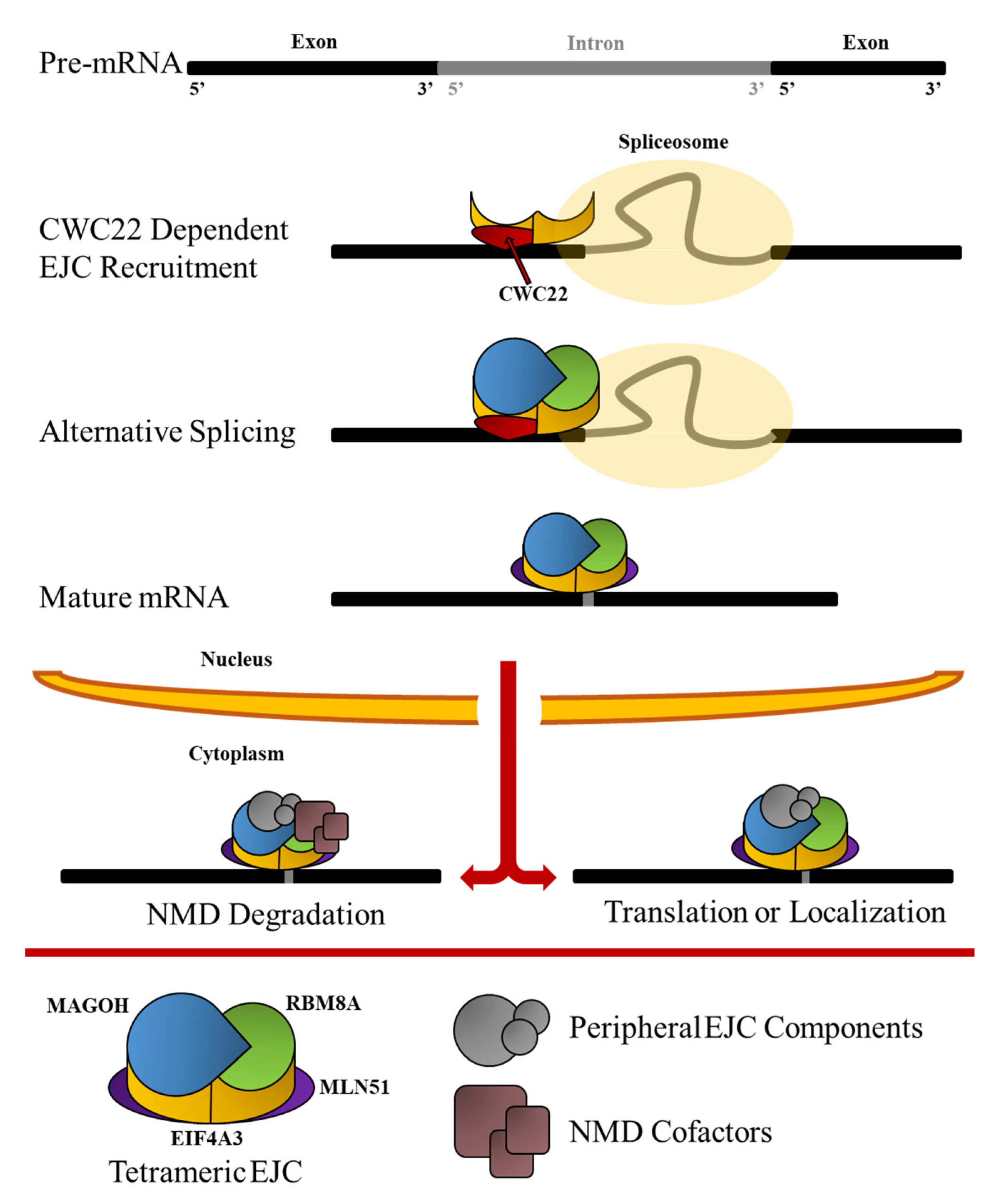
Table 1.
Common aliases used to describe each of the major EJC components discussed in this paper as RBM8A, MAGOH, EIF4A3, and MLN51. Included are the model organisms used to find in which species the alias was first identified or used with corresponding references.
Table 1.
Common aliases used to describe each of the major EJC components discussed in this paper as RBM8A, MAGOH, EIF4A3, and MLN51. Included are the model organisms used to find in which species the alias was first identified or used with corresponding references.
| EJC Component | Common Aliases | Model Organism | Reference |
|---|---|---|---|
| RBM8A | Y14 | Homo sapiens | [37] |
| Tsunagi | Drosophila melanogaster | [38] | |
| BOV 1 | Saccharomyces cerevisiae | [39] | |
| MAGOH | Mago-Nashi | Drosophila melanogaster | [40] |
| EIF4A3 | DEAD Box Protein | Mus muculus | [13] |
| NMP 265 | Homo sapiens | [41] | |
| DDX48 | Homo sapiens | [42] | |
| MLN51 | CASC3 | Homo sapiens | [43] |
| BTZ | Homo sapiens |
Table 2.
Core EJC Components and associated human disorders. Methodology in which the association was made between the EJC core component, and the human disorder is summarized accordingly as the tool employed in discovery. References are provided for the publications establishing the relevant association.
| EJC Component | Implicated Human Disorders | Tools Employed in Discovery | References |
|---|---|---|---|
| RBM8A | Thrombocytopenia-Absent Radius Syndrome (TAR) | Genetics analysis | [80,81] |
| Autism Spectrum Disorder | Mouse RNAi/Over-expression | [76] | |
| Mayer–Rokitansky–Küster–Hauser syndrome | Genetic variation analyses | [151] | |
| West Nile Virus | RNAi | [143] | |
| Glioblastoma | RNAseq Analysis | [152] | |
| Hepatocellular Carcinoma | Gene Expression Profiling Interactive Analysis | [153,154] | |
| Cervical Cancer | Microarray Gene Analysis | [155] | |
| Alzheimer’s Disease | GSEA and Identification of D.E.G.’s | [116] | |
| MAGOH 1 | |||
| EIF4A3 | Richieri-Costa-Pereira syndrome (RCP) | Genetics analysis | [82] |
| Anogenital Cancers | Gene expression analysis and RT-PCR | [156] | |
| Hepatocellular Carcinoma | Gene Expression Profiling Interactive Analysis | [157] | |
| MLN51 | HER2+ Breast Cancer | Immunofluorescence analysis | [63] |
1 MAGOH has not been substantively linked to a human disorder in the academic literature.
Publisher’s Note: MDPI stays neutral with regard to jurisdictional claims in published maps and institutional affiliations. |
© 2022 by the authors. Licensee MDPI, Basel, Switzerland. This article is an open access article distributed under the terms and conditions of the Creative Commons Attribution (CC BY) license (https://creativecommons.org/licenses/by/4.0/).
Share and Cite
MDPI and ACS Style
Asthana, S.; Martin, H.; Rupkey, J.; Patel, S.; Yoon, J.; Keegan, A.; Mao, Y. The Physiological Roles of the Exon Junction Complex in Development and Diseases. Cells 2022, 11, 1192. https://doi.org/10.3390/cells11071192
AMA Style
Asthana S, Martin H, Rupkey J, Patel S, Yoon J, Keegan A, Mao Y. The Physiological Roles of the Exon Junction Complex in Development and Diseases. Cells. 2022; 11(7):1192. https://doi.org/10.3390/cells11071192
Chicago/Turabian StyleAsthana, Shravan, Hannah Martin, Julian Rupkey, Shray Patel, Joy Yoon, Abiageal Keegan, and Yingwei Mao. 2022. "The Physiological Roles of the Exon Junction Complex in Development and Diseases" Cells 11, no. 7: 1192. https://doi.org/10.3390/cells11071192
Note that from the first issue of 2016, this journal uses article numbers instead of page numbers. See further details here.