WO2014028884A2 - Cancer diagnostics using biomarkers - Google Patents
Cancer diagnostics using biomarkers Download PDFInfo
- Publication number
- WO2014028884A2 WO2014028884A2 PCT/US2013/055429 US2013055429W WO2014028884A2 WO 2014028884 A2 WO2014028884 A2 WO 2014028884A2 US 2013055429 W US2013055429 W US 2013055429W WO 2014028884 A2 WO2014028884 A2 WO 2014028884A2
- Authority
- WO
- WIPO (PCT)
- Prior art keywords
- cancer
- targets
- target
- subject
- sample
- Prior art date
Links
Classifications
-
- C—CHEMISTRY; METALLURGY
- C12—BIOCHEMISTRY; BEER; SPIRITS; WINE; VINEGAR; MICROBIOLOGY; ENZYMOLOGY; MUTATION OR GENETIC ENGINEERING
- C12Q—MEASURING OR TESTING PROCESSES INVOLVING ENZYMES, NUCLEIC ACIDS OR MICROORGANISMS; COMPOSITIONS OR TEST PAPERS THEREFOR; PROCESSES OF PREPARING SUCH COMPOSITIONS; CONDITION-RESPONSIVE CONTROL IN MICROBIOLOGICAL OR ENZYMOLOGICAL PROCESSES
- C12Q1/00—Measuring or testing processes involving enzymes, nucleic acids or microorganisms; Compositions therefor; Processes of preparing such compositions
- C12Q1/68—Measuring or testing processes involving enzymes, nucleic acids or microorganisms; Compositions therefor; Processes of preparing such compositions involving nucleic acids
- C12Q1/6876—Nucleic acid products used in the analysis of nucleic acids, e.g. primers or probes
- C12Q1/6883—Nucleic acid products used in the analysis of nucleic acids, e.g. primers or probes for diseases caused by alterations of genetic material
- C12Q1/6886—Nucleic acid products used in the analysis of nucleic acids, e.g. primers or probes for diseases caused by alterations of genetic material for cancer
-
- C—CHEMISTRY; METALLURGY
- C12—BIOCHEMISTRY; BEER; SPIRITS; WINE; VINEGAR; MICROBIOLOGY; ENZYMOLOGY; MUTATION OR GENETIC ENGINEERING
- C12Q—MEASURING OR TESTING PROCESSES INVOLVING ENZYMES, NUCLEIC ACIDS OR MICROORGANISMS; COMPOSITIONS OR TEST PAPERS THEREFOR; PROCESSES OF PREPARING SUCH COMPOSITIONS; CONDITION-RESPONSIVE CONTROL IN MICROBIOLOGICAL OR ENZYMOLOGICAL PROCESSES
- C12Q2600/00—Oligonucleotides characterized by their use
- C12Q2600/118—Prognosis of disease development
-
- C—CHEMISTRY; METALLURGY
- C12—BIOCHEMISTRY; BEER; SPIRITS; WINE; VINEGAR; MICROBIOLOGY; ENZYMOLOGY; MUTATION OR GENETIC ENGINEERING
- C12Q—MEASURING OR TESTING PROCESSES INVOLVING ENZYMES, NUCLEIC ACIDS OR MICROORGANISMS; COMPOSITIONS OR TEST PAPERS THEREFOR; PROCESSES OF PREPARING SUCH COMPOSITIONS; CONDITION-RESPONSIVE CONTROL IN MICROBIOLOGICAL OR ENZYMOLOGICAL PROCESSES
- C12Q2600/00—Oligonucleotides characterized by their use
- C12Q2600/158—Expression markers
Definitions
- Cancer is the uncontrolled growth of abnormal cells anywhere in a body.
- the abnormal cells are termed cancer cells, malignant cells, or tumor cells.
- Many cancers and the abnormal cells that compose the cancer tissue are further identified by the name of the tissue that the abnormal cells originated from (for example, breast cancer, lung cancer, colon cancer, prostate cancer, pancreatic cancer, thyroid cancer).
- Cancer is not confined to humans; animals and other living organisms can get cancer.
- Cancer cells can proliferate uncontrollably and form a mass of cancer cells. Cancer cells can break away from this original mass of cells, travel through the blood and lymph systems, and lodge in other organs where they can again repeat the uncontrolled growth cycle.
- This process of cancer cells leaving an area and growing in another body area is often termed metastatic spread or metastatic disease. For example, if breast cancer cells spread to a bone (or anywhere else), it can mean that the individual has metastatic breast cancer.
- TAE microenvironment
- diagnostic, prognostic and therapeutic biomarkers to characterize the biology of each tumor may assist clinicians in making important decisions with regard to individual patient care and treatment.
- disclosed herein are methods, compositions and systems for the analysis of coding and non-coding targets for the diagnosis, prognosis, and monitoring of a cancer.
- a method of diagnosing, prognosing, determining progression the cancer, predicting a therapeutic regimen or predicting benefit from therapy in a subject comprising (a) assaying an expression level in a sample from the subject for a plurality of targets, wherein the plurality of targets comprises more than one target selected from Tables 2, 4, 11 or 55; and (b) diagnosing, prognosing, determining progression the cancer, predicting a therapeutic regimen or predicting benefit from therapy in a subject based on the expression levels of the plurality of targets.
- the cancer is selected from the group consisting of a carcinoma, sarcoma, leukemia, lymphoma, myeloma, and a CNS tumor.
- cancer is selected from the group consisting of bladder cancer, skin cancer, lung cancer, colon cancer, pancreatic cancer, prostate cancer, liver cancer, thyroid cancer, ovarian cancer, uterine cancer, breast cancer, cervical cancer, kidney cancer, epithelial carcinoma, squamous carcinoma, basal cell carcinoma, melanoma, papilloma, and adenomas.
- the cancer is a prostate cancer.
- the cancer is a pancreatic cancer.
- the cancer is a thyroid cancer. In some embodiments, the cancer is a bladder cancer. In some embodiments, the plurality of targets comprises a coding target. In some embodiments, the coding target is a coding antisense sequence an exonic sequence. In some embodiments, the plurality of targets comprises a non-coding target. In some embodiments, the non-coding target comprises an intronic sequence or partially overlaps an intronic sequence. In some embodiments, the non-coding target comprises an intronic sequence or partially overlaps an intronic sequence. In some embodiments, the non-coding target comprises a sequence within the UTR or partially overlaps with a UTR sequence. In some embodiments, the non-coding target comprises an antisense sequence or partially overlaps with an antisense sequence.
- the non-coding target comprises an intergenic sequence.
- the target comprises a nucleic acid sequence.
- the nucleic acid sequence is a DNA sequence.
- the nucleic acid sequence is an RNA sequence.
- the plurality of targets comprises at least 5 targets selected from Tables 2, 4, 11 or 55. In some embodiments, the plurality of targets comprises at least 10 targets selected from Tables 2, 4, 1 1 or 55. In some embodiments, the plurality of targets comprises at least 15 targets selected from Tables 2, 4, 11 or 55. In some embodiments, the plurality of targets comprises at least 20 targets selected from Tables 2, 4, 11 or 55. In some embodiments, the plurality of targets comprises at least 22 targets selected from Tables 2, 11 or 55.
- the plurality of targets comprises at least 30 targets selected from Tables 2, 11 or 55. In some embodiments, the plurality of targets comprises at least 35 targets selected from Tables 2, 11 or 55. In some embodiments, the plurality of targets comprises at least 40 targets selected from Tables 2, 11 or 55. In some embodiments, the plurality of targets comprises at least 5 targets selected from Tables 2, 4, 11 or 55. In some embodiments, the plurality of targets comprises 2, 3, 4, 5, 6, 7, 8, 9, 10, 11, 12, 13, 14, 15, 16, 17, 18, 19, 20, 21, 22, 23, 24, 25, 26, 27, 28, 29, 30, 31, 32, 33, 34, 35, 36, 37, 38, 39, 40, 41, 42, 43, 44, 45, 46, 47, 48, 49, or 50 targets selected from Tables 2, 4, 11 or 55.
- the plurality of targets is selected from SEQ ID NOs: l-43. In some embodiments, the plurality of targets is selected from SEQ ID NOs: l-22. In certain embodiments, the plurality of targets is selected from the group consisting of: PBX1, MYBPCl, LASPl, CAMK2N1, RABGAPl, UBE2C, PCAT-32, NFIB, TNFRSF19, PCDH7, AN07, GLYATL1P4/PCAT-80, C6orfl0, IQGAP3, THBS2, EPPK1, NUSAP1, ZWILCH, S1P4, SENP7, ANLN, C10orfl l6, PPP6R3, PDS5B, TOP2A, IL1RAP, CENPE, GALNT8, KCNQ1, C6orfl55, LUZP2, HEATR3, TMEM108, CFBXXbac-BPGl 16M5.17, ECHDC2, and ARN
- the plurality of targets is selected from the group consisting of: PBX1, MYBPCl, LASPl, CAMK2N1, RABGAPl, UBE2C, PCAT-32, NFIB, TNFRSF19, PCDH7, AN07,
- the diagnosing, prognosing, determining progression the cancer, predicting a therapeutic regimen or predicting benefit from therapy includes determining the malignancy of the cancer. In some embodiments, the diagnosing, prognosing, determining progression the cancer, predicting a therapeutic regimen or predicting benefit from therapy includes determining the stage of the cancer. In some embodiments, the diagnosing, prognosing, determining progression the cancer, predicting a therapeutic regimen or predicting benefit from therapy includes assessing the risk of cancer recurrence.
- the diagnosing, prognosing, determining progression the cancer, predicting a therapeutic regimen or predicting benefit from therapy includes assessing the grade of the cancer. In some embodiments, determining the treatment for the cancer includes determining the efficacy of treatment. In some embodiments, the method further comprises sequencing the plurality of targets. In some embodiments, the method further comprises hybridizing the plurality of targets to a solid support. In some embodiments, the solid support is a bead or array. In some embodiments, assaying the expression level of a plurality of targets may comprise the use of a probe set. In some embodiments, assaying the expression level may comprise the use of a classifier. The classifier may comprise a probe selection region (PSR). In some embodiments, the classifier may comprise the use of an algorithm. The algorithm may comprise a machine learning algorithm. In some embodiments, assaying the expression level may also comprise sequencing the plurality of targets.
- PSR probe selection region
- a method of determining a treatment for a cancer in a subject comprising (a) assaying an expression level in a sample from the subject for a plurality of targets, wherein the plurality of targets comprises more than one target selected from Tables 2, 4, 11 or 55; and (b) determining the treatment for the cancer based on the expression level of the plurality of targets.
- the cancer is selected from the group consisting of a carcinoma, sarcoma, leukemia, lymphoma, myeloma, and a CNS tumor.
- cancer is selected from the group consisting of bladder cancer, skin cancer, lung cancer, colon cancer, pancreatic cancer, prostate cancer, liver cancer, thyroid cancer, ovarian cancer, uterine cancer, breast cancer, cervical cancer, kidney cancer, epithelial carcinoma, squamous carcinoma, basal cell carcinoma, melanoma, papilloma, and adenomas.
- the cancer is a prostate cancer.
- the cancer is a bladder cancer.
- the cancer is a pancreatic cancer.
- the cancer is a thyroid cancer.
- the plurality of targets comprises a coding target.
- the coding target is a coding antisense sequence or an exonic sequence.
- the plurality of targets comprises a non-coding target.
- the non-coding target comprises an intronic sequence or partially overlaps an intronic sequence.
- the non-coding target comprises an intergenic sequence.
- the non-coding target comprises a sequence within the UTR or partially overlaps with a UTR sequence.
- the target comprises a nucleic acid sequence.
- the nucleic acid sequence is a DNA sequence.
- the nucleic acid sequence is an RNA sequence.
- the plurality of targets comprises at least 5 targets selected from Tables 2, 4, 11 or 55. In some embodiments, the plurality of targets comprises at least 10 targets selected from Tables 2, 4, 11 or 55. In some embodiments, the plurality of targets comprises at least 15 targets selected from Tables 2, 4, 11 or 55. In some embodiments, the plurality of targets comprises at least 20 targets selected from Tables 2, 4, 11 or 55.
- the plurality of targets comprises at least 22 targets selected from Tables 2, 11 or 55. In some embodiments, the plurality of targets comprises at least 30 targets selected from Tables 2, 11 or 55. In some embodiments, the plurality of targets comprises at least 35 targets selected from Tables 2, 11 or 55. In some embodiments, the plurality of targets comprises at least 40 targets selected from Tables 2, 11 or 55. In some embodiments, the plurality of targets comprises at least 5 targets selected from Tables 2, 4, 11 or 55.
- the plurality of targets comprises 2, 3, 4, 5, 6, 7, 8, 9, 10, 11, 12, 13, 14, 15, 16, 17, 18, 19, 20, 21, 22, 23, 24, 25, 26, 27, 28, 29, 30, 31, 32, 33, 34, 35, 36, 37, 38, 39, 40, 41, 42, 43, 44, 45, 46, 47, 48, 49, or 50 targets selected from Tables 2, 4, 11 or 55.
- the plurality of targets is selected from SEQ ID NOs: l-43. In some embodiments, the plurality of targets is selected from SEQ ID NOs: 1-22.
- the plurality of targets is selected from the group consisting of: PBX1, MYBPC1, LASP1, CAMK2N1, RABGAP1, UBE2C, PCAT-32, NF1B, TNFRSF19, PCDH7, AN07, GLYATL1P4/PCAT-80, C6orfl0, IQGAP3, THBS2, EPPK1,
- the plurality of targets is selected from the group consisting of: PBX1, MYBPC1, LASP1, CAMK2N1, RABGAP1, UBE2C, PCAT-32, NF1B, TNFRSF19, PCDH7, AN07, GLYATL1P4/PCAT-80, C6orfl0, IQGAP3, THBS2, EPPK1,
- the diagnosing, prognosing, determining progression the cancer, predicting a therapeutic regimen or predicting benefit from therapy includes determining the malignancy of the cancer. In some embodiments, the diagnosing, prognosing, determining progression the cancer, predicting a therapeutic regimen or predicting benefit from therapy includes determining the stage of the cancer. In some embodiments, the diagnosing, prognosing, determining progression the cancer, predicting a therapeutic regimen or predicting benefit from therapy includes assessing the risk of cancer recurrence. In some embodiments, determining the treatment for the cancer includes determining the efficacy of treatment. In some embodiments, the method further comprises sequencing the plurality of targets.
- the method further comprises hybridizing the plurality of targets to a solid support.
- the solid support is a bead or array.
- assaying the expression level of a plurality of targets may comprise the use of a probe set.
- assaying the expression level may comprise the use of a classifier.
- the classifier may comprise a probe selection region (PSR).
- the classifier may comprise the use of an algorithm.
- the algorithm may comprise a machine learning algorithm.
- assaying the expression level may also comprise amplifying the plurality of targets.
- assaying the expression level may also comprise quantifying the plurality of targets.
- a probe set for assessing a cancer status of a subject comprising a plurality of probes, wherein the probes in the set are capable of detecting an expression level of a plurality of targets selected from Tables 2, 4, 11 or 55, wherein the expression level determines the cancer status of the subject with at least 40% specificity.
- the plurality of targets comprises at least 5 targets selected from Tables 2, 4, 11 or 55.
- the plurality of targets comprises at least 10 targets selected from Tables 2, 4, 11 or 55.
- the plurality of targets comprises at least 15 targets selected from Tables 2, 4, 11 or 55.
- the plurality of targets comprises at least 20 targets selected from Tables 2, 4, 1 1 or 55.
- the plurality of targets comprises at least 22 targets selected from Tables 2, 11 or 55. In some embodiments, the plurality of targets comprises at least 30 targets selected from Tables 2, 11 or 55. In some embodiments, the plurality of targets comprises at least 35 targets selected from Tables 2, 11 or 55. In some embodiments, the plurality of targets comprises at least 40 targets selected from Tables 2, 11 or 55. In some embodiments, the plurality of targets comprises at least 5 targets selected from Tables 2, 4, 11 or 55.
- the plurality of targets comprises 2, 3, 4, 5, 6, 7, 8, 9, 10, 11, 12, 13, 14, 15, 16, 17, 18, 19, 20, 21, 22, 23, 24, 25, 26, 27, 28, 29, 30, 31, 32, 33, 34, 35, 36, 37, 38, 39, 40, 41, 42, 43, 44, 45, 46, 47, 48, 49, or 50 targets selected from Tables 2, 4, 11 or 55.
- the plurality of targets is selected from SEQ ID NOs: l-43.
- the plurality of targets is selected from SEQ ID NOs: l-22.
- the plurality of targets is selected from the group consisting of: PBX1, MYBPC1, LASP1, CAMK2N1, RABGAP1, UBE2C, PCAT-32, NF1B, TNFRSF19, PCDH7, AN07,
- GLYATL1P4/PCAT-80 C6orfl0, IQGAP3, THBS2, EPPK1, NUSAP1, ZWILCH, S1P4, SENP7, ANLN, C10orfl l6, PPP6R3, PDS5B, TOP2A, IL1RAP, CENPE, GALNT8, KCNQ1, C6orfl55, LUZP2, HEATR3, TMEM108, CFBXXbac-BPGl 16M5.17, ECHDC2, and ARNTL.
- the plurality of targets is selected from the group consisting of: PBX1, MYBPC1, LASP1, CAMK2N1, RABGAP1, UBE2C, PCAT-32, NF1B, TNFRSF19, PCDH7, AN07,
- the cancer is selected from the group consisting of a carcinoma, sarcoma, leukemia, lymphoma, myeloma, and a CNS tumor.
- the cancer is selected from the group consisting of bladder cancer, skin cancer, lung cancer, colon cancer, pancreatic cancer, prostate cancer, liver cancer, thyroid cancer, ovarian cancer, uterine cancer, breast cancer, cervical cancer, kidney cancer, epithelial carcinoma, squamous carcinoma, basal cell carcinoma, melanoma, papilloma, and adenomas.
- the cancer is a prostate cancer. In some embodiments, the cancer is a bladder cancer. In some embodiments, the cancer is a pancreatic cancer. In some embodiments, the cancer is a thyroid cancer. In some embodiments, the probe set further comprises a probe capable of detecting an expression level of more than one coding target. In some
- the coding target is a coding antisence sequence or an exonic sequence.
- the probe set further comprises a probe capable of detecting an expression level of more than one non-coding target.
- the non-coding target is an intronic sequence or partially overlaps with an intronic sequence.
- the non-coding target is an intergenic sequence.
- the non-coding target is a UTR sequence or partially overlaps with a UTR sequence.
- assessing the cancer status includes assessing cancer recurrence risk.
- assessing the cancer status includes determining a treatment modality.
- assessing the cancer status includes determining the efficacy of treatment.
- the target is a nucleic acid sequence.
- the nucleic acid sequence is a DNA sequence.
- the nucleic acid sequence is an RNA sequence.
- the probes are between about 15 nucleotides and about 500 nucleotides in length. In some embodiments, the probes are between about 15 nucleotides and about 450 nucleotides in length. In some embodiments, the probes are between about 15 nucleotides and about 400 nucleotides in length. In some embodiments, the probes are between about 15 nucleotides and about 350 nucleotides in length. In some embodiments, the probes are between about 15 nucleotides and about 300 nucleotides in length.
- the probes are between about 15 nucleotides and about 250 nucleotides in length. In some embodiments, the probes are between about 15 nucleotides and about 200 nucleotides in length. In some embodiments, the probes are at least 15 nucleotides in length. In some embodiments, the probes are at least 25 nucleotides in length. In some embodiments, the expression level determines the cancer status of the subject with at least 50% specificity. In some embodiments, the expression level determines the cancer status of the subject with at least 60% specificity. In some embodiments, the expression level determines the cancer status of the subject with at least 65 % specificity. In some embodiments, the expression level determines the cancer status of the subject with at least 70% specificity. In some embodiments, the expression level determines the cancer status of the subject with at least 75% specificity. In some embodiments, the expression level determines the cancer status of the subject with at least 80% specificity. In some embodiments, the expression level determines the cancer status of the subject with at least a
- the expression level determines the cancer status of the subject with at least 85% specificity.
- the non-coding target is a non-coding RNA transcript and the non-coding RNA transcript is non-polyadenylated.
- a system for analyzing a cancer comprising: (a) a probe set comprising a plurality of target sequences, wherein (i) the plurality of target sequences hybridizes to more than one target selected from Tables 2 or 4; or (ii) the plurality of target sequences comprises more than one target sequences selected from Table 11 ; and (b) a computer model or algorithm for analyzing an expression level and/or expression profile of the target hybridized to the probe in a sample from a subject suffering from a cancer.
- the system further comprises an electronic memory for capturing and storing an expression profile.
- the system further comprises a computer-processing device, optionally connected to a computer network.
- system further comprises a software module executed by the computer-processing device to analyze an expression profile. In some embodiments, the system further comprises a software module executed by the computer-processing device to compare the expression profile to a standard or control. In some embodiments, the system further comprises a software module executed by the computer-processing device to determine the expression level of the target. In some embodiments, the system further comprises a machine to isolate the target or the probe from the sample. In some
- the system further comprises a machine to sequence the target or the probe. In some embodiments, the system further comprises a machine to amplify the target or the probe. In some embodiments, the system further comprises a label that specifically binds to the target, the probe, or a combination thereof. In some embodiments, the system further comprises a software module executed by the computer-processing device to transmit an analysis of the expression profile to the individual or a medical professional treating the individual. In some embodiments, the system further comprises a software module executed by the computer-processing device to transmit a diagnosis or prognosis to the individual or a medical professional treating the individual. In some embodiments, the plurality of targets comprises at least 5 targets selected from Tables 2, 4, 11 or 55.
- the plurality of targets comprises at least 10 targets selected from Tables 2, 4, 1 1 or 55. In some embodiments, the plurality of targets comprises at least 15 targets selected from Tables 2, 4, 11 or 55. In some embodiments, the plurality of targets comprises at least 20 targets selected from Tables 2, 4, 11 or 55. In some embodiments, the plurality of targets comprises at least 30 targets selected from Tables 2, 11 or 55. In some embodiments, the plurality of targets comprises at least 35 targets selected from Tables 2, 11 or 55. In some embodiments, the plurality of targets comprises at least 40 targets selected from Tables 2, 11 or 55. In some embodiments, the plurality of targets comprises at least 22 targets selected from Tables 2, 11 or 55.
- the plurality of targets comprises at least 30 targets selected from Tables 2, 11 or 55. In some embodiments, the plurality of targets comprises at least 35 targets selected from Tables 2, 11 or 55. In some embodiments, the plurality of targets comprises at least 40 targets selected from Tables 2, 11 or 55. In some embodiments, the plurality of targets comprises at least 5 targets selected from Tables 2, 4, 11 or 55. In some embodiments, the plurality of targets comprises 2, 3, 4, 5, 6, 7, 8, 9, 10, 11, 12, 13, 14, 15, 16, 17, 18, 19, 20, 21, 22, 23, 24, 25, 26, 27, 28, 29, 30, 31, 32, 33, 34, 35, 36, 37, 38, 39, 40, 41, 42, 43, 44, 45, 46, 47, 48, 49, or 50 targets selected from Tables 2, 4, 11 or 55. In some embodiments, the plurality of targets is selected from SEQ ID NOs: l-43. In some embodiments, the plurality of targets is selected from SEQ ID NOs: l-43. In some embodiments, the plurality of targets is selected from SEQ ID NOs: l-43.
- the plurality of targets is selected from SEQ ID NOs: l-22.
- the plurality of targets is selected from the group consisting of: PBX1, MYBPC1, LASP1, CAMK2N1, RABGAP1, UBE2C, PCAT-32, NF1B, TNFRSF19, PCDH7, AN07,
- GLYATL1P4/PCAT-80 C6orfl0, IQGAP3, THBS2, EPPK1, NUSAP1, ZWILCH, S1P4, SENP7, ANLN, C10orfl l6, PPP6R3, PDS5B, TOP2A, IL1RAP, CENPE, GALNT8, KCNQ1, C6orfl55, LUZP2, HEATR3, TMEM108, CFBXXbac-BPGl 16M5.17, ECHDC2, and ARNTL.
- the plurality of targets is selected from the group consisting of: PBX1, MYBPC1, LASP1, CAMK2N1, RABGAP1, UBE2C, PCAT-32, NF1B, TNFRSF19, PCDH7, AN07,
- the cancer is selected from the group consisting of a carcinoma, sarcoma, leukemia, lymphoma, myeloma, and a CNS tumor.
- the cancer is selected from the group consisting of bladder, skin cancer, lung cancer, colon cancer, pancreatic cancer, prostate cancer, liver cancer, thyroid cancer, ovarian cancer, uterine cancer, breast cancer, cervical cancer, kidney cancer, epithelial carcinoma, squamous carcinoma, basal cell carcinoma, melanoma, papilloma, and adenomas.
- the system further comprises a sequence for sequencing the plurality of targets. In some embodiments, the system further comprises an instrument for amplifying the plurality of targets. In some embodiments, the system further comprises a label for labeling the plurality of targets.
- a method of analyzing a cancer in an individual in need thereof comprising: (a) obtaining an expression profile from a sample obtained from the individual, wherein the expression profile comprises more than one target selected from Tables 2, 4, 11 or 55; and (b) comparing the expression profile from the sample to an expression profile of a control or standard.
- the plurality of targets comprises at least 5 targets selected from Tables 2, 4, 11 or 55.
- the plurality of targets comprises at least 10 targets selected from Tables 2, 4, 11 or 55.
- the plurality of targets comprises at least 15 targets selected from Tables 2, 4, 11 or 55.
- the plurality of targets comprises at least 20 targets selected from Tables 2, 4, 11 or 55.
- the plurality of targets comprises at least 30 targets selected from Tables 2, 11 or 55. In some embodiments, the plurality of targets comprises at least 35 targets selected from Tables 2, 11 or 55. In some embodiments, the plurality of targets comprises at least 40 targets selected from Tables 2, 11 or 55. In some embodiments, the plurality of targets comprises at least 22 targets selected from Tables 2, 11 or 55. In some embodiments, the plurality of targets comprises at least 30 targets selected from Tables 2, 11 or 55. In some embodiments, the plurality of targets comprises at least 35 targets selected from Tables 2, 11 or 55. In some embodiments, the plurality of targets comprises at least 40 targets selected from Tables 2, 11 or 55.
- the plurality of targets comprises at least 5 targets selected from Tables 2, 4, 11 or 55. In some embodiments, the plurality of targets comprises 2, 3, 4, 5, 6, 7, 8, 9, 10, 11, 12, 13, 14, 15, 16, 17, 18, 19, 20, 21, 22, 23, 24, 25, 26, 27, 28, 29, 30, 31, 32, 33, 34, 35, 36, 37, 38, 39, 40, 41, 42, 43, 44, 45, 46, 47, 48, 49, or 50 targets selected from Tables 2, 4, 11 or 55. In some embodiments, the plurality of targets is selected from SEQ ID NOs: l-43. In some
- the plurality of targets is selected from SEQ ID NOs: l-22.
- the plurality of targets is selected from the group consisting of: PBX1, MYBPC1, LASP1, CAMK2N1, RABGAP1, UBE2C, PCAT-32, NF1B, TNFRSF19, PCDH7, AN07,
- GLYATL1P4/PCAT-80 C6orfl0, IQGAP3, THBS2, EPPK1, NUSAP1, ZWILCH, S1P4, SENP7, ANLN, C10orfl l6, PPP6R3, PDS5B, TOP2A, IL1RAP, CENPE, GALNT8, KCNQ1, C6orfl55, LUZP2, HEATR3, TMEM108, CFBXXbac-BPGl 16M5.17, ECHDC2, and ARNTL.
- the plurality of targets is selected from the group consisting of: PBX1, MYBPC1, LASP1, CAMK2N1, RABGAP1, UBE2C, PCAT-32, NF1B, TNFRSF19, PCDH7, AN07,
- the cancer is selected from the group consisting of a carcinoma, sarcoma, leukemia, lymphoma, myeloma, and a CNS tumor.
- the cancer is selected from the group consisting of bladder cancer, skin cancer, lung cancer, colon cancer, pancreatic cancer, prostate cancer, liver cancer, thyroid cancer, ovarian cancer, uterine cancer, breast cancer, cervical cancer, kidney cancer, epithelial carcinoma, squamous carcinoma, basal cell carcinoma, melanoma, papilloma, and adenomas.
- the cancer is a prostate cancer. In some embodiments, the cancer is a pancreatic cancer. In some embodiments, the cancer is a breast cancer. In some embodiments, the cancer is a thyroid cancer. In some embodiments, the cancer is a lung cancer. In some embodiments, the method further comprises a software module executed by a computer- processing device to compare the expression profiles. In some embodiments, the method further comprises providing diagnostic or prognostic information to the individual about the cardiovascular disorder based on the comparison.
- the method further comprises diagnosing the individual with a cancer if the expression profile of the sample (a) deviates from the control or standard from a healthy individual or population of healthy individuals, or (b) matches the control or standard from an individual or population of individuals who have or have had the cancer.
- the method further comprises predicting the susceptibility of the individual for developing a cancer based on (a) the deviation of the expression profile of the sample from a control or standard derived from a healthy individual or population of healthy individuals, or (b) the similarity of the expression profiles of the sample and a control or standard derived from an individual or population of individuals who have or have had the cancer.
- the method further comprises prescribing a treatment regimen based on (a) the deviation of the expression profile of the sample from a control or standard derived from a healthy individual or population of healthy individuals, or (b) the similarity of the expression profiles of the sample and a control or standard derived from an individual or population of individuals who have or have had the cancer.
- the method further comprises altering a treatment regimen prescribed or administered to the individual based on (a) the deviation of the expression profile of the sample from a control or standard derived from a healthy individual or population of healthy individuals, or (b) the similarity of the expression profiles of the sample and a control or standard derived from an individual or population of individuals who have or have had the cancer.
- the method further comprises predicting the individual's response to a treatment regimen based on (a) the deviation of the expression profile of the sample from a control or standard derived from a healthy individual or population of healthy individuals, or (b) the similarity of the expression profiles of the sample and a control or standard derived from an individual or population of individuals who have or have had the cancer.
- the deviation is the expression level of more than one target from the sample is greater than the expression level of more than one target from a control or standard derived from a healthy individual or population of healthy individuals.
- the deviation is the expression level of more than one target from the sample is at least about 30% greater than the expression level of more than one target from a control or standard derived from a healthy individual or population of healthy individuals. In some embodiments, the deviation is the expression level of more than one target from the sample is less than the expression level of more than one target from a control or standard derived from a healthy individual or population of healthy individuals. In some embodiments, the deviation is the expression level of more than one target from the sample is at least about 30% less than the expression level of more than one target from a control or standard derived from a healthy individual or population of healthy individuals. In some embodiments, the method further comprises using a machine to isolate the target or the probe from the sample.
- the method further comprises contacting the sample with a label that specifically binds to the target, the probe, or a combination thereof. In some embodiments, the method further comprises contacting the sample with a label that specifically binds to a target selected from Tables 2, 4, 11 or 55, or a combination thereof. In some embodiments, the method further comprises amplifying the target, the probe, or any combination thereof. In some embodiments, the method further comprises sequencing the target, the probe, or any combination thereof. In some
- the method further comprises quantifying the expression level of the plurality of targets. In some embodiments, the method further comprises labeling the plurality of targets. In some embodiments, assaying the expression level of a plurality of targets may comprise the use of a probe set. In some embodiments, obtaining the expression level may comprise the use of a classifier.
- the classifier may comprise a probe selection region (PSR). In some embodiments, the classifier may comprise the use of an algorithm. The algorithm may comprise a machine learning algorithm. In some embodiments, obtaining the expression level may also comprise sequencing the plurality of targets.
- a method of diagnosing cancer in an individual in need thereof comprising (a) obtaining an expression profile from a sample obtained from the individual, wherein the expression profile comprises more than one target selected from Tables 2, 4, 11 or 55; (b) comparing the expression profile from the sample to an expression profile of a control or standard; and (c) diagnosing a cancer in the individual if the expression profile of the sample (i) deviates from the control or standard from a healthy individual or population of healthy individuals, or (ii) matches the control or standard from an individual or population of individuals who have or have had the cancer.
- the plurality of targets comprises at least 5 targets selected from Tables 2, 4, 11 or 55.
- the plurality of targets comprises at least 10 targets selected from Tables 2, 4, 11 or 55. In some embodiments, the plurality of targets comprises at least 15 targets selected from Tables 2, 4, 11 or 55. In some embodiments, the plurality of targets comprises at least 20 targets selected from Tables 2, 4, 11 or 55. In some embodiments, the plurality of targets comprises at least 30 targets selected from Tables 2, 11 or 55. In some embodiments, the plurality of targets comprises at least 35 targets selected from Tables 2, 11 or 55. In some embodiments, the plurality of targets comprises at least 40 targets selected from Tables 2, 11 or 55. In some embodiments, the plurality of targets comprises at least 22 targets selected from Tables 2, 11 or 55.
- the plurality of targets comprises at least 30 targets selected from Tables 2, 11 or 55. In some embodiments, the plurality of targets comprises at least 35 targets selected from Tables 2, 11 or 55. In some embodiments, the plurality of targets comprises at least 40 targets selected from Tables 2, 11 or 55. In some embodiments, the plurality of targets comprises at least 5 targets selected from Tables 2, 4, 11 or 55. In some embodiments, the plurality of targets comprises 2, 3, 4, 5, 6, 7, 8, 9, 10, 11, 12, 13, 14, 15, 16, 17, 18, 19, 20, 21, 22, 23, 24, 25, 26, 27, 28, 29, 30, 31, 32, 33, 34, 35, 36, 37, 38, 39, 40, 41, 42, 43, 44, 45, 46, 47, 48, 49, or 50 targets selected from Tables 2, 4, 11 or 55.
- the plurality of targets is selected from SEQ ID NOs: l-43. In some embodiments, the plurality of targets is selected from SEQ ID NOs: l-22. In certain embodiments, the plurality of targets is selected from the group consisting of: PBX1, MYBPCl, LASPl, CAMK2N1, RABGAPl, UBE2C, PCAT-32, NFIB, TNFRSF19, PCDH7, AN07, GLYATL1P4/PCAT-80, C6orfl0, IQGAP3, THBS2, EPPK1, NUSAP1, ZWILCH, S1P4, SENP7, ANLN, C10orfl l6, PPP6R3, PDS5B, TOP2A, IL1RAP, CENPE, GALNT8, KCNQ1, C6orfl55, LUZP2, HEATR3, TMEM108, CFBXXbac-BPGl 16M5.17, ECHDC2, and ARN
- the plurality of targets is selected from the group consisting of: PBX1, MYBPCl, LASP1, CAMK2N1, RABGAP1, UBE2C, PCAT-32, NF1B, TNFRSF19, PCDH7, AN07,
- the cancer is selected from the group consisting of a carcinoma, sarcoma, leukemia, lymphoma, myeloma, and a CNS tumor.
- the cancer is selected from the group consisting of bladder cancer, skin cancer, lung cancer, colon cancer, pancreatic cancer, prostate cancer, liver cancer, thyroid cancer, ovarian cancer, uterine cancer, breast cancer, cervical cancer, kidney cancer, epithelial carcinoma, squamous carcinoma, basal cell carcinoma, melanoma, papilloma, and adenomas.
- the cancer is a prostate cancer. In some embodiments, the cancer is a pancreatic cancer. In some embodiments, the cancer is a breast cancer. In some embodiments, the cancer is a thyroid cancer. In some embodiments, the cancer is a lung cancer. In some embodiments, the method further comprises a software module executed by a computer- processing device to compare the expression profiles. In some embodiments, the deviation is the expression level of more than one target targets from the sample is greater than the expression level of more than one target from a control or standard derived from a healthy individual or population of healthy individuals.
- the deviation is the expression level of more than one target from the sample is at least about 30% greater than the expression level of more than one target from a control or standard derived from a healthy individual or population of healthy individuals. In some embodiments, the deviation is the expression level of more than one target from the sample is less than the expression level of more than one target from a control or standard derived from a healthy individual or population of healthy individuals. In some embodiments, the deviation is the expression level of more than one target from the sample is at least about 30% less than the expression level of more than one target from a control or standard derived from a healthy individual or population of healthy individuals. In some embodiments, the method further comprises using a machine to isolate the target or the probe from the sample.
- the method further comprises contacting the sample with a label that specifically binds to the target, the probe, or a combination thereof. In some embodiments, the method further comprises contacting the sample with a label that specifically binds to a target selected from Tables 2, 4, 11 or 55, or a combination thereof. In some embodiments, the method further comprises amplifying the target, the probe, or any combination thereof. In some
- the method further comprises sequencing the target, the probe, or any combination thereof. In some embodiments, the method further comprises quantifying the expression level of the plurality of targets. In some embodiments, the method further comprises labeling the plurality of targets. In some embodiments, obtaining the expression level may comprise the use of a classifier.
- the classifier may comprise a probe selection region (PSR).
- the classifier may comprise the use of an algorithm.
- the algorithm may comprise a machine learning algorithm. In some embodiments, obtaining the expression level may also comprise sequencing the plurality of targets.
- a method of predicting whether an individual is susceptible to developing a cancer comprising (a) obtaining an expression profile from a sample obtained from the individual, wherein the expression profile comprises more than one target selected from Tables 2, 4, 11 or 55; (b) comparing the expression profile from the sample to an expression profile of a control or standard; and (c) predicting the susceptibility of the individual for developing a cancer based on (i) the deviation of the expression profile of the sample from a control or standard derived from a healthy individual or population of healthy individuals, or (ii) the similarity of the expression profiles of the sample and a control or standard derived from an individual or population of individuals who have or have had the cancer.
- the plurality of targets comprises at least 5 targets selected from Tables 2, 4, 11 or 55. In some embodiments, the plurality of targets comprises at least 10 targets selected from Tables 2, 4, 11 or 55. In some embodiments, the plurality of targets comprises at least 15 targets selected from Tables 2, 4, 1 1 or 55. In some embodiments, the plurality of targets comprises at least 20 targets selected from Tables 2, 4, 11 or 55. In some embodiments, the plurality of targets comprises at least 30 targets selected from Tables 2, 11 or 55. In some embodiments, the plurality of targets comprises at least 35 targets selected from Tables 2, 11 or 55. In some embodiments, the plurality of targets comprises at least 40 targets selected from Tables 2, 11 or 55.
- the plurality of targets comprises at least 22 targets selected from Tables 2, 11 or 55. In some embodiments, the plurality of targets comprises at least 30 targets selected from Tables 2, 11 or 55. In some embodiments, the plurality of targets comprises at least 35 targets selected from Tables 2, 11 or 55. In some embodiments, the plurality of targets comprises at least 40 targets selected from Tables 2, 11 or 55. In some embodiments, the plurality of targets comprises at least 5 targets selected from Tables 2, 4, 11 or 55.
- the plurality of targets comprises 2, 3, 4, 5, 6, 7, 8, 9, 10, 11, 12, 13, 14, 15, 16, 17, 18, 19, 20, 21, 22, 23, 24, 25, 26, 27, 28, 29, 30, 31, 32, 33, 34, 35, 36, 37, 38, 39, 40, 41, 42, 43, 44, 45, 46, 47, 48, 49, or 50 targets selected from Tables 2, 4, 11 or 55.
- the plurality of targets is selected from SEQ ID NOs: l-43. In some embodiments, the plurality of targets is selected from SEQ ID NOs: 1-22.
- the plurality of targets is selected from the group consisting of: PBX1, MYBPC1, LASP1, CAMK2N1, RABGAP1, UBE2C, PCAT-32, NF1B, TNFRSF19, PCDH7, AN07, GLYATL1P4/PCAT-80, C6orfl0, IQGAP3, THBS2, EPPK1, NUSAP1, ZWILCH, S1P4, SENP7, ANLN, C10orfl l6, PPP6R3, PDS5B, TOP2A, IL1RAP, CENPE, GALNT8, KCNQ1, C6orfl55, LUZP2, HEATR3, TMEM108, CFBXXbac-BPG116M5.17, ECHDC2, and ARNTL.
- PBX1, MYBPC1, LASP1, CAMK2N1, RABGAP1, UBE2C PCAT-32, NF1B, TNFRSF19, PCDH7, AN07
- the plurality of targets is selected from the group consisting of: PBX1, MYBPC1, LASP1, CAMK2N1, RABGAP1, UBE2C, PCAT-32, NF1B, TNFRSF19, PCDH7, AN07, GLYATL1P4/PCAT-80, C6orfl0, IQGAP3, THBS2, EPPK1,
- the cancer is selected from the group consisting of a carcinoma, sarcoma, leukemia, lymphoma, myeloma, and a CNS tumor.
- the cancer is selected from the group consisting of bladder cancer, skin cancer, lung cancer, colon cancer, pancreatic cancer, prostate cancer, liver cancer, thyroid cancer, ovarian cancer, uterine cancer, breast cancer, cervical cancer, kidney cancer, epithelial carcinoma, squamous carcinoma, basal cell carcinoma, melanoma, papilloma, and adenomas.
- the cancer is a prostate cancer.
- the cancer is a pancreatic cancer.
- the cancer is a breast cancer. In some embodiments, the cancer is a thyroid cancer. In some embodiments, the cancer is a lung cancer. In some embodiments, the method further comprises a software module executed by a computer-processing device to compare the expression profiles. In some embodiments, the deviation is the expression level of more than one target from the sample is greater than the expression level of more than one target from a control or standard derived from a healthy individual or population of healthy individuals. In some embodiments, the deviation is the expression level of more than one target from the sample is at least about 30% greater than the expression level of more than one target from a control or standard derived from a healthy individual or population of healthy individuals.
- the deviation is the expression level of more than one target from the sample is less than the expression level of more than one target from a control or standard derived from a healthy individual or population of healthy individuals. In some embodiments, the deviation is the expression level of more than one target from the sample is at least about 30% less than the expression level of more than one target from a control or standard derived from a healthy individual or population of healthy individuals. In some embodiments, the method further comprises using a machine to isolate the target or the probe from the sample. In some embodiments, the method further comprises contacting the sample with a label that specifically binds to the target, the probe, or a combination thereof.
- the method further comprises contacting the sample with a label that specifically binds to a target selected from Tables 2, 4, 11 or 55, or a combination thereof. In some embodiments, the method further comprises amplifying the target, the probe, or any combination thereof. In some
- the method further comprises sequencing the target, the probe, or any combination thereof.
- obtaining the expression level may comprise the use of a classifier.
- the classifier may comprise a probe selection region (PSR).
- the classifier may comprise the use of an algorithm.
- the algorithm may comprise a machine learning algorithm.
- obtaining the expression level may also comprise sequencing the plurality of targets.
- obtaining the expression level may also comprise amplifying the plurality of targets.
- obtaining the expression level may also comprise quantifying the plurality of targets.
- a method of predicting an individual's response to a treatment regimen for a cancer comprising (a) obtaining an expression profile from a sample obtained from the individual, wherein the expression profile comprises more than one target selected from Tables 2, 4, 11 or 55; (b) comparing the expression profile from the sample to an expression profile of a control or standard; and (c) predicting the individual's response to a treatment regimen based on (a) the deviation of the expression profile of the sample from a control or standard derived from a healthy individual or population of healthy individuals, or (b) the similarity of the expression profiles of the sample and a control or standard derived from an individual or population of individuals who have or have had the cancer.
- the plurality of targets comprises at least 5 targets selected from Tables 2, 4, 11 or 55. In some embodiments, the plurality of targets comprises at least 10 targets selected from Tables 2, 4, 11 or 55. In some embodiments, the plurality of targets comprises at least 15 targets selected from Tables 2, 4, 1 1 or 55. In some embodiments, the plurality of targets comprises at least 20 targets selected from Tables 2, 4, 11 or 55. In some embodiments, the plurality of targets comprises at least 30 targets selected from Tables 2, 11 or 55. In some embodiments, the plurality of targets comprises at least 35 targets selected from Tables 2, 11 or 55. In some embodiments, the plurality of targets comprises at least 40 targets selected from Tables 2, 11 or 55.
- the plurality of targets comprises at least 22 targets selected from Tables 2, 11 or 55. In some embodiments, the plurality of targets comprises at least 30 targets selected from Tables 2, 11 or 55. In some embodiments, the plurality of targets comprises at least 35 targets selected from Tables 2, 11 or 55. In some embodiments, the plurality of targets comprises at least 40 targets selected from Tables 2, 11 or 55. In some embodiments, the plurality of targets comprises at least 5 targets selected from Tables 2, 4, 11 or 55.
- the plurality of targets comprises 2, 3, 4, 5, 6, 7, 8, 9, 10, 11, 12, 13, 14, 15, 16, 17, 18, 19, 20, 21, 22, 23, 24, 25, 26, 27, 28, 29, 30, 31, 32, 33, 34, 35, 36, 37, 38, 39, 40, 41, 42, 43, 44, 45, 46, 47, 48, 49, or 50 targets selected from Tables 2, 4, 11 or 55.
- the plurality of targets is selected from SEQ ID NOs: l-43. In some embodiments, the plurality of targets is selected from SEQ ID NOs: 1-22.
- the plurality of targets is selected from the group consisting of: PBX1, MYBPC1, LASP1, CAMK2N1, RABGAP1, UBE2C, PCAT-32, NF1B, TNFRSF19, PCDH7, AN07, GLYATL1P4/PCAT-80, C6orfl0, IQGAP3, THBS2, EPPKl, NUSAPl, ZWILCH, S1P4, SENP7, ANLN, C10orfl l6, PPP6R3, PDS5B, TOP2A, ILIRAP, CENPE, GALNT8, KCNQ1, C6orfl55, LUZP2, HEATR3, TMEM108, CFBXXbac-BPG116M5.17, ECHDC2, and ARNTL.
- PBX1, MYBPC1, LASP1, CAMK2N1, RABGAP1, UBE2C PCAT-32, NF1B, TNFRSF19, PCDH7, AN
- the plurality of targets is selected from the group consisting of: PBX1, MYBPC1, LASP1, CAMK2N1, RABGAPl, UBE2C, PCAT-32, NF1B, TNFRSF19, PCDH7, AN07, GLYATL1P4/PCAT-80, C6orfl0, IQGAP3, THBS2, EPPKl,
- the cancer is selected from the group consisting of a carcinoma, sarcoma, leukemia, lymphoma, myeloma, and a CNS tumor.
- the cancer is selected from the group consisting of bladder cancer, skin cancer, lung cancer, colon cancer, pancreatic cancer, prostate cancer, liver cancer, thyroid cancer, ovarian cancer, uterine cancer, breast cancer, cervical cancer, kidney cancer, epithelial carcinoma, squamous carcinoma, basal cell carcinoma, melanoma, papilloma, and adenomas.
- the cancer is a prostate cancer.
- the cancer is a pancreatic cancer.
- the cancer is a breast cancer. In some embodiments, the cancer is a thyroid cancer. In some embodiments, the cancer is a lung cancer. In some embodiments, the method further comprises a software module executed by a computer-processing device to compare the expression profiles. In some embodiments, the deviation is the expression level of more than one target from the sample is greater than the expression level of more than one target from a control or standard derived from a healthy individual or population of healthy individuals. In some embodiments, the deviation is the expression level of more than one target from the sample is at least about 30% greater than the expression level of more than one target from a control or standard derived from a healthy individual or population of healthy individuals.
- the deviation is the expression level of more than one target from the sample is less than the expression level of more than one target from a control or standard derived from a healthy individual or population of healthy individuals. In some embodiments, the deviation is the expression level of more than one target from the sample is at least about 30% less than the expression level of more than one target from a control or standard derived from a healthy individual or population of healthy individuals. In some embodiments, the method further comprises using a machine to isolate the target or the probe from the sample. In some embodiments, the method further comprises contacting the sample with a label that specifically binds to the target, the probe, or a combination thereof.
- the method further comprises contacting the sample with a label that specifically binds to a target selected from Tables 2, 4, 11 or 55, or a combination thereof. In some embodiments, the method further comprises amplifying the target, the probe, or any combination thereof. In some
- the method further comprises sequencing the target, the probe, or any combination thereof. In some embodiments, the method further comprises quantifying the target, the probe, or any combination thereof. In some embodiments, the method further comprises labeling the target, the probe, or any combination thereof.
- obtaining the expression level may comprise the use of a classifier.
- the classifier may comprise a probe selection region (PSR).
- the classifier may comprise the use of an algorithm.
- the algorithm may comprise a machine learning algorithm.
- obtaining the expression level may also comprise sequencing the plurality of targets. In some embodiments, obtaining the expression level may also comprise amplifying the plurality of targets. In some embodiments, obtaining the expression level may also comprise quantifying the plurality of targets.
- a method of prescribing a treatment regimen for a cancer to an individual in need thereof comprising (a) obtaining an expression profile from a sample obtained from the individual, wherein the expression profile comprises more than one target targets selected from Tables 2, 4, 11 or 55; (b) comparing the expression profile from the sample to an expression profile of a control or standard; and (c) prescribing a treatment regimen based on (i) the deviation of the expression profile of the sample from a control or standard derived from a healthy individual or population of healthy individuals, or (ii) the similarity of the expression profiles of the sample and a control or standard derived from an individual or population of individuals who have or have had the cancer.
- the plurality of targets comprises at least 5 targets selected from Tables 2, 4, 11 or 55. In some embodiments, the plurality of targets comprises at least 10 targets selected from Tables 2, 4, 11 or 55. In some embodiments, the plurality of targets comprises at least 15 targets selected from Tables 2, 4, 11 or 55. In some embodiments, the plurality of targets comprises at least 20 targets selected from Tables 2, 4, 11 or 55. In some embodiments, the plurality of targets comprises at least 30 targets selected from Tables 2, 11 or 55. In some embodiments, the plurality of targets comprises at least 35 targets selected from Tables 2, 11 or 55. In some embodiments, the plurality of targets comprises at least 40 targets selected from Tables 2, 11 or 55.
- the plurality of targets comprises at least 22 targets selected from Tables 2, 11 or 55. In some embodiments, the plurality of targets comprises at least 30 targets selected from Tables 2, 11 or 55. In some embodiments, the plurality of targets comprises at least 35 targets selected from Tables 2, 11 or 55. In some embodiments, the plurality of targets comprises at least 40 targets selected from Tables 2, 11 or 55. In some embodiments, the plurality of targets comprises at least 5 targets selected from Tables 2, 4, 11 or 55.
- the plurality of targets comprises 2, 3, 4, 5, 6, 7, 8, 9, 10, 11, 12, 13, 14, 15, 16, 17, 18, 19, 20, 21, 22, 23, 24, 25, 26, 27, 28, 29, 30, 31, 32, 33, 34, 35, 36, 37, 38, 39, 40, 41, 42, 43, 44, 45, 46, 47, 48, 49, or 50 targets selected from Tables 2, 4, 11 or 55.
- the plurality of targets is selected from SEQ ID NOs: l-43. In some embodiments, the plurality of targets is selected from SEQ ID NOs: l-22.
- the plurality of targets is selected from the group consisting of: PBX1, MYBPCl, LASPl, CAMK2N1, RABGAPl, UBE2C, PCAT-32, NFIB, TNFRSF19, PCDH7, AN07, GLYATL1P4/PCAT-80, C6orfl0, IQGAP3, THBS2, EPPK1, NUSAP1, ZWILCH, S1P4, SENP7, ANLN, C10orfl l6, PPP6R3, PDS5B, TOP2A, IL1RAP, CENPE, GALNT8, KCNQ1, C6orfl55, LUZP2, HEATR3, TMEM108, CFBXXbac-BPGl 16M5.17, ECHDC2, and ARNTL.
- the plurality of targets is selected from the group consisting of: PBX1, MYBPCl, LASPl, CAMK2N1, RABGAPl, UBE2C, PCAT-32, NFIB, TNFRSF19, PCDH7, AN07,
- the cancer is selected from the group consisting of a carcinoma, sarcoma, leukemia, lymphoma, myeloma, and a CNS tumor.
- the cancer is selected from the group consisting of bladder cancer, skin cancer, lung cancer, colon cancer, pancreatic cancer, prostate cancer, liver cancer, thyroid cancer, ovarian cancer, uterine cancer, breast cancer, cervical cancer, kidney cancer, epithelial carcinoma, squamous carcinoma, basal cell carcinoma, melanoma, papilloma, and adenomas.
- the cancer is a prostate cancer. In some embodiments, the cancer is a pancreatic cancer. In some embodiments, the cancer is a breast cancer. In some embodiments, the cancer is a thyroid cancer. In some embodiments, the cancer is a lung cancer. In some embodiments, the method further comprises a software module executed by a computer- processing device to compare the expression profiles. In some embodiments, the deviation is the expression level of more than one target targets from the sample is greater than the expression level of more than one target from a control or standard derived from a healthy individual or population of healthy individuals.
- the deviation is the expression level of more than one target from the sample is at least about 30% greater than the expression level of more than one target from a control or standard derived from a healthy individual or population of healthy individuals. In some embodiments, the deviation is the expression level of more than one target from the sample is less than the expression level of more than one target from a control or standard derived from a healthy individual or population of healthy individuals. In some embodiments, the deviation is the expression level of more than one target from the sample is at least about 30% less than the expression level of more than one target from a control or standard derived from a healthy individual or population of healthy individuals. In some embodiments, the method further comprises using a machine to isolate the target or the probe from the sample.
- the method further comprises contacting the sample with a label that specifically binds to the target, the probe, or a combination thereof. In some embodiments, the method further comprises contacting the sample with a label that specifically binds to a target selected from Tables 2, 4, 11 or 55, or a combination thereof. In some embodiments, the method further comprises amplifying the target, the probe, or any combination thereof. In some
- the method further comprises sequencing the target, the probe, or any combination thereof. In some embodiments, the method further comprises converting the expression levels of the target sequences into a likelihood score that indicates the probability that a biological sample is from a patient who will exhibit no evidence of disease, who will exhibit systemic cancer, or who will exhibit biochemical recurrence. In some embodiments, the method further comprises quantifying the expression level of the plurality of targets. In some embodiments, the method further comprises labeling the plurality of targets. In some embodiments, the target sequences are differentially expressed the cancer. In some embodiments, the differential expression is dependent on aggressiveness. In some
- the expression profile is determined by a method selected from the group consisting of RT-PCR, Northern blotting, ligase chain reaction, array hybridization, and a combination thereof.
- obtaining the expression level may comprise the use of a classifier.
- the classifier may comprise a probe selection region (PSR).
- the classifier may comprise the use of an algorithm.
- the algorithm may comprise a machine learning algorithm.
- obtaining the expression level may also comprise sequencing the plurality of targets.
- obtaining the expression level may also comprise amplifying the plurality of targets.
- obtaining the expression level may also comprise quantifying the plurality of targets.
- the classifier for analyzing a cancer, wherein the classifier has an AUC value of at least about 0.60.
- the AUC of the classifier may be at least about 0.60, 0.61, 0.62, 0.63, 0.64, 0.65, 0.66, 0.67, 0.68, 0.69, 0.70 or more.
- the AUC of the classifier may be at least about 0.71, 0.72, 0.73, 0.74, 0.75, 0.76, 0.77, 0.78, 0.79, 0.80 or more.
- the AUC of the classifier may be at least about 0.81, 0.82, 0.83, 0.84, 0.85, 0.86, 0.87, 0.88, 0.89, 0.90 or more.
- the AUC of the classifier may be at least about 0.91, 0.92, 0.93, 0.94, 0.95, 0.96, 0.97, 0.98, 0.99 or more.
- the 95% CI of a classifier or biomarker may be between about 1.10 to 1.70. In some instances, the difference in the range of the 95% CI for a biomarker or classifier is between about 0.25 to about 0.50, between about 0.27 to about 0.47, or between about 0.30 to about 0.45.
- a method for analyzing a cancer comprising use of more than one classifier, wherein the significance of the of more than one classifier is based on one or more metrics selected from the group comprising AUC, AUC P-value (Auc.pvalue), Wilcoxon Test P-value, Median Fold Difference (MFD), Kaplan Meier (KM) curves, survival AUC (survAUC), Kaplan Meier P-value (KM P-value), Univariable Analysis Odds Ratio P- value (uvaORPval ), multivariable analysis Odds Ratio P-value (mvaORPval ), Univariable Analysis Hazard Ratio P-value (uvaHRPval) and Multivariable Analysis Hazard Ratio P- value (mvaHRPval).
- the significance of the of more than one classifier may be based on two or more metrics selected from the group comprising AUC, AUC P-value (Auc.pvalue), Wilcoxon Test P-value, Median Fold Difference (MFD), Kaplan Meier (KM) curves, survival AUC (survAUC), Univariable Analysis Odds Ratio P-value (uvaORPval ), multivariable analysis Odds Ratio P-value (mvaORPval ), Kaplan Meier P-value (KM P-value),
- the significance of the of more than one classifier may be based on three or more metrics selected from the group comprising AUC, AUC P-value
- the one or more metrics may comprise AUC.
- the one or more metrics may comprise AUC and AUC P-value.
- the one or more metrics may comprise AUC P-value and Wilcoxon Test P-value.
- the one or more metrics may comprise Wilcoxon Test P-value.
- the one or more metrics may comprise AUC and Univariable Analysis Odds Ratio P-value (uvaORPval ).
- the one or more metrics may comprise multivariable analysis Odds Ratio P-value
- the one or more metrics may comprise AUC and Multivariable Analysis Hazard Ratio P-value
- the one or more metrics may comprise Wilcoxon Test P-value and
- the clinical significance of the classifier may be based on the AUC value.
- the AUC of the classifier may be at least about about 0.60, 0.61, 0.62, 0.63, 0.64, 0.65, 0.66, 0.67, 0.68, 0.69, 0.70 or more.
- the AUC of the classifier may be at least about 0.71, 0.72, 0.73, 0.74, 0.75, 0.76, 0.77, 0.78, 0.79, 0.80 or more.
- the AUC of the classifier may be at least about 0.81, 0.82, 0.83, 0.84, 0.85, 0.86, 0.87, 0.88, 0.89, 0.90 or more.
- the AUC of the classifier may be at least about 0.91, 0.92, 0.93, 0.94, 0.95, 0.96, 0.97, 0.98, 0.99 or more.
- the 95% CI of a classifier or biomarker may be between about 1.10 to 1.70. In some instances, the difference in the range of the 95% CI for a biomarker or classifier is between about 0.25 to about 0.50, between about 0.27 to about 0.47, or between about 0.30 to about 0.45.
- the clinical significance of the classifier may be based on Univariable Analysis Odds Ratio P-value (uvaORPval ).
- the Univariable Analysis Odds Ratio P-value (uvaORPval ) of the classifier may be between about 0-0.4.
- the Univariable Analysis Odds Ratio P-value (uvaORPval ) of the classifier may be between about 0-0.3.
- the Univariable Analysis Odds Ratio P-value (uvaORPval ) of the classifier may be between about 0-0.2.
- the Univariable Analysis Odds Ratio P-value (uvaORPval ) of the classifier may be less than or equal to 0.25, 0.22, 0.21, 0.20, 0.19, 0.18, 0.17, 0.16, 0.15, 0.14, 0.13, 0.12, 0.11.
- the Univariable Analysis Odds Ratio P-value (uvaORPval ) of the classifier may be less than or equal to 0.10, 0.09, 0.08, 0.07, 0.06, 0.05, 0.04, 0.03, 0.02, 0.01.
- the Univariable Analysis Odds Ratio P-value (uvaORPval ) of the classifier may be less than or equal to 0.009, 0.008, 0.007, 0.006, 0.005, 0.004, 0.003, 0.002, 0.001.
- the clinical significance of the classifier may be based on multivariable analysis Odds Ratio P-value (mvaORPval ).
- the multivariable analysis Odds Ratio P-value (mvaORPval ) of the classifier may be between about 0-1.
- the multivariable analysis Odds Ratio P-value (mvaORPval ) of the classifier may be between about 0-0.9.
- the multivariable analysis Odds Ratio P-value (mvaORPval ) of the classifier may be between about 0-0.8.
- the multivariable analysis Odds Ratio P-value (mvaORPval ) of the classifier may be less than or equal to 0.90, 0.88, 0.86, 0.84, 0.82, 0.80.
- the multivariable analysis Odds Ratio P-value (mvaORPval ) of the classifier may be less than or equal to 0.78, 0.76, 0.74, 0.72, 0.70, 0.68, 0.66, 0.64, 0.62, 0.60, 0.58, 0.56, 0.54, 0.52, 0.50.
- (mvaORPval ) of the classifier may be less than or equal to 0.48, 0.46, 0.44, 0.42, 0.40, 0.38, 0.36, 0.34, 0.32, 0.30, 0.28, 0.26, 0.25, 0.22, 0.21, 0.20, 0.19, 0.18, 0.17, 0.16, 0.15, 0.14, 0.13, 0.12, 0.11.
- the multivariable analysis Odds Ratio P-value (mvaORPval ) of the classifier may be less than or equal to 0.10, 0.09, 0.08, 0.07, 0.06, 0.05, 0.04, 0.03, 0.02, 0.01.
- the multivariable analysis Odds Ratio P-value (mvaORPval ) of the classifier may be less than or equal to 0.009, 0.008, 0.007, 0.006, 0.005, 0.004, 0.003, 0.002, 0.001.
- the clinical significance of the classifier may be based on the Kaplan Meier P-value (KM P-value).
- the Kaplan Meier P-value (KM P-value) of the classifier may be between about 0-0.8.
- the Kaplan Meier P-value (KM P-value) of the classifier may be between about 0-0.7.
- the Kaplan Meier P-value (KM P-value) of the classifier may be less than or equal to 0.80, 0.78, 0.76, 0.74, 0.72, 0.70, 0.68, 0.66, 0.64, 0.62, 0.60, 0.58, 0.56, 0.54, 0.52, 0.50.
- the Kaplan Meier P-value (KM P-value) of the classifier may be less than or equal to 0.48, 0.46, 0.44, 0.42, 0.40, 0.38, 0.36, 0.34, 0.32, 0.30, 0.28, 0.26, 0.25, 0.22, 0.21, 0.20, 0.19, 0.18, 0.17, 0.16, 0.15, 0.14, 0.13, 0.12, 0.11.
- the Kaplan Meier P-value (KM P-value) of the classifier may be less than or equal to 0.10, 0.09, 0.08, 0.07, 0.06, 0.05, 0.04, 0.03, 0.02, 0.01.
- the Kaplan Meier P-value (KM P-value) of the classifier may be less than or equal to 0.009, 0.008, 0.007, 0.006, 0.005, 0.004, 0.003, 0.002, 0.001.
- the clinical significance of the classifier may be based on the survival AUC value (survAUC).
- the survival AUC value (survAUC) of the classifier may be between about 0-1.
- the survival AUC value (survAUC) of the classifier may be between about 0-0.9.
- the survival AUC value (survAUC) of the classifier may be less than or equal to 1, 0.98, 0.96, 0.94, 0.92, 0.90, 0.88, 0.86, 0.84, 0.82, 0.80.
- the survival AUC value (survAUC) of the classifier may be less than or equal to 0.80, 0.78, 0.76, 0.74, 0.72, 0.70, 0.68, 0.66, 0.64, 0.62, 0.60, 0.58, 0.56, 0.54, 0.52, 0.50.
- the survival AUC value (survAUC) of the classifier may be less than or equal to 0.48, 0.46, 0.44, 0.42, 0.40, 0.38, 0.36, 0.34, 0.32, 0.30, 0.28, 0.26, 0.25, 0.22, 0.21, 0.20, 0.19, 0.18, 0.17, 0.16, 0.15, 0.14, 0.13, 0.12, 0.11.
- the survival AUC value (survAUC) of the classifier may be less than or equal to 0.10, 0.09, 0.08, 0.07, 0.06, 0.05, 0.04, 0.03, 0.02, 0.01.
- the survival AUC value (survAUC) of the classifier may be less than or equal to 0.009, 0.008, 0.007, 0.006, 0.005, 0.004, 0.003, 0.002, 0.001.
- the clinical significance of the classifier may be based on the Univariable Analysis Hazard Ratio P-value (uvaHRPval).
- the Univariable Analysis Hazard Ratio P-value uvaHRPval
- the Univariable Analysis Hazard Ratio P-value (uvaHRPval) of the classifier may be between about 0-0.4.
- the Univariable Analysis Hazard Ratio P-value (uvaHRPval) of the classifier may be between about 0-0.3.
- the Univariable Analysis Hazard Ratio P-value (uvaHRPval) of the classifier may be less than or equal to 0.40, 0.38, 0.36, 0.34, 0.32.
- the Univariable Analysis Hazard Ratio P-value (uvaHRPval) of the classifier may be less than or equal to 0.30, 0.29, 0.28, 0.27, 0.26, 0.25, 0.24, 0.23, 0.22, 0.21, 0.20.
- the Univariable Analysis Hazard Ratio P-value (uvaHRPval) of the classifier may be less than or equal to 0.19, 0.18, 0.17, 0.16, 0.15, 0.14, 0.13, 0.12, 0.11.
- the Univariable Analysis Hazard Ratio P-value (uvaHRPval) of the classifier may be less than or equal to 0.10, 0.09, 0.08, 0.07, 0.06, 0.05, 0.04, 0.03, 0.02, 0.01.
- the Univariable Analysis Hazard Ratio P-value (uvaHRPval) of the classifier may be less than or equal to 0.009, 0.008, 0.007, 0.006, 0.005, 0.004, 0.003, 0.002, 0.001.
- the clinical significance of the classifier may be based on the Multivariable Analysis Hazard Ratio P-value (mvaHRPval)mva HRPval.
- the Multivariable Analysis Hazard Ratio P-value (mvaHRPval)mva HRPval of the classifier may be between about 0-1.
- Multivariable Analysis Hazard Ratio P-value (mvaHRPval)mva HRPval of the classifier may be between about 0-0.9.
- the Multivariable Analysis Hazard Ratio P-value (mvaHRPval)mva HRPval of the classifier may be less than or equal to 1, 0.98, 0.96, 0.94, 0.92, 0.90, 0.88, 0.86, 0.84, 0.82, 0.80.
- the Multivariable Analysis Hazard Ratio P-value (mvaHRPval)mva HRPval of the classifier may be less than or equal to 0.80, 0.78, 0.76, 0.74, 0.72, 0.70, 0.68, 0.66, 0.64, 0.62, 0.60, 0.58, 0.56, 0.54, 0.52, 0.50.
- the Multivariable Analysis Hazard Ratio P-value (mvaHRPval)mva HRPval of the classifier may be less than or equal to 0.48, 0.46, 0.44, 0.42, 0.40, 0.38, 0.36, 0.34, 0.32, 0.30, 0.28, 0.26, 0.25, 0.22, 0.21, 0.20, 0.19, 0.18, 0.17, 0.16, 0.15, 0.14, 0.13, 0.12, 0.11.
- the Multivariable Analysis Hazard Ratio P-value (mvaHRPval)mva HRPval of the classifier may be less than or equal to 0.10, 0.09, 0.08, 0.07, 0.06, 0.05, 0.04, 0.03, 0.02, 0.01.
- the Multivariable Analysis Hazard Ratio P-value (mvaHRPval)mva HRPval of the classifier may be less than or equal to 0.009, 0.008, 0.007, 0.006, 0.005, 0.004, 0.003, 0.002, 0.001.
- the clinical significance of the classifier may be based on the Multivariable Analysis Hazard Ratio P-value (mvaHRPval).
- the Multivariable Analysis Hazard Ratio P-value (mvaHRPval) of the classifier may be between about 0 to about 0.60. significance of the classifier may be based on the Multivariable Analysis Hazard Ratio P-value (mvaHRPval).
- the Multivariable Analysis Hazard Ratio P-value (mvaHRPval) of the classifier may be between about 0 to about 0.50. significance of the classifier may be based on the
- the Multivariable Analysis Hazard Ratio P-value (mvaHRPval) of the classifier may be less than or equal to 0.50, 0.47, 0.45, 0.43, 0.40, 0.38, 0.35, 0.33, 0.30, 0.28, 0.25, 0.22, 0.20, 0.18, 0.16, 0.15, 0.14, 0.13, 0.12, 0.11, 0.10.
- the Multivariable Analysis Hazard Ratio P-value (mvaHRPval) of the classifier may be less than or equal to 0.10, 0.09, 0.08, 0.07, 0.06, 0.05, 0.04, 0.03, 0.02, 0.01.
- the Multivariable Analysis Hazard Ratio P-value (mvaHRPval) of the classifier may be less than or equal to 0.01, 0.009, 0.008, 0.007, 0.006, 0.005, 0.004, 0.003, 0.002, 0.001.
- the method may further comprise determining an expression profile based on the more than one classifier.
- the method may further comprise providing a sample from a subject.
- the subject may be a healthy subject.
- the subject may be suffering from a cancer or suspected of suffering from a cancer.
- the method may further comprise diagnosing a cancer in a subject based on the expression profile or classifier.
- the method may further comprise treating a cancer in a subject in need thereof based on the expression profile or classifier.
- the method may further comprise determining a treatment regimen for a cancer in a subject in need thereof based on the expression profile or classifier.
- the method may further comprise prognosing a cancer in a subject based on the expression profile or classifier.
- kits for analyzing a cancer comprising (a) a probe set comprising a plurality of target sequences, wherein the plurality of target sequences comprises more than one target sequence listed in Table 11 ; and (b) a computer model or algorithm for analyzing an expression level and/or expression profile of the target sequences in a sample.
- the kit further comprises a computer model or algorithm for correlating the expression level or expression profile with disease state or outcome.
- the kit further comprises a computer model or algorithm for designating a treatment modality for the individual.
- the kit further comprises a computer model or algorithm for normalizing expression level or expression profile of the target sequences.
- the kit further comprises a computer model or algorithm comprising a robust multichip average (RMA), probe logarithmic intensity error estimation (PLIER), non-linear fit (NLFIT) quantile-based, nonlinear normalization, or a combination thereof.
- RMA robust multichip average
- PIER probe logarithmic intensity error estimation
- NLFIT non-linear fit
- the plurality of target sequences comprises at least 5 target sequences selected from Table 11.
- the plurality of target sequences comprises at least 10 target sequences selected from Table 11.
- the plurality of target sequences comprises at least 15 target sequences selected from Table 11. In some embodiments, the plurality of target sequences comprises at least 20 target sequences selected from Table 11. In some embodiments, the plurality of target sequences comprises at least 30 target sequences selected from Table 11. In some
- the plurality of target sequences comprises at least 35 target sequences selected from Table 11. In some embodiments, the plurality of targets comprises at least 40 target sequences selected from Table 11.
- the cancer is selected from the group consisting of a carcinoma, sarcoma, leukemia, lymphoma, myeloma, and a CNS tumor. In some embodiments, the cancer is selected from the group consisting of skin cancer, lung cancer, colon cancer, pancreatic cancer, prostate cancer, liver cancer, thyroid cancer, ovarian cancer, uterine cancer, breast cancer, cervical cancer, kidney cancer, epithelial carcinoma, squamous carcinoma, basal cell carcinoma, melanoma, papilloma, and adenomas.
- the cancer is a prostate cancer. In some embodiments, the cancer is a pancreatic cancer. In some embodiments, the cancer is a breast cancer. In some embodiments, the cancer is a thyroid cancer. In some embodiments, the cancer is a lung cancer.
- kits for analyzing a cancer comprising (a) a probe set comprising a plurality of target sequences, wherein the plurality of target sequences hybridizes to more than one target selected from Tables 2, 4, 11 or 55; and (b) a computer model or algorithm for analyzing an expression level and/or expression profile of the target sequences in a sample.
- the kit further comprises a computer model or algorithm for correlating the expression level or expression profile with disease state or outcome.
- the kit further comprises a computer model or algorithm for designating a treatment modality for the individual.
- the kit further comprises a computer model or algorithm for normalizing expression level or expression profile of the target sequences.
- the kit further comprises a computer model or algorithm comprising a robust multichip average (RMA), probe logarithmic intensity error estimation (PLIER), non-linear fit (NLFIT) quantile-based, nonlinear normalization, or a combination thereof.
- RMA robust multichip average
- PLIER probe logarithmic intensity error estimation
- NLFIT non-linear fit quantile-based, nonlinear normalization, or a combination thereof.
- the targets comprise at least 5 targets selected from Tables 2, 4, 11 or 55.
- the targets comprise at least 10 targets selected from Tables 2, 4, 11 or 55.
- the targets comprise at least 15 targets selected from Tables 2, 4, 11 or 55.
- the targets comprise at least 20 targets selected from Tables 2, 4, 11 or 55.
- the targets comprise at least 30 targets selected from Tables 2, 11 or 55.
- the targets comprise at least 35 targets selected from Tables 2, 11 or 55. In some embodiments, the targets comprise comprises at least 40 targets selected from Tables 2, 11 or 55. In some embodiments, the plurality of targets comprises at least 22 targets selected from Tables 2, 11 or 55. In some embodiments, the plurality of targets comprises at least 30 targets selected from Tables 2, 11 or 55. In some embodiments, the plurality of targets comprises at least 35 targets selected from Tables 2, 11 or 55. In some embodiments, the plurality of targets comprises at least 40 targets selected from Tables 2, 11 or 55. In some embodiments, the plurality of targets comprises at least 5 targets selected from Tables 2, 4, 11 or 55.
- the plurality of targets comprises 2, 3, 4, 5, 6, 7, 8, 9, 10, 11, 12, 13, 14, 15, 16, 17, 18, 19, 20, 21, 22, 23, 24, 25, 26, 27, 28, 29, 30, 31, 32, 33, 34, 35, 36, 37, 38, 39, 40, 41, 42, 43, 44, 45, 46, 47, 48, 49, or 50 targets selected from Tables 2, 4, 11 or 55.
- the plurality of targets is selected from SEQ ID NOs: l-43.
- the plurality of targets is selected from SEQ ID NOs: l-22.
- the targets are selected from Table 2. In some embodiments, the targets are selected from Table 4. In some embodiments, the targets are selected from Table 11. In certain embodiments, the plurality of targets is selected from the group consisting of: PBX1, MYBPC1, LASP1, CAMK2N1, RABGAP1, UBE2C, PCAT-32, NF1B, TNFRSF19, PCDH7, AN07,
- GLYATL1P4/PCAT-80 C6orfl0, IQGAP3, THBS2, EPPK1, NUSAP1, ZWILCH, S1P4, SENP7, ANLN, C10orfl l6, PPP6R3, PDS5B, TOP2A, IL1RAP, CENPE, GALNT8, KCNQ1, C6orfl55, LUZP2, HEATR3, TMEM108, CFBXXbac-BPGl 16M5.17, ECHDC2, and ARNTL.
- the plurality of targets is selected from the group consisting of: PBX1, MYBPC1, LASP1, CAMK2N1, RABGAP1, UBE2C, PCAT-32, NF1B, TNFRSF19, PCDH7, AN07,
- the cancer is selected from the group consisting of a carcinoma, sarcoma, leukemia, lymphoma, myeloma, and a CNS tumor.
- the cancer is selected from the group consisting of bladder cancer, skin cancer, lung cancer, colon cancer, pancreatic cancer, prostate cancer, liver cancer, thyroid cancer, ovarian cancer, uterine cancer, breast cancer, cervical cancer, kidney cancer, epithelial carcinoma, squamous carcinoma, basal cell carcinoma, melanoma, papilloma, and adenomas.
- the cancer is a prostate cancer. In some embodiments, the cancer is a pancreatic cancer. In some embodiments, the cancer is a breast cancer. In some embodiments, the cancer is a thyroid cancer. In some embodiments, the cancer is a lung cancer.
- a system for analyzing cancer comprising a computer processing device for determining an expression profile for a probe set; a computer model or algorithm for analyzing an expression level and/or expression profile of the target hybridized to the probe in a sample from a subject suffering from a cancer; and an output providing the analysis.
- the probe set comprises a plurality of target sequences, wherein the plurality of target sequences hybridizes to more than one target sequence selected from Tables 2 or 4; or the plurality of target sequences comprises more than one target sequence selected from Table 11.
- the system further comprises an electronic memory device for capturing and storing an expression profile; a software module; a machine to isolate target or the probe from the sample; a machine to sequence the target or the probe; and/or a machine to amplify the target or the probe.
- the software module executed by the computer-processing device analyzes an expression profile.
- the software compare executed by the computer-processing devices the expression profile to a standard or control.
- the software module executed by the computer-processing device determines the expression level of the target.
- the software module executed by the computer-processing device transmits an analysis of the expression profile to the subject or a medical professional treating the subject.
- the system further comprises a label that specifically binds to the target, the probe, or a combination thereof.
- the target sequences comprise at least 5 targets selected from Tables 2, 4, 11 or 55. In some embodiments, the target sequences comprise at least 10 targets selected from Tables 2, 4, 11 or 55. In some embodiments, the target sequences comprise at least 15 targets selected from Tables 2, 4, 11 or 55. In some embodiments, the target sequences comprise at least 20 targets selected from Tables 2, 4, 11 or 55. In some embodiments, the target sequences comprise at least 30 targets selected from Tables 2, 11 or 55. In some embodiments, the target sequences comprise at least 35 targets selected from Tables 2, 11 or 55.
- the target sequences comprise comprises at least 40 targets selected from Tables 2, 1 1 or 55. In some embodiments, the plurality of target sequences comprises at least 22 targets selected from Tables 2, 11 or 55. In some embodiments, the plurality of target sequences comprises at least 30 targets selected from Tables 2, 11 or 55. In some embodiments, the plurality of target sequences comprises at least 35 targets selected from Tables 2, 11 or 55. In some embodiments, the plurality of target sequences comprises at least 40 targets selected from Tables 2, 11 or 55.
- the plurality of target sequences comprises 2, 3, 4, 5, 6, 7, 8, 9, 10, 11, 12, 13, 14, 15, 16, 17, 18, 19, 20, 21, 22, 23, 24, 25, 26, 27, 28, 29, 30, 31, 32, 33, 34, 35, 36, 37, 38, 39, 40, 41, 42, 43, 44, 45, 46, 47, 48, 49, or 50 targets selected from Tables 2, 4, 11 or 55.
- the plurality of target sequences is selected from SEQ ID NOs: l-43. In some embodiments, the plurality of target sequences is selected from SEQ ID NOs: l-22.
- the plurality of targets is selected from the group consisting of: PBX1, MYBPC1, LASP1, CAMK2N1, RABGAPl, UBE2C, PCAT-32, NF1B, TNFRSF19, PCDH7, AN07, GLYATL1P4/PCAT-80, C6orfl0, IQGAP3, THBS2, EPPK1, NUSAP1, ZWILCH, S1P4, SENP7, ANLN, C10orfl l6, PPP6R3, PDS5B, TOP2A, IL1RAP, CENPE, GALNT8, KCNQ1, C6orfl55, LUZP2, HEATR3, TMEM108, CFBXXbac-BPGl 16M5.17, ECHDC2, and ARNTL.
- the plurality of targets is selected from the group consisting of: PBX1, MYBPC1, LASP1, CAMK2N1,
- the cancer is selected from the group consisting of a carcinoma, sarcoma, leukemia, lymphoma, myeloma, and a CNS tumor.
- the cancer is selected from the group consisting of bladder cancer, skin cancer, lung cancer, colon cancer, pancreatic cancer, prostate cancer, liver cancer, thyroid cancer, ovarian cancer, uterine cancer, breast cancer, cervical cancer, kidney cancer, epithelial carcinoma, squamous carcinoma, basal cell carcinoma, melanoma, papilloma, and adenomas.
- a method for analyzing cancer comprising a computer processing device for determining an expression profile for a probe set; a computer model or algorithm for analyzing an expression level and/or expression profile of the target hybridized to the probe in a sample from a subject suffering from a cancer; and an output providing the analysis.
- the probe set comprises a plurality of target sequences, wherein the plurality of target sequences hybridizes to more than one target sequence selected from Tables 2 or 4; or the plurality of target sequences comprises more than one target sequence selected from Table 11.
- the method further comprises an electronic memory device for capturing and storing an expression profile; a software module; a machine to isolate target or the probe from the sample; a machine to sequence the target or the probe; and/or a machine to amplify the target or the probe.
- the software module executed by the computer-processing device analyzes an expression profile.
- the software compare executed by the computer-processing devices the expression profile to a standard or control.
- the software module executed by the computer-processing device determines the expression level of the target.
- the software module executed by the computer-processing device transmits an analysis of the expression profile to the subject or a medical professional treating the subject.
- the method further comprises a label that specifically binds to the target, the probe, or a combination thereof.
- the target sequences comprise at least 5 targets selected from Tables 2, 4, 11 or 55. In some embodiments, the target sequences comprise at least 10 targets selected from Tables 2, 4, 11 or 55. In some embodiments, the target sequences comprise at least 15 targets selected from Tables 2, 4, 11 or 55. In some embodiments, the target sequences comprise at least 20 targets selected from Tables 2, 4, 11 or 55. In some embodiments, the target sequences comprise at least 30 targets selected from Tables 2, 11 or 55. In some embodiments, the target sequences comprise at least 35 targets selected from Tables 2, 11 or 55.
- the target sequences comprise comprises at least 40 targets selected from Tables 2, 1 1 or 55. In some embodiments, the plurality of target sequences comprises at least 22 targets selected from Tables 2, 11 or 55. In some embodiments, the plurality of target sequences comprises at least 30 targets selected from Tables 2, 11 or 55. In some embodiments, the plurality of target sequences comprises at least 35 targets selected from Tables 2, 11 or 55. In some embodiments, the plurality of target sequences comprises at least 40 targets selected from Tables 2, 11 or 55.
- the plurality of target sequences comprises 2, 3, 4, 5, 6, 7, 8, 9, 10, 11, 12, 13, 14, 15, 16, 17, 18, 19, 20, 21, 22, 23, 24, 25, 26, 27, 28, 29, 30, 31, 32, 33, 34, 35, 36, 37, 38, 39, 40, 41, 42, 43, 44, 45, 46, 47, 48, 49, or 50 targets selected from Tables 2, 4, 11 or 55.
- the plurality of target sequences is selected from SEQ ID NOs: l-43. In some embodiments, the plurality of target sequences is selected from SEQ ID NOs: l-22.
- the plurality of targets is selected from the group consisting of: PBX1, MYBPC1, LASP1, CAMK2N1, RABGAP1, UBE2C, PCAT-32, NF1B, TNFRSF19, PCDH7, AN07, GLYATL1P4/PCAT-80, C6orfl0, IQGAP3, THBS2, EPPK1, NUSAP1, ZWILCH, S1P4, SENP7, ANLN, C10orfl l6, PPP6R3, PDS5B, TOP2A, IL1RAP, CENPE, GALNT8, KCNQ1, C6orfl55, LUZP2, HEATR3, TMEM108, CFBXXbac-BPGl 16M5.17, ECHDC2, and ARNTL.
- the plurality of targets is selected from the group consisting of: PBX1, MYBPC1, LASP1, CAMK2N1,
- the cancer is selected from the group consisting of a carcinoma, sarcoma, leukemia, lymphoma, myeloma, and a CNS tumor.
- the cancer is selected from the group consisting of bladder cancer, skin cancer, lung cancer, colon cancer, pancreatic cancer, prostate cancer, liver cancer, thyroid cancer, ovarian cancer, uterine cancer, breast cancer, cervical cancer, kidney cancer, epithelial carcinoma, squamous carcinoma, basal cell carcinoma, melanoma, papilloma, and adenomas.
- FIG. 1 Overview of the studies. CONSORT diagram illustrating the design for the training and independent validation studies.
- FIG. 2 KM curves for BCR, METS, PCSM and Overall Survival events for NED,
- FIG. 4. PSR Annotation of the 43-Biomarker Set
- FIG. 5 Biomarker variable mean squared error for selection of 22-biomarker signature
- FIG. 6 Biomarker signature variable importance plot
- FIG. 7 Development of Genomic Classifier (GC).
- FIG. 8 Survival ROC to compare the accuracy of predicting metastatic disease
- survival ROC evaluates the ability of a marker measured at baseline (in this case RP) to discriminate between patients who develop CP from those who do not over a follow-up interval of 5 years. C discrimination index with 95% confidence intervals are shown for each prognostic classifier.
- FIG. 9 Standard ROC and Discrimination plot for GC, CC and GCC.
- FIG. 10 Calibration plots for probability of METS
- FIG. 11. Discrimination plots for CC, GC, and GCC models (for METS) [0040] FIG. 12. Survival decision curve analysis for the prognostic models at 5 years following radical prostatectomy. Performance of models is compared to extremes of classifying all patients as CP (thus potentially treating all patients, light gray line), against classifying no patients as CP (this treating none, horizontal dark gray line).
- a decision-to- treat threshold is a cutoff used to classify a patient as having CP or not. In decision curve analysis, this threshold varies from 0 to 1 , and the sensitivity and specificity are calculated at each threshold, to determine the net-benefit.
- a model with a high net-benefit that does not overlap the "Treat All" line is optimal.
- the x-axis is the threshold probabilities while the y- axis is the net-benefit.
- FIG. 13 Comparison of CC, GC and GCC cumulative Incidence (for METS)
- FIG. 14 Cumulative incidence removing patients with adjuvant hormones: GPSM and GCC
- FIG. 15 Cumulative incidence removing patients with adjuvant hormones: GPSM and GC.
- FIG. 17 Cumulative Incidence: D'Amico and GC
- FIG. 18 Cumulative incidence of GPSM and GC groups. A) Patients are segregated into low ( ⁇ 5), intermediate (5-9) and high risk (>10) GPSM groups as suggested in
- FIG. 26 Multidimensional scaling plot of (A) the training and (B) the testing sets. Controls are indicated as '+' and cases are indicated as circles. In both the training and validation sets the controls tend to cluster on the left of the plot and the cases on the right of the plot. In this manner, most of the biological differences are expressed in the first dimension of the scaling. Random forest proximity [http://www.stat.berkeley.edu/ ⁇ breiman ] was used to measure the 22 marker distance between samples.
- FIG. 27 Performance of external signatures in training and testing sets. For each signature, the institution associated to it, year of publication, lead author, the AUC obtained in the training and testing sets, as well as the 95% Confidence Interval for this metric is shown.
- FIG. 28 Performance of single genes in training and testing sets. For each gene, the AUC obtained in the training and testing sets, as well as the 95% Confidence Interval for this metric is shown.
- FIG. 29 ROC curve and AUC with 95% confidence interval for classifiers and individual clinicopatho logic variables in training and testing sets.
- CC clinical-only classifier.
- GC genomic classifier.
- GCC combined genomic-clinical classifier.
- FIG. 30 ROC curve of multivariable models and individual clinicopathologic variables. A) ROC curves in Training B) ROC curves in testing.
- FIG. 31 Metastasis-Free 5 -year Survival ROC of GC an individual clinicopathologic variables in the independent validation set.
- FIG. 32 Metastasis-Free 5-year Decision Curve of GC an individual
- FIG. 33 Distribution of GC scores among pathologic GS categories in testing. GC scores are plotted with a jitter so as to more easily differentiate the patients among each Pathologic GS (x-axis) groups. Cases (black) and controls patients (gray) are shown for each category. The dashed black line indicates the GC cutoff of 0.5. Trends show the patients with high GC scores tend to have high Gleason Scores as well.
- FIG. 34 Distribution of GC scores among pathologic GS categories in the independent validation set.
- FIG. 35A-C Prostate Cancer Specific Mortality Kaplan-Meier Plots on Training and Testing Sets.
- FIG 35A -Gleason Score 7;
- FIG 35B -Gleason Score 8;
- FIG 35C -Gleason Score > 9.
- FIG. 36 Kaplan Meier estimates for all PSA Controls with metastasis endpoint. PSA controls were separated into two groups based on high (>0.5) or low risk according to GC.
- FIG. 37 Survival decision curve analysis of GC and CAPRA-S.
- FIG. 38 Distribution of GC scores among CAPRA-S score categories.
- FIG. 39 The cumulative incidence plot for the CAPRA-S high risk group (A) and stratified by GC score (B).
- FIG. 40A-C Prediction Curve for GC, CAPRA-S and an integrated genomic-clinical model.
- A CAPRA-S;
- B GC;
- C Integrated Genomic-Clinical model.
- FIG. 41A-B Breakdown of treatment recommendations pre and post-GC for Low and High GC Risk groups in the Adjuvant setting.
- A pre-GC;
- B post-GC.
- FIG. 42 Proportion of recommendations for treatment for the indicated values of clinical variables (eg: Presence / Absence) Pre-GC and the resulting proportion recommended for treatment post-GC in High and Low GC Risk groups in the Adjuvant setting.
- clinical variables eg: Presence / Absence
- FIG. 43 Breakdown of treatment recommendations pre and post-GC for Low
- FIG. 44 Proportion of recommendations for treatment for the indicated values of clinical variables (eg: Presence / Absence) Pre-GC and the resulting proportion recommended for treatment post-GC in High and Low GC Risk groups in the Salvage setting.
- clinical variables eg: Presence / Absence
- FIG. 45 Urologists confidence in treatment recommendations made post GC test results.
- FIG. 46 GC Score distribution among METS (right -light grey circles) and No- METS patients (left -dark grey circles).
- FIG. 47 3-year Survival ROC comparing GC and clinicopathologic features. Values within legend indicate AUC and its corresponding 95% Confidence Interval.
- FIG. 48 3 -year Survival ROC of clinical-only and genomic-based models. Values within legend indicate AUC and its corresponding 95% Confidence.
- FIG. 49 3 -year Survival ROC of clinical-only and genomic-based models after excluding patients with Adjuvant therapy. Values within legend indicate AUC and its corresponding 95% Confidence Interval.
- FIG. 50 Distribution of GC scores among pathological Gleason Score categories for patients with and without metastasis after Biochemical Recurrence. METS (triangle); No- METS (circle).
- FIG. 51 Cumulative Incidence of metastasis after BCR with a GC cut-off of 0.4
- FIG. 52 Cumulative Incidence of metastasis after BCR with a GC cut-off of 0.5
- FIG. 53 Cumulative Incidence of metastasis after BCR after excluding patients with Adjuvant Treatment
- FIG. 54 Reclassification of BCR patients by GC
- FIG. 55 3-year Decision Curve Analysis
- FIG 56 GC prediction of metastasis in in 962 patients from multiple cohorts in the meta-validation study. All 5 cohorts with metastasis information show good separation in Met probability between no mets patients and met patients. 6% Met probability at 5 years is indicated by a dash line. Medians of each group indicated by black lines.
- FIG 57 Proportion of low, intermediate and high risk patients as determined by GC shows GC probabilities relationship to cohort risk in the meta-validation study. In cohorts with high risk (right), there are a higher proportion of intermediate and high risk patients compared to cohorts with lower risk (left). Cohort risk were determined based on prevalence of biochemical recurrence, pT3 and Gleason 8+.
- FIG 58 GC probabilities and Gleason Score in 997 patients from 7 cohorts in the meta-validation study. Patients with adverse outcome tend to have higher probabilities than patients with no Mets in each Gleason Score subset. Median lines of Mets vs no Mets patients are shown.
- FIG 59 GC probabilities in 414 no BCR patients in the meta-validation study.
- Tx patients with any treatment are denoted as Tx while patients with no treatment are denoted as no Tx. Patients with no treatment information are denoted as unknown.
- FIG 60 5-year survival ROC for prediction of metastasis after RP comparing
- GC with standard clinicopathologic variables in the NCCN/AUA high risk cohort.
- SVI seminal vesicle invasion
- ECE Extra capsular extension
- SM surgical margin
- PathGS pathological Gleason score
- Pre-op PSA pre -operative prostate specific antigen.
- FIG 61 AUC values for previously reported prognostic biomarkers in prostate cancer based on 5-year ROC for prediction of metastasis after RP in the NCCN/AUA high risk cohort. Survival AUC (95% CI) for predicting clinical metastasis by RNA expression analysis for several genes previously reported to be prognostic in the literature. Expression of each gene was evaluated using high-density microarray analysis of primary tumor from 648 men treated at Mayo Clinic with radical prostatectomy for localized prostate cancer with pT3 or pT2/SM disease.
- FIG 62 5-year survival ROC for prediction of metastasis after RP comparing GC with clinical-only models and genomic-clinical models in the NCCN/AUA high risk cohort.
- CC clinical-only classifier
- GCC genomic-clinical classifier
- Stephenson
- Figure 63 Decision curve analysis for GC in the NCCN/AUA high risk cohort. Net clinical benefit of GC was compared to (A) other clinicopathologic factors and (B) clinical-only nomograms.
- FIG 64 Reclassification of patients classified as high risk by AUA/NCCN criteria based on GC scores. Patients were categorized to low risk (GC ⁇ 0.4) and high risk (GC > 0.4) groups based on their GC scores.
- FIG 65 Reclassification of patients classified as high risk by AUA/NCCN criteria based on GC scores after excluding those who received adjuvant treatments.
- FIG 66 Cumulative incidence probability of metastasis after RP in the
- NCCN/AUA high risk cohort Patients were categorized to low risk (GC ⁇ 0.4) and high risk (GC > 0.4) groups based on their GC scores.
- FIG 67 ROC curves and AUC values for prediction of metastatic disease progression across both cohorts in the APF subset study.
- FIG 68 ROC curves and AUC values for prediction of metastatic disease progression in the SVI APF subset.
- FIG 69 ROC curves and AUC values for prediction of metastatic disease progression in the ECE APF subset.
- FIG 70 ROC curves and AUC values for prediction of metastatic disease progression in the SM+ APF subset.
- FIG 71 ROC curves and AUC values for prediction of metastatic disease progression in the GS APF subset. [0100] Table 1. Clinical characteristics of Discovery and Validation data set
- Table 15 Multivariable Cox proportional hazards modeling of decile risk groups of the genomic classifier (GC) after adjusting for treatment.
- Table 17 Reclassification by GC of Gleason Risk categories among cases and controls in the testing set.
- Table 18 Number of metastasis and PCSM events for different GC score risk groups among pathologic GS categories in the independent validation set.
- Table 20 Number of patients at risk of metastasis at various time points after BCR
- Table 21 Clinical characteristics of the cohort in Example 9.
- Table 22 Survival ROC AUCs and associated 95% confidence intervals for GC, CAPRA-S and other individual clinical variables.
- Table 23 Reclassification of GC low risk ( GC Score ⁇ 0.4) and GC high risk (GC Score > 0.4) for CAPRA-S low, intermediate and higk risk scores.
- Table 25 Characteristics of urologists participating in study.
- Table 28 Change in treatment intensity by initial Perceived and GC risks.
- Table 33 Clinical and pathologic characteristics of patient cohort.
- Table 34 Number of patients at risk of developing metastasis at various time points after BCR .
- Table 35 Number of patients at risk of developing metastasis at various time points after BCR.
- Table 36 Number of patients at risk of developing metastasis at various time points after BCR.
- Table 38 Survival Analysis for GC, Stephenson and CAPRA-S. Multivariable analyses are adjusted for adjuvant treatment, GC and Stephenson reported for 10%> unit increase.
- Table 39 biomarkers from the 43 biomarker panel with significance for Wilcoxon P-value (auc.pvalue) and other metrics for the BCR event endpoint.
- Table 40 biomarkers from the 43 biomarker panel with significance for Wilcoxon P-value (auc.pvalue) and other metrics for the ECE endpoint.
- Table 41 biomarkers from the 43 biomarker panel with significance for Wilcoxon P-value (auc.pvalue) and other metrics for the LCR event endpoint.
- Table 42 biomarkers from the 43 biomarker panel with significance for Wilcoxon P-value (auc.pvalue) and other metrics for the LNI endpoint.
- Table 43 biomarkers from the 43 biomarker panel with significance for Wilcoxon P-value (auc.pvalue) and other metrics for the MET event endpoint.
- Table 44 biomarkers from the 43 biomarker panel with significance for Wilcoxon P-value (auc.pvalue) and other metrics for the OS event endpoint.
- Table 45 biomarkers from the 43 biomarker panel with significance for Wilcoxon
- Table 46 biomarkers from the 43 biomarker panel with significance for Wilcoxon
- Table 48 biomarkers from the 43 biomarker panel with significance for Wilcoxon P-value (auc.pvalue) and other metrics for the SVI endpoint.
- Table 55 2,040 biomarker library. For each feature, genomic category, associated Affymetrix probeset ID, Associated Gene, Kolmogorov Smirnov Test P-value demonstrating statistical significance at 0.05 level is shown, together with Mean Decrease in Gini and Mean Decrease in Accuracy.
- Table 56 Study characteristics and GC performance in the meta-validation study. Study characteristics of 5 cohorts with Metastasis information available and GC performance in predicting Metastasis. Overall accuracy, sensitivity and specificity reflect 6% Metastatic probability cutoff. NCCN high risk as defined by either pathologic tumor stage pT3 or positive surgical margins (SM).
- SM positive surgical margins
- Table 57 Percentage of treated patients in each study in relation to the cutoff in the meta-validation Study.
- Table 58 Clinical and pathological characteristics of the cohort in the NCCN/AUA high risk cohort.
- the set of patients on this cohort correspond to a subset of patients reported elsewhere (Example 1) that have pT3N0M0 or pT2N0M0 with positive margins.
- Table 59 Hazard ratio for GC and clinicopathological features in the NCCN/AUA high risk cohort.
- A Univariable setting.
- B Multivariable setting, by Cox proportional hazards modeling.
- Table 60 Hazard ratio for GC and clinicopathological features considering primary and secondary pathological Gleason grade separately in the NCCN/AUA high risk cohort.
- A Univariable setting.
- B Multivariable setting, by Cox proportional hazards modeling.
- Table 61 Hazard ratio for GC and clinical-only nomograms in the NCCN/AUA high risk cohort.
- A Univariable setting.
- B Multivariable setting, by Cox proportional hazards modeling.
- Table 62 Clinical and Pathological Characteristics of the cohorts used for the assessment of GC within different APF sets of patients.
- Table 63 Multivariable Analysis of GC and different clinical-pathological variables across the two cohorts in the APF subset study. Odds Ratios for GC were calculated for 10% unit increase in score. Pathological Gleason score was dichotomized : > 8. Log2 of pPSA was calculated.
- Table 64 Multivariable analysis for metastatic disease progression endpoint within APF subsets. GC is the most significant predictor of metastatic disease in APF patients after adjusting for other variables.
- Table 65 Reclassification of APF subsets with the Genomic Classifier. For each APF subset, most of the prostate cancer deaths occur in patients with high GC scores. DETAILED DESCRIPTION OF THE INVENTION
- the present invention discloses systems and methods for diagnosing, predicting, and/or monitoring the status or outcome of a cancer in a subject using expression-based analysis of a plurality of targets.
- the method comprises (a) optionally providing a sample from a subject suffering from a cancer; (b) assaying the expression level for a plurality of targets in the sample; and (c) diagnosing, predicting and/or monitoring the status or outcome of the cancer based on the expression level of the plurality of targets.
- Assaying the expression level for a plurality of targets in the sample may comprise applying the sample to a microarray. In some instances, assaying the expression level may comprise the use of an algorithm. The algorithm may be used to produce a classifier.
- the classifier may comprise a probe selection region.
- assaying the expression level for a plurality of targets comprises detecting and/or quantifying the plurality of targets.
- assaying the expression level for a plurality of targets comprises sequencing the plurality of targets.
- assaying the expression level for a plurality of targets comprises amplifying the plurality of targets.
- assaying the expression level for a plurality of targets comprises quantifying the plurality of targets.
- assaying the expression level for a plurality of targets comprises conducting a multiplexed reaction on the plurality of targets.
- the plurality of targets comprises more than one target selected from Tables 2, 4, 11 or 55. In some instances, the plurality of targets comprises at least about 2, at least about 3, at least about 4, at least about 5, at least about 6, at least about 7, at least about 8, at least about 9, or at least about 10 targets selected from Table 2, 4, and 11. In other instances, the plurality of targets comprises at least aboutl2, at least about 15, at least about
- the targets are selected from Table 2.
- the plurality of targets comprises 2, 3, 4, 5, 6, 7, 8, 9, 10, 11, 12, 13, 14, 15, 16, 17,
- the plurality of targets is selected from SEQ ID NOs: l-43. In some instances, the plurality of targets is selected from SEQ ID NOs: l-22. In some instances, the targets are selected from Table 4. In some instances, the targets are selected from Table 11. In some instances, the plurality of targets comprises a coding target, non-coding target, or any combination thereof. In some instances, the coding target comprises an exonic sequence. In other instances, the non-coding target comprises a non-exonic sequence.
- the non-exonic sequence comprises an untranslated region (e.g., UTR), intronic region, intergenic region, or any combination thereof.
- the plurality of targets comprises an anti-sense sequence.
- the plurality of targets comprises a non-coding RNA transcript.
- the probe set comprises a plurality of probes capable of detecting an expression level of more than one target selected from Tables 2, 4, 11 or 55, wherein the expression level determines the cancer status of the subject with at least about 45% specificity.
- detecting an expression level comprise detecting gene expression, protein expression, or any combination thereof.
- the plurality of targets comprises more than one target selected from Tables 2, 4, 11 or 55.
- the plurality of targets comprises at least about 2, at least about 3, at least about 4, at least about 5, at least about 6, at least about 7, at least about 8, at least about 9, or at least about 10 targets selected from Table 2, 4, and 11.
- the plurality of targets comprises at least aboutl2, at least about 15, at least about 17, at least about 20, at least about 22, at least about 25, at least about 27, at least about 30, at least about 32, at least about 35, at least about 37, or at least about 40 targets selected from Tables 2, 4, 11 or 55.
- the targets are selected from Table 2.
- the targets are selected from Table 4.
- the targets are selected from Table 11. .
- the plurality of targets comprises 2, 3, 4, 5, 6, 7, 8, 9, 10, 11, 12, 13, 14, 15, 16, 17, 18, 19, 20, 21, 22, 23, 24, 25, 26, 27, 28, 29, 30, 31, 32, 33, 34, 35, 36, 37, 38, 39, 40, 41, 42, 43, 44, 45, 46, 47, 48, 49, or 50 targets selected from Tables 2, 4, 11 or 55.
- the plurality of targets is selected from SEQ ID NOs: l-43.
- the plurality of targets is selected from SEQ ID NOs: l-22.
- the plurality of targets comprises a coding target, non- coding target, or any combination thereof.
- the coding target comprises an exonic sequence.
- the non-coding target comprises a non-exonic sequence.
- the non-exonic sequence comprises an untranslated region (e.g., UTR), intronic region, intergenic region, or any combination thereof.
- the plurality of targets comprises an anti-sense sequence.
- the plurality of targets comprises a non-coding RNA transcript.
- the method comprises: (a) providing a sample from a subject; (b) assaying the expression level for a plurality of targets in the sample; and (c) characterizing the subject based on the expression level of the plurality of targets.
- the plurality of targets comprises more than one target selected from Tables 2, 4, 11 or 55.
- the plurality of targets comprises at least about 2, at least about 3, at least about 4, at least about 5, at least about 6, at least about 7, at least about 8, at least about 9, or at least about 10 targets selected from Tables 2, 4, 11 or 55.
- the plurality of targets comprises at least aboutl2, at least about 15, at least about 17, at least about 20, at least about 22, at least about 25, at least about 27, at least about 30, at least about 32, at least about 35, at least about 37, or at least about 40 targets selected from Tables 2, 4, 11 or 55.
- the targets are selected from Table 2.
- the targets are selected from Table 4.
- the targets are selected from Table 11. .
- the plurality of targets comprises 2, 3, 4, 5, 6, 7, 8, 9, 10, 11, 12, 13, 14, 15, 16, 17, 18, 19, 20, 21, 22, 23, 24, 25, 26, 27, 28, 29, 30, 31, 32, 33, 34, 35, 36, 37, 38, 39, 40, 41, 42, 43, 44, 45, 46, 47, 48, 49, or 50 targets selected from Tables 2, 4, 11 or 55.
- the plurality of targets is selected from SEQ ID NOs: l-43.
- the plurality of targets is selected from SEQ ID NOs: l-22.
- the plurality of targets comprises a coding target, non-coding target, or any combination thereof.
- the coding target comprises an exonic sequence.
- the non-coding target comprises a non-exonic sequence.
- the non-exonic sequence comprises an untranslated region (e.g., UTR), intronic region, intergenic region, or any combination thereof.
- the plurality of targets comprises an anti-sense sequence.
- the plurality of targets comprises a non-coding RNA transcript.
- characterizing the subject comprises determining whether the subject would respond to an anti-cancer therapy. Alternatively, characterizing the subject comprises identifying the subject as a non-responder to an anti-cancer therapy. Optionally, characterizing the subject comprises identifying the subject as a responder to an anti-cancer therapy.
- polynucleotide refers to a polymer of greater than one nucleotide in length of ribonucleic acid (RNA), deoxyribonucleic acid (DNA), hybrid R A/DNA, modified RNA or DNA, or RNA or DNA mimetics, including peptide nucleic acids (PNAs).
- the polynucleotides may be single- or double-stranded.
- the term includes polynucleotides composed of naturally-occurring nucleobases, sugars and covalent internucleoside (backbone) linkages as well as polynucleotides having non-naturally- occurring portions which function similarly.
- backbone backbone linkages
- Complementary or “substantially complementary” refers to the ability to hybridize or base pair between nucleotides or nucleic acids, such as, for instance, between a sensor peptide nucleic acid or polynucleotide and a target polynucleotide.
- Complementary nucleotides are, generally, A and T (or A and U), or C and G.
- Two single-stranded polynucleotides or PNAs are said to be substantially complementary when the bases of one strand, optimally aligned and compared and with appropriate insertions or deletions, pair with at least about 80% of the bases of the other strand, usually at least about 90% to 95%, and more preferably from about 98 to 100%.
- substantial complementarity exists when a polynucleotide may hybridize under selective hybridization conditions to its complement.
- selective hybridization may occur when there is at least about 65% complementarity over a stretch of at least 14 to 25 bases, for example at least about 75%, or at least about 90%
- Hybridization conditions may typically include salt concentrations of less than about 1M, more usually less than about 500 mM, for example less than about 200 mM. In the case of hybridization between a peptide nucleic acid and a polynucleotide, the hybridization can be done in solutions containing little or no salt. Hybridization temperatures can be as low as 5° C, but are typically greater than 22° C, and more typically greater than about 30° C, for example in excess of about 37° C.
- hybridization conditions which may be controlled include buffer type and concentration, solution pH, presence and concentration of blocking reagents to decrease background binding such as repeat sequences or blocking protein solutions, detergent type(s) and concentrations, molecules such as polymers which increase the relative concentration of the polynucleotides, metal ion(s) and their concentration(s), chelator(s) and their concentrations, and other conditions known in the art.
- Multiplexing herein refers to an assay or other analytical method in which multiple analytes are assayed. In some instances, the multiple analytes are from the same sample. In some instances, the multiple analytes are assayed simultaneously. Alternatively, the multiple analytes are assayed sequentially. In some instances, assaying the multiple analytes occurs in the same reaction volume. Alternatively, assaying the multiple analytes occurs in separate or multiple reaction volumes.
- a "target sequence” as used herein refers to a region of the genome against which one or more probes can be designed.
- a “target sequence” may be a coding target or a non-coding target.
- a “target sequence” may comprise exonic and/or non-exonic sequences.
- a “target sequence” may comprise an ultraconserved region.
- An ultraconserved region is generally a sequence that is at least 200 base pairs and is conserved across multiple specieis.
- An ultraconserved region may be exonic or non-exonic. Exonic sequences may comprise regions on a protein-coding gene, such as an exon, UTR, or a portion thereof.
- Non-exonic sequences may comprise regions on a protein-coding, non protein-coding gene, or a portion thereof.
- non-exonic sequences may comprise intronic regions, promoter regions, intergenic regions, a non-coding transcript, an exon anti-sense region, an intronic anti-sense region, UTR anti-sense region, non-coding transcript anti-sense region, or a portion thereof.
- a probe is any polynucleotide capable of selectively hybridizing to a target sequence or its complement, or to an RNA version of either.
- a probe may comprise ribonucleotides, deoxyribonucleotides, peptide nucleic acids, and combinations thereof.
- a probe may optionally comprise one or more labels.
- a probe may be used to amplify one or both strands of a target sequence or an RNA form thereof, acting as a sole primer in an amplification reaction or as a member of a set of primers.
- a non-coding target may comprise a nucleotide sequence.
- the nucleotide sequence is a DNA or RNA sequence.
- a non-coding target may include a UTR sequence, an intronic sequence, or a non-coding RNA transcript.
- a non-coding target also includes sequences which partially overlap with a UTR sequence or an intronic sequence.
- a non-coding target also includes non-exonic transcripts.
- a coding target includes nucleotide sequences that encode for a protein and peptide sequences.
- the nucleotide sequence is a DNA or RNA sequence.
- the coding target includes protein-coding sequence.
- Protein-coding sequences include exon- coding sequences (e.g., exonic sequences).
- diagnosis of cancer may include the identification of cancer in a subject, determining the malignancy of the cancer, or determining the stage of the cancer.
- prognosis of cancer may include predicting the clinical outcome of the patient, assessing the risk of cancer recurrence, determining treatment modality, or determining treatment efficacy.
- 'NED' describes a clinically distinct disease state in which patients show no evidence of disease (NED') at least 5 years after surgery
- 'PSA' describes a clinically distinct disease state in which patients show biochemical relapse only (two successive increases in prostate-specific antigen levels but no other symptoms of disease with at least 5 years follow up after surgery
- 'SYS' describes a clinically distinct disease state in which patients develop biochemical relapse and present with systemic cancer disease or metastases ('SYS') within five years after the initial treatment with radical prostatectomy.
- METS cardiovascular disease
- SYS biological reccurrence
- RP radial prostectomy
- metastases within 5 years of BCR.
- METS may experience BCR after 5 years of RP.
- the term "about” refers to approximately a +/-10% variation from a given value. It is to be understood that such a variation is always included in any given value provided herein, whether or not it is specifically referred to.
- the methods disclosed herein often comprise assaying the expression level of a plurality of targets.
- the plurality of targets may comprise coding targets and/or non-coding targets of a protein-coding gene or a non protein-coding gene.
- a protein-coding gene structure may comprise an exon and an intron.
- the exon may further comprise a coding sequence (CDS) and an untranslated region (UTR).
- CDS coding sequence
- UTR untranslated region
- the protein-coding gene may be transcribed to produce a pre-mRNA and the pre-mRNA may be processed to produce a mature mRNA.
- the mature mRNA may be translated to produce a protein.
- a non protein-coding gene structure may comprise an exon and intron. Usually, the exon region of a non protein-coding gene primarily contains a UTR. The non protein-coding gene may be transcribed to produce a pre-mRNA and the pre-mRNA may be processed to produce a non-coding RNA (ncRNA).
- ncRNA non-coding RNA
- a coding target may comprise a coding sequence of an exon.
- a non-coding target may comprise a UTR sequence of an exon, intron sequence, intergenic sequence, promoter sequence, non-coding transcript, CDS antisense, intronic antisense, UTR antisense, or non- coding transcript antisense.
- a non-coding transcript may comprise a non-coding RNA (ncRNA).
- the plurality of targets may be differentially expressed.
- a plurality of probe selection regions (PSRs) is differentially expressed.
- the plurality of targets comprises more than one target selected from Tables 2, 4, 11 or 55. In some instances, the plurality of targets comprises at least about 2, at least about 3, at least about 4, at least about 5, at least about 6, at least about 7, at least about 8, at least about 9, or at least about 10 targets selected from Tables 2, 4, 11 or 55. In other instances, the plurality of targets comprises at least aboutl2, at least about 15, at least about 17, at least about 20, at least about 22, at least about 25, at least about 27, at least about 30, at least about 32, at least about 35, at least about 37, or at least about 40 targets selected from Tables 2, 4, 11 or 55. In some instances, the targets are selected from Table 2. In some instances, the targets are selected from Table 4.
- the targets are selected from Table 11. .
- the plurality of targets comprises 2, 3, 4, 5, 6, 7, 8, 9, 10, 11, 12, 13, 14, 15, 16, 17, 18, 19, 20, 21, 22, 23, 24, 25, 26, 27, 28, 29, 30, 31, 32, 33, 34, 35, 36, 37, 38, 39, 40, 41, 42, 43, 44, 45, 46, 47, 48, 49, or 50 targets selected from Tables 2, 4, 11 or 55.
- the plurality of targets is selected from SEQ ID NOs: l-43.
- the plurality of targets is selected from SEQ ID NOs: l-22.
- the plurality of targets comprises a coding target, non-coding target, or any combination thereof.
- the coding target comprises an exonic sequence.
- the non-coding target comprises a non-exonic sequence.
- a non-coding target comprises a UTR sequence, an intronic sequence, or a non-coding RNA transcript.
- a non-coding target comprises sequences which partially overlap with a UTR sequence or an intronic sequence.
- a non-coding target also includes non- exonic transcripts. Exonic sequences may comprise regions on a protein-coding gene, such as an exon, UTR, or a portion thereof. Non-exonic sequences may comprise regions on a protein-coding, non protein-coding gene, or a portion thereof.
- non-exonic sequences may comprise intronic regions, promoter regions, intergenic regions, a non-coding transcript, an exon anti-sense region, an intronic anti-sense region, UTR anti-sense region, non-coding transcript anti-sense region, or a portion thereof.
- the plurality of targets comprises a non-coding RNA transcript.
- the plurality of targets is at least about 70% identical to a sequence selected from SEQ ID NOs 1-43. Alternatively, the plurality of targets is at least about 80% identical to a sequence selected from SEQ ID NOs 1-43. In some instances, the plurality of targets is at least about 85%> identical to a sequence selected from SEQ ID NOs 1- 43. In some instances, the plurality of targets is at least about 90%> identical to a sequence selected from SEQ ID NOs 1-43. Alternatively, the plurality of targets is at least about 95% identical to a sequence selected from SEQ ID NOs 1-43.
- the present invention provides for a probe set for diagnosing, monitoring and/or predicting a status or outcome of a cancer in a subject comprising a plurality of probes, wherein (i) the probes in the set are capable of detecting an expression level of more than one non-coding target; and (ii) the expression level determines the cancer status of the subject with at least about 40%> specificity.
- the probe set may comprise more than one polynucleotide probe. Individual polynucleotide probes comprise a nucleotide sequence derived from the nucleotide sequence of the target sequences or complementary sequences thereof.
- the nucleotide sequence of the polynucleotide probe is designed such that it corresponds to, or is complementary to the target sequences.
- the polynucleotide probe can specifically hybridize under either stringent or lowered stringency hybridization conditions to a region of the target sequences, to the complement thereof, or to a nucleic acid sequence (such as a cDNA) derived therefrom.
- polynucleotide probe sequences and determination of their uniqueness may be carried out in silico using techniques known in the art, for example, based on a BLASTN search of the polynucleotide sequence in question against gene sequence databases, such as the Human Genome Sequence, UniGene, dbEST or the non-redundant database at NCBI.
- the polynucleotide probe is complementary to a region of a target mR A derived from a target sequence in the probe set.
- Computer programs can also be employed to select probe sequences that may not cross hybridize or may not hybridize non-specifically.
- microarray hybridization of R A may yield a dataset that is then summarized and normalized by the fRMA technique.
- the 5,362,207 raw expression probes are summarized and normalized into 1,411,399 probe selection regions ("PSRs").
- PSRs probe selection regions
- the data can be decomposed into its principal components and an analysis of variance model is used to determine the extent to which a batch effect remains present in the first 10 principal components.
- nucleotide sequence of the polynucleotide probe need not be identical to its target sequence in order to specifically hybridize thereto.
- the polynucleotide probes of the present invention therefore, comprise a nucleotide sequence that is at least about 65% identical to a region of the coding target or non-coding target selected from Tables 2, 4, 11 or 55.
- nucleotide sequence of the polynucleotide probe is at least about 70% identical a region of the coding target or non- coding target from Tables 2, 4, 11 or 55.
- nucleotide sequence of the polynucleotide probe is at least about 75% identical a region of the coding target or non- coding target from Tables 2, 4, 11 or 55. In another embodiment, the nucleotide sequence of the polynucleotide probe is at least about 80% identical a region of the coding target or non- coding target from Tables 2, 4, 11 or 55. In another embodiment, the nucleotide sequence of the polynucleotide probe is at least about 85% identical a region of the coding target or non- coding target from Tables 2, 4, 11 or 55.
- the nucleotide sequence of the polynucleotide probe is at least about 90% identical a region of the coding target or non- coding target from Tables 2, 4, 11 or 55. In a further embodiment, the nucleotide sequence of the polynucleotide probe is at least about 95% identical to a region of the coding target or non-coding target from Tables 2, 4, 11 or 55.
- the targets are selected from Table 2. In some instances, the targets are selected from Table 4. In some instances, the targets are selected from Table 11.
- nucleotide sequence of the polynucleotide probes of the present invention may exhibit variability by differing (e.g. by nucleotide substitution, including transition or transversion) at one, two, three, four or more nucleotides from the sequence of the coding target or non-coding target.
- the probes can be designed to have ⁇ 50%> G content and/or between about 25% and about 70%> G+C content.
- Strategies to optimize probe hybridization to the target nucleic acid sequence can also be included in the process of probe selection.
- Hybridization under particular pH, salt, and temperature conditions can be optimized by taking into account melting temperatures and by using empirical rules that correlate with desired hybridization behaviors.
- Computer models may be used for predicting the intensity and concentration-dependence of probe hybridization.
- the polynucleotide probes of the present invention may range in length from about 15 nucleotides to the full length of the coding target or non-coding target. In one embodiment of the invention, the polynucleotide probes are at least about 15 nucleotides in length. In another embodiment, the polynucleotide probes are at least about 20 nucleotides in length. In a further embodiment, the polynucleotide probes are at least about 25 nucleotides in length. In another embodiment, the polynucleotide probes are between about 15 nucleotides and about 500 nucleotides in length.
- the polynucleotide probes are between about 15 nucleotides and about 450 nucleotides, about 15 nucleotides and about 400 nucleotides, about 15 nucleotides and about 350 nucleotides, about 15 nucleotides and about 300 nucleotides, about 15 nucleotides and about 250 nucleotides, about 15 nucleotides and about 200 nucleotides in length.
- the probes are at least 15 nucleotides in length. In some embodiments, the probes are at least 15 nucleotides in length.
- the probes are at least 20 nucleotides, at least 25 nucleotides, at least 50 nucleotides, at least 75 nucleotides, at least 100 nucleotides, at least 125 nucleotides, at least 150 nucleotides, at least 200 nucleotides, at least 225 nucleotides, at least 250 nucleotides, at least 275 nucleotides, at least 300 nucleotides, at least 325 nucleotides, at least 350 nucleotides, at least 375 nucleotides in length.
- the polynucleotide probes of a probe set can comprise RNA, DNA, RNA or DNA mimetics, or combinations thereof, and can be single-stranded or double-stranded.
- the polynucleotide probes can be composed of naturally-occurring nucleobases, sugars and covalent internucleoside (backbone) linkages as well as polynucleotide probes having non- naturally-occurring portions which function similarly.
- Such modified or substituted polynucleotide probes may provide desirable properties such as, for example, enhanced affinity for a target gene and increased stability.
- the probe set may comprise a coding target and/or a non-coding target.
- the probe set comprises a combination of a coding target and non-coding target.
- the probe set comprise a plurality of target sequences that hybridize to at least about 5 coding targets and/or non-coding targets selected from Tables 2, 4, 11 or 55.
- the probe set comprise a plurality of target sequences that hybridize to at least about 10 coding targets and/or non-coding targets selected from Tables 2, 4, 11 or 55.
- the probe set comprise a plurality of target sequences that hybridize to at least about 15 coding targets and/or non-coding targets selected from Tables 2, 4, 11 or 55.
- the probe set comprise a plurality of target sequences that hybridize to at least about 20 coding targets and/or non-coding targets selected from Tables 2, 4, 11 or 55.
- the probe set comprise a plurality of target sequences that hybridize to at least about 30 coding targets and/or non-coding targets selected from Tables 2, 4, 11 or 55.
- the targets are selected from Table 2.
- the targets are selected from Table 4.
- the targets are selected from Table 11.
- the probe set comprises a plurality of target sequences that hybridize to a plurality of targets, wherein the at least about 20% of the plurality of targets are targets selected from Tables 2, 4, 11 or 55. In some embodiments, the probe set comprises a plurality of target sequences that hybridize to a plurality of targets, wherein the at least about 25% of the plurality of targets are targets selected from Tables 2, 4, 11 or 55. In some embodiments, the probe set comprise a plurality of target sequences that hybridize to a plurality of targets, wherein the at least about 30% of the plurality of targets are targets selected from Tables 2, 4, 11 or 55.
- the probe set comprise a plurality of target sequences that hybridize to a plurality of targets, wherein the at least about 35% of the plurality of targets are targets selected from Tables 2, 4, 11 or 55. In some embodiments, the probe set comprise a plurality of target sequences that hybridize to a plurality of targets, wherein the at least about 40% of the plurality of targets are targets selected from Tables 2, 4, 11 or 55. In some embodiments, the probe set comprise a plurality of target sequences that hybridize to a plurality of targets, wherein the at least about 45% of the plurality of targets are targets selected from Tables 2, 4, 11 or 55.
- the probe set comprise a plurality of target sequences that hybridize to a plurality of targets, wherein the at least about 50%) of the plurality of targets are targets selected from Tables 2, 4, 11 or 55. In some embodiments, the probe set comprise a plurality of target sequences that hybridize to a plurality of targets, wherein the at least about 60% of the plurality of targets are targets selected from Tables 2, 4, 11 or 55. In some embodiments, the probe set comprise a plurality of target sequences that hybridize to a plurality of targets, wherein the at least about 70% of the plurality of targets are targets selected from Tables 2, 4, 11 or 55. In some instances, the targets are selected from Table 2. In some instances, the targets are selected from Table 4. In some instances, the targets are selected from Table 11.
- the system of the present invention further provides for primers and primer pairs capable of amplifying target sequences defined by the probe set, or fragments or subsequences or complements thereof.
- the nucleotide sequences of the probe set may be provided in computer-readable media for in silico applications and as a basis for the design of appropriate primers for amplification of one or more target sequences of the probe set.
- Primers based on the nucleotide sequences of target sequences can be designed for use in amplification of the target sequences.
- a pair of primers can be used.
- the exact composition of the primer sequences is not critical to the invention, but for most applications the primers may hybridize to specific sequences of the probe set under stringent conditions, particularly under conditions of high stringency, as known in the art.
- the pairs of primers are usually chosen so as to generate an amplification product of at least about 50 nucleotides, more usually at least about 100 nucleotides.
- primer sequences are generally known, and are available in commercial software packages. These primers may be used in standard quantitative or qualitative PCR-based assays to assess transcript expression levels of RNAs defined by the probe set. Alternatively, these primers may be used in combination with probes, such as molecular beacons in amplifications using real-time PCR.
- the primers or primer pairs when used in an amplification reaction, specifically amplify at least a portion of a nucleic acid sequence of a target selected from any of Tables 2, 4, 11 or 55 (or subgroups thereof as set forth herein), an RNA form thereof, or a complement to either thereof.
- the targets are selected from Table 2.
- the targets are selected from Table 4.
- the targets are selected from Table 11.
- a nucleoside is a base-sugar combination and a nucleotide is a nucleoside that further includes a phosphate group covalently linked to the sugar portion of the nucleoside.
- the phosphate groups covalently link adjacent nucleosides to one another to form a linear polymeric compound, with the normal linkage or backbone of RNA and DNA being a 3' to 5' phosphodiester linkage.
- polynucleotide probes or primers useful in this invention include oligonucleotides containing modified backbones or non-natural internucleoside linkages.
- oligonucleotides having modified backbones include both those that retain a phosphorus atom in the backbone and those that lack a phosphorus atom in the backbone.
- modified oligonucleotides that do not have a phosphorus atom in their internucleoside backbone can also be considered to be oligonucleotides.
- Exemplary polynucleotide probes or primers having modified oligonucleotide backbones include, for example, those with one or more modified internucleotide linkages that are phosphorothioates, chiral phosphorothioates, phosphorodithioates, phosphotriesters, aminoalkylphosphotriesters, methyl and other alkyl phosphonates including 3'-alkylene phosphonates and chiral phosphonates, phosphinates, phosphoramidates including 3 'amino phosphoramidate and aminoalkylphosphoramidates, thionophosphoramidates, thionoalkyl- phosphonates, thionoalkylphosphotriesters, and boranophosphates having normal 3'-5' linkages, 2'-5' linked analogs of these, and those having inverted polarity wherein the adjacent pairs of nucleoside units are linked 3 -5' to 5'-3' or 2 -5' to 5
- Exemplary modified oligonucleotide backbones that do not include a phosphorus atom are formed by short chain alkyl or cycloalkyl internucleoside linkages, mixed heteroatom and alkyl or cycloalkyl internucleoside linkages, or one or more short chain heteroatomic or heterocyclic internucleoside linkages.
- Such backbones include morpholino linkages (formed in part from the sugar portion of a nucleoside); siloxane backbones; sulfide, sulfoxide and sulphone backbones; formacetyl and thioformacetyl backbones; methylene formacetyl and thioformacetyl backbones; alkene containing backbones; sulphamate backbones; methyleneimino and methylenehydrazino backbones; sulphonate and sulfonamide backbones; amide backbones; and others having mixed N, 0, S and CH 2 component parts.
- the present invention also contemplates oligonucleotide mimetics in which both the sugar and the internucleoside linkage of the nucleotide units are replaced with novel groups.
- the base units are maintained for hybridization with an appropriate nucleic acid target compound.
- PNA peptide nucleic acid
- the sugar-backbone of an oligonucleotide is replaced with an amide containing backbone, in particular an aminoethylglycine backbone.
- the nucleobases are retained and are bound directly or indirectly to aza-nitrogen atoms of the amide portion of the backbone.
- LNAs locked nucleic acids
- LNA and LNA analogues may display very high duplex thermal stabilities with complementary DNA and RNA, stability towards 3'-exonuclease degradation, and good solubility properties.
- LNAs may form duplexes with complementary DNA or RNA or with
- LNA complementary LNA
- the universality of LNA-mediated hybridization has been emphasized by the formation of exceedingly stable LNA:LNA duplexes.
- LNA:LNA hybridization was shown to be the most thermally stable nucleic acid type duplex system, and the RNA-mimicking character of LNA was established at the duplex level.
- Introduction of three LNA monomers (T or A) resulted in significantly increased melting points toward DNA complements.
- Modified polynucleotide probes or primers may also contain one or more substituted sugar moieties.
- oligonucleotides may comprise sugars with one of the following substituents at the 2' position: OH; F; 0-, S-, or N-alkyl; 0-, S-, or N-alkenyl; 0-, S- or N-alkynyl; or O-alkyl-O-alkyl, wherein the alkyl, alkenyl and alkynyl may be substituted or unsubstituted Ci to Cio alkyl or C2 to Cio alkenyl and alkynyl.
- Examples of such groups are:0[(CH 2 ) consent0] m CH 3 , 0(CH 2 ) complicat OCH 3 , 0(CH 2 ) n NH 2 , 0(CH 2 ) n CH 3 ONH 2 , and 0(CH 2 ) n ON[((CH 2 ) n CH 3 )] 2 , where n and m are from 1 to about 10.
- the oligonucleotides may comprise one of the following substituents at the 2' position: Ci to Cio lower alkyl, substituted lower alkyl, alkaryl, aralkyl, O-alkaryl or O-aralkyl, SH, SCH 3 , OCN, CI, Br, CN, CF 3 , OCF 3 , SOCH 3 , S0 2 CH 3 , ON0 2 , N0 2 , N 3 , NH 2 , heterocycloalkyl, heterocycloalkaryl, aminoalkylamino, polyalkylamino, substituted silyl, an RNA cleaving group, a reporter group, an intercalator, a group for improving the pharmacokinetic properties of an oligonucleotide, or a group for improving the pharmacodynamic properties of an oligonucleotide, and other substituents having similar properties.
- 2'-methoxyethoxy (2'-0 ⁇ CH 2 CH 2 OCH 3 , also known as 2'-0-(2-methoxyethyl) or 2'-MOE), 2'-dimethylaminooxyethoxy (0(CH2)2 ON(CH 3 ) 2 group, also known as 2'- DMAOE), 2'- methoxy (2 * -0 ⁇ CH 3 ), 2 * -aminopropoxy (2 * -OCH 2 CH 2 CH 2 NH 2 ) and 2 * -fluoro (2 * -F).
- polynucleotide probes or primers may also have sugar mimetics such as cyclobutyl moieties in place of the pentofuranosyl sugar.
- Polynucleotide probes or primers may also include modifications or substitutions to the nucleobase.
- "unmodified” or “natural” nucleobases include the purine bases adenine (A) and guanine (G), and the pyrimidine bases thymine (T), cytosine (C) and uracil (U).
- Modified nucleobases include other synthetic and natural nucleobases such as 5- methylcytosine (5-me-C), 5- hydroxymethyl cytosine, xanthine, hypoxanthine, 2- aminoadenine, 6-methyl and other alkyl derivatives of adenine and guanine, 2-propyl and other alkyl derivatives of adenine and guanine, 2-thiouracil, 2-thiothymine and 2- thiocytosine, 5-halouracil and cytosine, 5- propynyl uracil and cytosine, 6-azo uracil, cytosine and thymine, 5-uracil (pseudouracil), 4-thiouracil, 8-halo, 8-amino, 8-thiol, 8-thioalkyl, 8- hydroxyl and other 8-substituted adenines and guanines, 5-halo particularly 5-bromo, 5- trifluoromethyl and other nucleo
- nucleobases include those disclosed in U.S. Pat. No. 3,687,808; The Concise Encyclopedia Of Polymer Science And Engineering, (1990) pp 858-859, Kroschwitz, J. I., ed. John Wiley & Sons; Englisch et ah, Angewandte Chemie, Int. Ed., 30:613 (1991); and Sanghvi, Y. S., (1993) Antisense Research and Applications, pp 289- 302, Crooke, S. T. and Lebleu, B., ed., CRC Press. Certain of these nucleobases are particularly useful for increasing the binding affinity of the polynucleotide probes of the invention.
- 5-substituted pyrimidines 6-azapyrimidines and N-2, N-6 and 0-6 substituted purines, including 2-aminopropyl adenine, 5- propynyluracil and 5- propynyl cytosine. 5-methylcytosine substitutions have been shown to increase nucleic acid duplex stability by 0.6-1.2°C.
- nucleotide sequence of the entire length of the polynucleotide probe or primer does not need to be derived from the target sequence.
- the polynucleotide probe may comprise nucleotide sequences at the 5' and/or 3' termini that are not derived from the target sequences.
- Nucleotide sequences which are not derived from the nucleotide sequence of the target sequence may provide additional functionality to the polynucleotide probe. For example, they may provide a restriction enzyme recognition sequence or a "tag" that facilitates detection, isolation, purification or immobilization onto a solid support.
- the additional nucleotides may provide a self-complementary sequence that allows the primer/probe to adopt a hairpin configuration.
- Such configurations are necessary for certain probes, for example, molecular beacon and Scorpion probes, which can be used in solution hybridization techniques.
- the polynucleotide probes or primers can incorporate moieties useful in detection, isolation, purification, or immobilization, if desired.
- moieties are well-known in the art (see, for example, Ausubel et ah, (1997 & updates) Current Protocols in Molecular Biology, Wiley & Sons, New York) and are chosen such that the ability of the probe to hybridize with its target sequence is not affected.
- Suitable moieties are detectable labels, such as radioisotopes, fluorophores, chemiluminophores, enzymes, colloidal particles, and fluorescent
- microparticles as well as antigens, antibodies, haptens, avidin/streptavidin, biotin, haptens, enzyme cofactors / substrates, enzymes, and the like.
- a label can optionally be attached to or incorporated into a probe or primer polynucleotide to allow detection and/or quantitation of a target polynucleotide representing the target sequence of interest.
- the target polynucleotide may be the expressed target sequence RNA itself, a cDNA copy thereof, or an amplification product derived therefrom, and may be the positive or negative strand, so long as it can be specifically detected in the assay being used.
- an antibody may be labeled.
- labels used for detecting different targets may be distinguishable.
- the label can be attached directly (e.g., via covalent linkage) or indirectly, e.g., via a bridging molecule or series of molecules (e.g., a molecule or complex that can bind to an assay component, or via members of a binding pair that can be incorporated into assay components, e.g. biotin-avidin or streptavidin).
- a bridging molecule or series of molecules e.g., a molecule or complex that can bind to an assay component, or via members of a binding pair that can be incorporated into assay components, e.g. biotin-avidin or streptavidin.
- Many labels are commercially available in activated forms which can readily be used for such conjugation (for example through amine acylation), or labels may be attached through known or determinable conjugation schemes, many of which are known in the art.
- Labels useful in the invention described herein include any substance which can be detected when bound to or incorporated into the biomolecule of interest. Any effective detection method can be used, including optical, spectroscopic, electrical, piezoelectrical, magnetic, Raman scattering, surface plasmon resonance, colorimetric, calorimetric, etc.
- a label is typically selected from a chromophore, a lumiphore, a fluorophore, one member of a quenching system, a chromogen, a hapten, an antigen, a magnetic particle, a material exhibiting nonlinear optics, a semiconductor nanocrystal, a metal nanoparticle, an enzyme, an antibody or binding portion or equivalent thereof, an aptamer, and one member of a binding pair, and combinations thereof.
- Quenching schemes may be used, wherein a quencher and a fluorophore as members of a quenching pair may be used on a probe, such that a change in optical parameters occurs upon binding to the target introduce or quench the signal from the fluorophore.
- a molecular beacon is a molecular beacon. Suitable
- a polynucleotide may comprise a biotin-binding species, and an optically detectable label may be conjugated to biotin and then bound to the labeled polynucleotide.
- a polynucleotide sensor may comprise an immunological species such as an antibody or fragment, and a secondary antibody containing an optically detectable label may be added.
- Chromophores useful in the methods described herein include any substance which can absorb energy and emit light.
- a plurality of different signaling chromophores can be used with detectably different emission spectra.
- the chromophore can be a lumophore or a fluorophore.
- Typical fluorophores include fluorescent dyes,
- semiconductor nanocrystals semiconductor nanocrystals, lanthanide chelates, polynucleotide-specific dyes and green fluorescent protein.
- Coding schemes may optionally be used, comprising encoded particles and/or encoded tags associated with different polynucleotides of the invention.
- a variety of different coding schemes are known in the art, including fluorophores, including SCNCs, deposited metals, and RF tags.
- Polynucleotides from the described target sequences may be employed as probes for detecting target sequences expression, for ligation amplification schemes, or may be used as primers for amplification schemes of all or a portion of a target sequences. When amplified, either strand produced by amplification may be provided in purified and/or isolated form.
- polynucleotides of the invention include (a) a nucleic acid depicted in Tables 2, 4, 1 1 or 55; (b) an RNA form of any one of the nucleic acids depicted in Tables 2, 4, 11 or 55; (c) a peptide nucleic acid form of any of the nucleic acids depicted in Tables 2, 4, 11 or 55; (d) a nucleic acid comprising at least 20 consecutive bases of any of (a- c); (e) a nucleic acid comprising at least 25 bases having at least 90% sequenced identity to any of (a-c); and (f) a complement to any of (a-e).
- Complements may take any polymeric form capable of base pairing to the species recited in (a)-(e), including nucleic acid such as RNA or DNA, or may be a neutral polymer such as a peptide nucleic acid.
- Polynucleotides of the invention can be selected from the subsets of the recited nucleic acids described herein, as well as their complements.
- polynucleotides of the invention comprise at least 20 consecutive bases of the nucleic acid sequence of a target selected from any of Tables 2, 4, 11 or 55 or a complement thereto.
- the polynucleotides may comprise at least 21, 22, 23, 24, 25, 27, 30, 32, 35 or more consecutive bases of the nucleic acids sequence of a target selected from any of Tables 2, 4, 11 or 55, as applicable.
- the targets are selected from Table 2.
- the targets are selected from Table 4.
- the targets are selected from Table 11.
- the polynucleotides may be provided in a variety of formats, including as solids, in solution, or in an array.
- the polynucleotides may optionally comprise one or more labels, which may be chemically and/or enzymatically incorporated into the polynucleotide.
- solutions comprising polynucleotide and a solvent are also provided.
- the solvent may be water or may be predominantly aqueous.
- the solution may comprise at least two, three, four, five, six, seven, eight, nine, ten, twelve, fifteen, seventeen, twenty or more different polynucleotides, including primers and primer pairs, of the invention. Additional substances may be included in the solution, alone or in combination, including one or more labels, additional solvents, buffers, biomolecules, polynucleotides, and one or more enzymes useful for performing methods described herein, including polymerases and ligases.
- the solution may further comprise a primer or primer pair capable of amplifying a polynucleotide of the invention present in the solution.
- one or more polynucleotides provided herein can be provided on a substrate.
- the substrate can comprise a wide range of material, either biological, nonbiological, organic, inorganic, or a combination of any of these.
- the substrate may be a polymerized Langmuir Blodgett film, functionalized glass, Si, Ge, GaAs, GaP, Si0 2 , SiN 4 , modified silicon, or any one of a wide variety of gels or polymers such as (poly)tetrafluoroethylene, (poly)vinylidenedifluoride, polystyrene, cross-linked polystyrene, polyacrylic, polylactic acid, polyglycolic acid, poly(lactide coglycolide), polyanhydrides, poly(methyl methacrylate), poly(ethylene-co-vinyl acetate), polysiloxanes, polymeric silica, latexes, dextran polymers, epoxies,
- Substrates can be planar crystalline substrates such as silica based substrates (e.g. glass, quartz, or the like), or crystalline substrates used in, e.g., the semiconductor and microprocessor industries, such as silicon, gallium arsenide, indium doped GaN and the like, and include semiconductor nanocrystals.
- silica based substrates e.g. glass, quartz, or the like
- crystalline substrates used in, e.g., the semiconductor and microprocessor industries, such as silicon, gallium arsenide, indium doped GaN and the like, and include semiconductor nanocrystals.
- the substrate can take the form of an array, a photodiode, an optoelectronic sensor such as an optoelectronic semiconductor chip or optoelectronic thin-film semiconductor, or a biochip.
- the location(s) of probe(s) on the substrate can be addressable; this can be done in highly dense formats, and the location(s) can be microaddressable or nanoaddressable.
- Silica aerogels can also be used as substrates, and can be prepared by methods known in the art. Aerogel substrates may be used as free standing substrates or as a surface coating for another substrate material.
- the substrate can take any form and typically is a plate, slide, bead, pellet, disk, particle, microparticle, nanoparticle, strand, precipitate, optionally porous gel, sheets, tube, sphere, container, capillary, pad, slice, film, chip, multiwell plate or dish, optical fiber, etc.
- the substrate can be any form that is rigid or semi-rigid.
- the substrate may contain raised or depressed regions on which an assay component is located.
- the surface of the substrate can be etched using known techniques to provide for desired surface features, for example trenches, v-grooves, mesa structures, or the like.
- Surfaces on the substrate can be composed of the same material as the substrate or can be made from a different material, and can be coupled to the substrate by chemical or physical means.
- Such coupled surfaces may be composed of any of a wide variety of materials, for example, polymers, plastics, resins, polysaccharides, silica or silica-based materials, carbon, metals, inorganic glasses, membranes, or any of the above-listed substrate materials.
- the surface can be optically transparent and can have surface Si-OH
- the substrate and/or its optional surface can be chosen to provide appropriate characteristics for the synthetic and/or detection methods used.
- the substrate and/or surface can be transparent to allow the exposure of the substrate by light applied from multiple directions.
- the substrate and/or surface may be provided with reflective "mirror" structures to increase the recovery of light.
- the substrate and/or its surface is generally resistant to, or is treated to resist, the conditions to which it is to be exposed in use, and can be optionally treated to remove any resistant material after exposure to such conditions.
- the substrate or a region thereof may be encoded so that the identity of the sensor located in the substrate or region being queried may be determined.
- Any suitable coding scheme can be used, for example optical codes, RFID tags, magnetic codes, physical codes, fluorescent codes, and combinations of codes.
- polynucleotide probes or primers of the present invention can be prepared by conventional techniques well-known to those skilled in the art. For example, the
- polynucleotide probes can be prepared using solid-phase synthesis using commercially available equipment. As is well-known in the art, modified oligonucleotides can also be readily prepared by similar methods. The polynucleotide probes can also be synthesized directly on a solid support according to methods standard in the art. This method of synthesizing polynucleotides is particularly useful when the polynucleotide probes are part of a nucleic acid array.
- Polynucleotide probes or primers can be fabricated on or attached to the substrate by any suitable method, for example the methods described in U.S. Pat. No. 5,143,854, PCT Publ. No. WO 92/10092, U.S. Patent Application Ser. No. 07/624,120, filed Dec. 6, 1990 (now abandoned), Fodor et al, Science, 251 : 767-777 (1991), and PCT Publ. No. WO 90/15070). Techniques for the synthesis of these arrays using mechanical synthesis strategies are described in, e.g., PCT Publication No. WO 93/09668 and U.S. Pat. No. 5,384,261.
- Still further techniques include bead based techniques such as those described in PCT Appl. No. PCT/US93/04145 and pin based methods such as those described in U.S. Pat. No. 5,288,514. Additional flow channel or spotting methods applicable to attachment of sensor
- the polynucleotide probes of the present invention can be prepared by enzymatic digestion of the naturally occurring target gene, or mRNA or cDNA derived therefrom, by methods known in the art.
- Diagnostic samples for use with the systems and in the methods of the present invention comprise nucleic acids suitable for providing RNAs expression information.
- the biological sample from which the expressed RNA is obtained and analyzed for target sequence expression can be any material suspected of comprising cancer tissue or cells.
- the diagnostic sample can be a biological sample used directly in a method of the invention.
- the diagnostic sample can be a sample prepared from a biological sample.
- the sample or portion of the sample comprising or suspected of comprising cancer tissue or cells can be any source of biological material, including cells, tissue or fluid, including bodily fluids.
- the source of the sample include an aspirate, a needle biopsy, a cytology pellet, a bulk tissue preparation or a section thereof obtained for example by surgery or autopsy, lymph fluid, blood, plasma, serum, tumors, and organs.
- the sample is from urine.
- the sample is from blood, plasma or serum.
- the sample is from saliva.
- the samples may be archival samples, having a known and documented medical outcome, or may be samples from current patients whose ultimate medical outcome is not yet known.
- the sample may be dissected prior to molecular analysis.
- the sample may be prepared via macrodissection of a bulk tumor specimen or portion thereof, or may be treated via microdissection, for example via Laser Capture Microdissection (LCM).
- LCD Laser Capture Microdissection
- the sample may initially be provided in a variety of states, as fresh tissue, fresh frozen tissue, fine needle aspirates, and may be fixed or unfixed. Frequently, medical laboratories routinely prepare medical samples in a fixed state, which facilitates tissue storage.
- fixatives can be used to fix tissue to stabilize the morphology of cells, and may be used alone or in combination with other agents. Exemplary fixatives include crosslinking agents, alcohols, acetone, Bouin's solution, Zenker solution, Hely solution, osmic acid solution and Carnoy solution.
- Crosslinking fixatives can comprise any agent suitable for forming two or more covalent bonds, for example an aldehyde.
- Sources of aldehydes typically used for fixation include formaldehyde, paraformaldehyde, glutaraldehyde or formalin.
- the crosslinking agent comprises formaldehyde, which may be included in its native form or in the form of paraformaldehyde or formalin.
- formaldehyde which may be included in its native form or in the form of paraformaldehyde or formalin.
- One of skill in the art would appreciate that for samples in which crosslinking fixatives have been used special preparatory steps may be necessary including for example heating steps and proteinase-k digestion; see methods.
- One or more alcohols may be used to fix tissue, alone or in combination with other fixatives. Exemplary alcohols used for fixation include methanol, ethanol and isopropanol.
- Formalin fixation is frequently used in medical laboratories.
- Formalin comprises both an alcohol, typically methanol, and formaldehyde, both of which can act to fix a biological sample.
- the biological sample may optionally be embedded in an embedding medium.
- embedding media used in histology including paraffin, Tissue-Tek® V.I.P.TM, Paramat, Paramat Extra, Paraplast, Paraplast X-tra, Paraplast Plus, Peel Away Paraffin Embedding Wax, Polyester Wax, Carbowax Polyethylene Glycol, PolyfinTM, Tissue Freezing Medium TFMFM, Cryo-GefTM, and OCT Compound (Electron Microscopy Sciences, Hatfield, PA).
- the embedding material may be removed via any suitable techniques, as known in the art.
- the embedding material may be removed by extraction with organic solvent(s), for example xylenes.
- Kits are commercially available for removing embedding media from tissues. Samples or sections thereof may be subjected to further processing steps as needed, for example serial hydration or dehydration steps.
- the sample is a fixed, wax-embedded biological sample.
- samples from medical laboratories are provided as fixed, wax-embedded samples, most commonly as formalin-fixed, paraffin embedded (FFPE) tissues.
- FFPE formalin-fixed, paraffin embedded
- the target polynucleotide that is ultimately assayed can be prepared synthetically (in the case of control sequences), but typically is purified from the biological source and subjected to one or more preparative steps.
- the RNA may be purified to remove or diminish one or more undesired components from the biological sample or to concentrate it. Conversely, where the RNA is too concentrated for the particular assay, it may be diluted.
- RNA can be extracted and purified from biological samples using any suitable technique.
- a number of techniques are known in the art, and several are commercially available (e.g., FormaPure nucleic acid extraction kit, Agencourt Biosciences, Beverly MA, High Pure FFPE RNA Micro Kit, Roche Applied Science, Indianapolis, IN).
- RNA can be extracted from frozen tissue sections using TRIzol (Invitrogen, Carlsbad, CA) and purified using RNeasy Protect kit (Qiagen, Valencia, CA). RNA can be further purified using DNAse I treatment (Ambion, Austin, TX) to eliminate any contaminating DNA.
- RNA concentrations can be made using a Nanodrop ND-1000 spectrophotometer (Nanodrop Technologies, Rockland, DE).
- RNA can be further purified to eliminate contaminants that interfere with cDNA synthesis by cold sodium acetate precipitation.
- RNA integrity can be evaluated by running electropherograms, and RNA integrity number (RIN, a correlative measure that indicates intactness of mRNA) can be determined using the RNA 6000 PicoAssay for the Bioanalyzer 2100 (Agilent Technologies, Santa Clara, CA).
- Kits for performing the desired method(s) comprise a container or housing for holding the components of the kit, one or more vessels containing one or more nucleic acid(s), and optionally one or more vessels containing one or more reagents.
- the reagents include those described in the composition of matter section above, and those reagents useful for performing the methods described, including amplification reagents, and may include one or more probes, primers or primer pairs, enzymes (including polymerases and ligases), intercalating dyes, labeled probes, and labels that can be incorporated into amplification products.
- the kit comprises primers or primer pairs specific for those subsets and combinations of target sequences described herein. At least two, three, four or five primers or pairs of primers suitable for selectively amplifying the same number of target sequence-specific polynucleotides can be provided in kit form. In some embodiments, the kit comprises from five to fifty primers or pairs of primers suitable for amplifying the same number of target sequence-representative polynucleotides of interest.
- the primers or primer pairs of the kit when used in an amplification reaction, specifically amplify a non-coding target, coding target, or non-exonic target described herein, at least a portion of a nucleic acid sequence depicted in one of SEQ ID NOs: 1-43, a nucleic acid sequence corresponding to a target selected from Tables 2, 4, 11 or 55, an RNA form thereof, or a complement to either thereof.
- the kit may include a plurality of such primers or primer pairs which can specifically amplify a corresponding plurality of different amplify a non-coding target, coding target, or non-exonic transcript described herein, nucleic acids depicted in one of SEQ ID NOs: 1-43, a nucleic acid sequence corresponding to a target selected from Tables 2, 4, 11 or 55, RNA forms thereof, or complements thereto. At least two, three, four or five primers or pairs of primers suitable for selectively amplifying the same number of target sequence-specific polynucleotides can be provided in kit form.
- the kit comprises from five to fifty primers or pairs of primers suitable for amplifying the same number of target sequence-representative polynucleotides of interest.
- the targets are selected from Table 2. In some instances, the targets are selected from Table 4. In some instances, the targets are selected from Table 11.
- the reagents may independently be in liquid or solid form.
- the reagents may be provided in mixtures.
- Control samples and/or nucleic acids may optionally be provided in the kit.
- Control samples may include tissue and/or nucleic acids obtained from or representative of tumor samples from patients showing no evidence of disease, as well as tissue and/or nucleic acids obtained from or representative of tumor samples from patients that develop systemic cancer.
- the nucleic acids may be provided in an array format, and thus an array or microarray may be included in the kit.
- the kit optionally may be certified by a government agency for use in prognosing the disease outcome of cancer patients and/or for designating a treatment modality.
- kit Instructions for using the kit to perform one or more methods of the invention can be provided with the container, and can be provided in any fixed medium.
- the instructions may be located inside or outside the container or housing, and/or may be printed on the interior or exterior of any surface thereof.
- a kit may be in multiplex form for concurrently detecting and/or quantitating one or more different target polynucleotides representing the expressed target sequences.
- the devices can comprise means for characterizing the expression level of a target sequence of the invention, for example components for performing one or more methods of nucleic acid extraction, amplification, and/or detection.
- Such components may include one or more of an amplification chamber (for example a thermal cycler), a plate reader, a spectrophotometer, capillary electrophoresis apparatus, a chip reader, and or robotic sample handling
- the devices may include an excitation and/or a detection means. Any instrument that provides a wavelength that can excite a species of interest and is shorter than the emission wavelength(s) to be detected can be used for excitation. Commercially available devices can provide suitable excitation wavelengths as well as suitable detection component.
- Exemplary excitation sources include a broadband UV light source such as a deuterium lamp with an appropriate filter, the output of a white light source such as a xenon lamp or a deuterium lamp after passing through a monochromator to extract out the desired wavelength(s), a continuous wave (cw) gas laser, a solid state diode laser, or any of the pulsed lasers. Emitted light can be detected through any suitable device or technique; many suitable approaches are known in the art. For example, a fluorimeter or spectrophotometer may be used to detect whether the test sample emits light of a wavelength characteristic of a label used in an assay.
- the devices typically comprise a means for identifying a given sample, and of linking the results obtained to that sample.
- Such means can include manual labels, barcodes, and other indicators which can be linked to a sample vessel, and/or may optionally be included in the sample itself, for example where an encoded particle is added to the sample.
- the results may be linked to the sample, for example in a computer memory that contains a sample designation and a record of expression levels obtained from the sample. Linkage of the results to the sample can also include a linkage to a particular sample receptacle in the device, which is also linked to the sample identity.
- the devices also comprise a means for correlating the expression levels of the target sequences being studied with a prognosis of disease outcome.
- a means for correlating the expression levels of the target sequences being studied with a prognosis of disease outcome comprises one or more of a variety of correlative techniques, including lookup tables, algorithms, multivariate models, and linear or nonlinear combinations of expression models or algorithms.
- the expression levels may be converted to one or more likelihood scores, reflecting likelihood that the patient providing the sample may exhibit a particular disease outcome.
- the models and/or algorithms can be provided in machine readable format and can optionally further designate a treatment modality for a patient or class of patients.
- the device also comprises output means for outputting the disease status, prognosis and/or a treatment modality.
- output means can take any form which transmits the results to a patient and/or a healthcare provider, and may include a monitor, a printed format, or both.
- the device may use a computer system for performing one or more of the steps provided.
- the method, systems, and kits disclosed herein further comprise the transmission of data/information.
- data/information derived from the detection and/or quantification of the target may be transmitted to another device and/or instrument.
- the information obtained from an algorithm is transmitted to another device and/or instrument.
- Transmission of the data/information may comprise the transfer of data/information from a first source to a second source.
- the first and second sources may be in the same approximate location (e.g., within the same room, building, block, campus). Alternatively, first and second sources may be in multiple locations (e.g., multiple cities, states, countries, continents, etc).
- transmission of the data/information comprises digital transmission or analog transmission.
- Digital transmission may comprise the physical transfer of data (a digital bit stream) over a point-to-point or point-to-multipoint communication channel. Examples of such channels are copper wires, optical fibers, wireless communication channels, and storage media.
- the data is represented as an
- electromagnetic signal such as an electrical voltage, radiowave, microwave, or infrared signal.
- Analog transmission may comprise the transfer of a continuously varying analog signal.
- the messages can either be represented by a sequence of pulses by means of a line code (baseband transmission), or by a limited set of continuously varying wave forms (passband transmission), using a digital modulation method.
- the passband modulation and corresponding demodulation also known as detection
- modem equipment According to the most common definition of digital signal, both baseband and passband signals representing bit-streams are considered as digital transmission, while an alternative definition only considers the baseband signal as digital, and passband transmission of digital data as a form of digital-to-analog conversion.
- the nucleic acid portion of the sample comprising RNA that is or can be used to prepare the target polynucleotide(s) of interest can be subjected to one or more preparative reactions.
- These preparative reactions can include in vitro transcription (IVT), labeling, fragmentation, amplification and other reactions.
- mR A can first be treated with reverse transcriptase and a primer to create cDNA prior to detection, quantitation and/or amplification; this can be done in vitro with purified mRNA or in situ, e.g., in cells or tissues affixed to a slide.
- amplification is meant any process of producing at least one copy of a nucleic acid, in this case an expressed RNA, and in many cases produces multiple copies.
- An amplification product can be RNA or DNA, and may include a complementary strand to the expressed target sequence.
- DNA amplification products can be produced initially through reverse translation and then optionally from further amplification reactions.
- the amplification product may include all or a portion of a target sequence, and may optionally be labeled.
- a variety of amplification methods are suitable for use, including polymerase-based methods and ligation-based methods.
- Exemplary amplification techniques include the polymerase chain reaction method (PCR), the lipase chain reaction (LCR), ribozyme-based methods, self sustained sequence replication (3SR), nucleic acid sequence-based amplification (NASBA), the use of Q Beta replicase, reverse transcription, nick translation, and the like.
- Asymmetric amplification reactions may be used to preferentially amplify one strand representing the target sequence that is used for detection as the target polynucleotide.
- the presence and/or amount of the amplification product itself may be used to determine the expression level of a given target sequence. In other instances, the
- amplification product may be used to hybridize to an array or other substrate comprising sensor polynucleotides which are used to detect and/or quantitate target sequence expression.
- the first cycle of amplification in polymerase-based methods typically forms a primer extension product complementary to the template strand.
- the template is single- stranded RNA
- a polymerase with reverse transcriptase activity is used in the first amplification to reverse transcribe the RNA to DNA, and additional amplification cycles can be performed to copy the primer extension products.
- the primers for a PCR must, of course, be designed to hybridize to regions in their corresponding template that can produce an amplifiable segment; thus, each primer must hybridize so that its 3' nucleotide is paired to a nucleotide in its complementary template strand that is located 3' from the 3' nucleotide of the primer used to replicate that complementary template strand in the PCR.
- the target polynucleotide can be amplified by contacting one or more strands of the target polynucleotide with a primer and a polymerase having suitable activity to extend the primer and copy the target polynucleotide to produce a full-length complementary polynucleotide or a smaller portion thereof.
- Any enzyme having a polymerase activity that can copy the target polynucleotide can be used, including DNA polymerases, RNA polymerases, reverse transcriptases, enzymes having more than one type of polymerase or enzyme activity.
- the enzyme can be thermolabile or thermostable. Mixtures of enzymes can also be used.
- Exemplary enzymes include: DNA polymerases such as DNA Polymerase I ("Pol I"), the Klenow fragment of Pol I, T4, T7, Sequenase® T7, Sequenase® Version 2.0 T7, Tub, Taq, Tth, Pfic, Pfu, Tsp, Tfl, Tli and Pyrococcus sp GB-D DNA polymerases; RNA polymerases such as E. coil, SP6, T3 and T7 R A polymerases; and reverse transcriptases such as AMV, M-MuLV, MMLV, R Ase H MMLV (Superscript®), Superscript® II, ThermoScript®, HIV-1, and RAV2 reverse transcriptases.
- DNA polymerases such as DNA Polymerase I ("Pol I"), the Klenow fragment of Pol I, T4, T7, Sequenase® T7, Sequenase® Version 2.0 T7, Tub, Taq, Tth, Pfic, Pfu, Tsp
- Exemplary polymerases with multiple specificities include RAV2 and Tli (exo-) polymerases.
- Exemplary thermostable polymerases include Tub, Taq, Tth, Pfic, Pfu, Tsp, Tfl, Tli and Pyrococcus sp.
- GB-D DNA polymerases are commercially available.
- Suitable reaction conditions are chosen to permit amplification of the target polynucleotide, including pH, buffer, ionic strength, presence and concentration of one or more salts, presence and concentration of reactants and cofactors such as nucleotides and magnesium and/or other metal ions (e.g., manganese), optional cosolvents, temperature, thermal cycling profile for amplification schemes comprising a polymerase chain reaction, and may depend in part on the polymerase being used as well as the nature of the sample.
- Cosolvents include formamide (typically at from about 2 to about 10 %), glycerol (typically at from about 5 to about 10 %), and DMSO (typically at from about 0.9 to about 10 %).
- Techniques may be used in the amplification scheme in order to minimize the production of false positives or artifacts produced during amplification. These include "touchdown" PCR, hot-start techniques, use of nested primers, or designing PCR primers so that they form stem- loop structures in the event of primer-dimer formation and thus are not amplified. Techniques to accelerate PCR can be used, for example centrifugal PCR, which allows for greater convection within the sample, and comprising infrared heating steps for rapid heating and cooling of the sample. One or more cycles of amplification can be performed. An excess of one primer can be used to produce an excess of one primer extension product during PCR; preferably, the primer extension product produced in excess is the amplification product to be detected. A plurality of different primers may be used to amplify different target
- polynucleotides or different regions of a particular target polynucleotide within the sample are polynucleotides or different regions of a particular target polynucleotide within the sample.
- An amplification reaction can be performed under conditions which allow an optionally labeled sensor polynucleotide to hybridize to the amplification product during at least part of an amplification cycle.
- an optionally labeled sensor polynucleotide to hybridize to the amplification product during at least part of an amplification cycle.
- real-time detection of this hybridization event can take place by monitoring for light emission or fluorescence during amplification, as known in the art.
- amplification product is to be used for hybridization to an array or microarray
- suitable commercially available amplification products include amplification kits available from NuGEN, Inc. (San Carlos, CA), including the WT-OvationTm System, WT-OvationTm System v2, WT-OvationTm Pico System, WT- Ovation'm FFPE Exon Module, WT-OvationTm FFPE Exon Module RiboAmp and
- RNAAmp plus RNA Amplification Kits MDS Analytical Technologies (formerly Arcturus) (Mountain View, CA), Genisphere, Inc. (Hatfield, PA), including the RampUp PlusTM and SenseAmpTM RNA Amplification kits, alone or in combination.
- Amplified nucleic acids may be subjected to one or more purification reactions after amplification and labeling, for example using magnetic beads (e.g., RNAClean magnetic beads, Agencourt Biosciences).
- RNA biomarkers can be analyzed using real-time quantitative multiplex RT-PCR platforms and other multiplexing technologies such as GenomeLab GeXP Genetic Analysis System (Beckman Coulter, Foster City, CA), SmartCycler® 9600 or GeneXpert(R) Systems (Cepheid, Sunnyvale, CA), ABI 7900 HT Fast Real Time PCR system (Applied Biosystems, Foster City, CA), LightCycler® 480 System (Roche Molecular Systems, Pleasanton, CA), xMAP 100 System (Luminex, Austin, TX) Solexa Genome Analysis System (Illumina, Hayward, CA), OpenArray Real Time qPCR (BioTrove, Woburn, MA) and BeadXpress System (Illumina, Hayward, CA).
- GenomeLab GeXP Genetic Analysis System Beckman Coulter, Foster City, CA
- SmartCycler® 9600 or GeneXpert(R) Systems (Cepheid, Sunnyvale, CA)
- any method of detecting and/or quantitating the expression of the encoded target sequences can in principle be used in the invention.
- the expressed target sequences can be directly detected and/or quantitated, or may be copied and/or amplified to allow detection of amplified copies of the expressed target sequences or its complement.
- Methods for detecting and/or quantifying a target can include Northern blotting, sequencing, array or microarray hybridization, by enzymatic cleavage of specific structures (e.g., an Invader® assay, Third Wave Technologies, e.g. as described in U.S. Pat. Nos. 5,846,717, 6,090,543; 6,001,567; 5,985,557; and 5,994,069) and amplification methods, e.g. RT-PCR, including in a TaqMan® assay (PE Biosystems, Foster City, Calif, e.g. as described in U.S. Pat. Nos.
- specific structures e.g., an Invader® assay, Third Wave Technologies, e.g. as described in U.S. Pat. Nos. 5,846,717, 6,090,543; 6,001,567; 5,985,557; and 5,994,069
- amplification methods e.g. RT-PCR, including in a Taq
- nucleic acids may be amplified, labeled and subjected to microarray analysis.
- target sequences may be detected by sequencing.
- Sequencing methods may comprise whole genome sequencing or exome sequencing. Sequencing methods such as Maxim-Gilbert, chain-termination, or high-throughput systems may also be used. Additional, suitable sequencing techniques include classic dideoxy sequencing reactions (Sanger method) using labeled terminators or primers and gel separation in slab or capillary, sequencing by synthesis using reversibly terminated labeled nucleotides, pyrosequencing, 454 sequencing, allele specific hybridization to a library of labeled oligonucleotide probes, sequencing by synthesis using allele specific hybridization to a library of labeled clones that is followed by ligation, real time monitoring of the
- Additional methods for detecting and/or quantifying a target include single- molecule sequencing (e.g., Helicos, PacBio), sequencing by synthesis (e.g., Illumina, Ion Torrent), sequencing by ligation (e.g., ABI SOLID), sequencing by hybridization (e.g., Complete Genomics), in situ hybridization, bead-array technologies (e.g., Luminex xMAP, Illumina BeadChips), branched DNA technology (e.g., Panomics, Genisphere). Sequencing methods may use fluorescent (e.g., Illumina) or electronic (e.g., Ion Torrent, Oxford
- Nanopore methods of detecting nucleotides.
- Reverse transcription can be performed by any method known in the art.
- reverse transcription may be performed using the Omniscript kit (Qiagen, Valencia, CA), Superscript III kit (Invitrogen, Carlsbad, CA), for RT-PCR.
- Target-specific priming can be performed in order to increase the sensitivity of detection of target sequences and generate target-specific cDNA.
- TaqMan ® RT-PCR can be performed using Applied Biosystems Prism (ABI) 7900 HT instruments in a 5 1.11 volume with target sequence-specific cDNA equivalent to 1 ng total RNA.
- Primers and probes concentrations for TaqMan analysis are added to amplify fluorescent amplicons using PCR cycling conditions such as 95°C for 10 minutes for one cycle, 95°C for 20 seconds, and 60°C for 45 seconds for 40 cycles.
- a reference sample can be assayed to ensure reagent and process stability.
- Negative controls e.g., no template should be assayed to monitor any exogenous nucleic acid contamination.
- a probe set or probes derived therefrom may be provided in an array format.
- an "array” is a spatially or logically organized collection of polynucleotide probes.
- An array comprising probes specific for a coding target, non-coding target, or a combination thereof may be used.
- an array comprising probes specific for two or more of transcripts of a target selected from any of Tables 2, 4, 11 or 55 or a product derived thereof can be used.
- an array may be specific for 5, 10, 15, 20, 25, 30, 50, 75, 100, 150, 200 or more of transcripts of a target selected from any of Tables 2, 4, 11 or 55.
- the target is selected from Table 2.
- the target is selected from Table 4.
- the target is selected from Table 11. Expression of these sequences may be detected alone or in combination with other transcripts.
- an array is used which comprises a wide range of sensor probes for prostate-specific expression products, along with appropriate control sequences.
- the array may comprise the Human Exon 1.0 ST Array (HuEx 1.0 ST, Affymetrix, Inc., Santa Clara, CA.).
- the polynucleotide probes are attached to a solid substrate and are ordered so that the location (on the substrate) and the identity of each are known.
- the polynucleotide probes can be attached to one of a variety of solid substrates capable of withstanding the reagents and conditions necessary for use of the array. Examples include, but are not limited to, polymers, such as (poly)tetrafluoroethylene, (poly)vinylidenedifluoride, polystyrene, polycarbonate, polypropylene and polystyrene; ceramic; silicon; silicon dioxide; modified silicon; (fused) silica, quartz or glass; functionalized glass; paper, such as filter paper;
- diazotized cellulose nitrocellulose filter
- nylon membrane nitrocellulose filter
- polyacrylamide gel pad nitrocellulose filter
- Substrates that are transparent to light are useful for arrays that may be used in an assay that involves optical detection.
- Examples of array formats include membrane or filter arrays (for example, nitrocellulose, nylon arrays), plate arrays (for example, multiwell, such as a 24-, 96-, 256-, 384-, 864- or 1536-well, microtitre plate arrays), pin arrays, and bead arrays (for example, in a liquid "slurry").
- Arrays on substrates such as glass or ceramic slides are often referred to as chip arrays or "chips.” Such arrays are well known in the art.
- the Cancer Prognosticarray is a chip.
- one or more pattern recognition methods can be used in analyzing the expression level of target sequences.
- the pattern recognition method can comprise a linear combination of expression levels, or a nonlinear combination of expression levels.
- expression measurements for RNA transcripts or combinations of RNA transcript levels are formulated into linear or non-linear models or algorithms (e.g., an 'expression signature') and converted into a likelihood score.
- This likelihood score indicates the probability that a biological sample is from a patient who may exhibit no evidence of disease, who may exhibit systemic cancer, or who may exhibit biochemical recurrence.
- the likelihood score can be used to distinguish these disease states.
- the models and/or algorithms can be provided in machine readable format, and may be used to correlate expression levels or an expression profile with a disease state, and/or to designate a treatment modality for a patient or class of patients.
- Assaying the expression level for a plurality of targets may comprise the use of an algorithm or classifier.
- Array data can be managed, classified, and analyzed using techniques known in the art.
- Assaying the expression level for a plurality of targets may comprise probe set modeling and data pre-processing.
- Probe set modeling and data pre-processing can be derived using the Robust Multi- Array (RMA) algorithm or variants GC-RMA,/RMA, Probe Logarithmic Intensity Error (PLIER) algorithm or variant iterPLIER.
- Variance or intensity filters can be applied to pre-process data using the RMA algorithm, for example by removing target sequences with a standard deviation of ⁇ 10 or a mean intensity of ⁇ 100 intensity units of a normalized data range, respectively.
- assaying the expression level for a plurality of targets may comprise the use of a machine learning algorithm.
- the machine learning algorithm may comprise a supervised learning algorithm.
- supervised learning algorithms may include Average One-Dependence Estimators (AODE), Artificial neural network (e.g.,
- Bayesian statistics e.g., Naive Bayes classifier, Bayesian network, Bayesian knowledge base
- Case-based reasoning Decision trees
- Supervised learning may comprise ordinal classification such as regression analysis and Information fuzzy networks (IFN).
- IFN Information fuzzy networks
- supervised learning methods may comprise statistical classification, such as AODE, Linear classifiers (e.g., Fisher's linear discriminant, Logistic regression, Naive Bayes classifier, Perceptron, and Support vector machine), quadratic classifiers, k-nearest neighbor, Boosting, Decision trees (e.g., C4.5, Random forests), Bayesian networks, and Hidden Markov models.
- AODE AODE
- Linear classifiers e.g., Fisher's linear discriminant, Logistic regression, Naive Bayes classifier, Perceptron, and Support vector machine
- quadratic classifiers e.g., k-nearest neighbor
- Boosting e.g., Boosting
- Decision trees e.g., C4.5, Random forests
- Bayesian networks e.g., Markov models
- the machine learning algorithms may also comprise an unsupervised learning algorithm.
- unsupervised learning algorithms may include artificial neural network, Data clustering, Expectation-maximization algorithm, Self-organizing map, Radial basis function network, Vector Quantization, Generative topographic map, Information bottleneck method, and IBSEAD.
- Unsupervised learning may also comprise association rule learning algorithms such as Apriori algorithm, Eclat algorithm and FP-growth algorithm.
- Hierarchical clustering such as Single-linkage clustering and Conceptual clustering, may also be used.
- unsupervised learning may comprise partitional clustering such as K- means algorithm and Fuzzy clustering.
- the machine learning algorithms comprise a reinforcement learning algorithm.
- reinforcement learning algorithms include, but are not limited to, temporal difference learning, Q-learning and Learning Automata.
- the machine learning algorithm may comprise Data Pre-processing.
- the machine learning algorithms may include, but are not limited to, Average One-Dependence Estimators (AODE), Fisher's linear discriminant, Logistic regression, , Perceptron, Multilayer Perceptron, Artificial Neural Networks, Support vector machines, Quadratic classifiers, Boosting, Decision trees, C4.5, Bayesian networks, Hidden Markov models, High-Dimensional Discriminant Analysis, and Gaussian Mixture Models.
- the machine learning algorithm may comprise support vector machines, Naive Bayes classifier, k-nearest neighbor, high-dimensional discriminant analysis, or Gaussian mixture models. In some instances, the machine learning algorithm comprises Random Forests.
- Factors known in the art for diagnosing and/or suggesting, selecting, designating, recommending or otherwise determining a course of treatment for a patient or class of patients suspected of having cancer can be employed in combination with measurements of the target sequence expression.
- the methods disclosed herein may include additional techniques such as cytology, histology, ultrasound analysis, MRI results, CT scan results, and measurements of PSA levels.
- Certified tests for classifying disease status and/or designating treatment modalities may also be used in diagnosing, predicting, and/or monitoring the status or outcome of a cancer in a subject.
- a certified test may comprise a means for characterizing the expression levels of one or more of the target sequences of interest, and a certification from a government regulatory agency endorsing use of the test for classifying the disease status of a biological sample.
- the certified test may comprise reagents for amplification reactions used to detect and/or quantitate expression of the target sequences to be
- An array of probe nucleic acids can be used, with or without prior target amplification, for use in measuring target sequence expression.
- test is submitted to an agency having authority to certify the test for use in distinguishing disease status and/or outcome.
- Results of detection of expression levels of the target sequences used in the test and correlation with disease status and/or outcome are submitted to the agency.
- a certification authorizing the diagnostic and/or prognostic use of the test is obtained.
- portfolios of expression levels comprising a plurality of normalized expression levels of the target selected from any of Tables 2, 4, 11 or 55.
- the target is selected from Table 2.
- the target is selected from Table 4.
- the target is selected from Table 11.
- Such portfolios may be provided by performing the methods described herein to obtain expression levels from an individual patient or from a group of patients.
- the expression levels can be normalized by any method known in the art; exemplary normalization methods that can be used in various embodiments include Robust Multichip Average (RMA), probe logarithmic intensity error estimation (PLIER), non-linear fit (NLFIT) quantile-based and nonlinear normalization, and combinations thereof.
- Background correction can also be performed on the expression data; exemplary techniques useful for background correction include mode of intensities, normalized using median polish probe modeling and sketch-normalization.
- portfolios are established such that the combination of genes in the portfolio exhibit improved sensitivity and specificity relative to known methods.
- a small standard deviation in expression measurements correlates with greater specificity.
- Other measurements of variation such as correlation coefficients can also be used in this capacity.
- the invention also encompasses the above methods where the expression level determines the status or outcome of a cancer in the subject with at least about 45% specificity. In some embodiments, the expression level determines the status or outcome of a cancer in the subject with at least about 50% specificity. In some embodiments, the expression level determines the status or outcome of a cancer in the subject with at least about 55% specificity.
- the expression level determines the status or outcome of a cancer in the subject with at least about 60%) specificity. In some embodiments, the expression level determines the status or outcome of a cancer in the subject with at least about 65% specificity. In some embodiments, the expression level determines the status or outcome of a cancer in the subject with at least about 70%) specificity. In some embodiments, the expression level determines the status or outcome of a cancer in the subject with at least about 75% specificity. In some embodiments, the expression level determines the status or outcome of a cancer in the subject with at least about 80%) specificity. In some embodiments, t the expression level determines the status or outcome of a cancer in the subject with at least about 85% specificity. In some embodiments, the expression level determines the status or outcome of a cancer in the subject with at least about 90%) specificity. In some embodiments, the expression level determines the status or outcome of a cancer in the subject with at least about 95% specificity.
- the invention also encompasses the any of the methods disclosed herein where the accuracy of diagnosing, monitoring, and/or predicting a status or outcome of a cancer is at least about 45%. In some embodiments, the accuracy of diagnosing, monitoring, and/or predicting a status or outcome of a cancer is at least about 50%. In some embodiments, the accuracy of diagnosing, monitoring, and/or predicting a status or outcome of a cancer is at least about 55%. In some embodiments, the accuracy of diagnosing, monitoring, and/or predicting a status or outcome of a cancer is at least about 60%. In some embodiments, the accuracy of diagnosing, monitoring, and/or predicting a status or outcome of a cancer is at least about 65%>.
- the accuracy of diagnosing, monitoring, and/or predicting a status or outcome of a cancer is at least about 70%. In some embodiments, the accuracy of diagnosing, monitoring, and/or predicting a status or outcome of a cancer is at least about 75%. In some embodiments, the accuracy of diagnosing, monitoring, and/or predicting a status or outcome of a cancer is at least about 80%. In some embodiments, the accuracy of diagnosing, monitoring, and/or predicting a status or outcome of a cancer is at least about 85%. In some embodiments, the accuracy of diagnosing, monitoring, and/or predicting a status or outcome of a cancer is at least about 90%. In some embodiments, the accuracy of diagnosing, monitoring, and/or predicting a status or outcome of a cancer is at least about 95%.
- the accuracy of a classifier or biomarker may be determined by the 95% confidence interval (CI). Generally, a classifier or biomarker is considered to have good accuracy if the 95% CI does not overlap 1. In some instances, the 95% CI of a classifier or biomarker is at least about 1.08, 1.10, 1.12, 1.14, 1.15, 1.16, 1.17, 1.18, 1.19, 1.20, 1.21 , 1.22, 1.23, 1.24, 1.25, 1.26, 1.27, 1.28, 1.29, 1.30, 1.31 , 1.32, 1.33, 1.34, or 1.35 or more.
- the 95% CI of a classifier or biomarker may be at least about 1.14, 1.15, 1.16, 1.20, 1.21 , 1.26, or 1.28.
- the 95% CI of a classifier or biomarker may be less than about 1.75, 1.74, 1.73, 1.72, 1.71 , 1.70, 1.69, 1.68, 1.67, 1.66, 1.65, 1.64, 1.63, 1.62, 1.61 , 1.60, 1.59, 1.58, 1.57, 1.56, 1.55, 1.54, 1.53, 1.52, 1.51 , 1.50 or less.
- the 95%> CI of a classifier or biomarker may be less than about
- the 95% CI of a classifier or biomarker may be between about 1.10 to 1.70, between about 1.12 to about 1.68, between about 1.14 to about
- the accuracy of a biomarker or classifier is dependent on the difference in range of the 95%> CI (e.g., difference in the high value and low value of the 95%> CI interval).
- biomarkers or classifiers with large differences in the range of the 95% CI interval have greater variability and are considered less accurate than biomarkers or classifiers with small differences in the range of the 95% CI intervals.
- a biomarker or classifier is considered more accurate if the difference in the range of the 95% CI is less than about 0.60, 0.55, 0.50, 0.49, 0.48, 0.47, 0.46, 0.45, 0.44, 0.43, 0.42, 0.41 , 0.40, 0.39, 0.38, 0.37, 0.36, 0.35, 0.34, 0.33, 0.32, 0.31 , 0.30, 0.29, 0.28, 0.27, 0.26, 0.25 or less.
- the difference in the range of the 95% CI of a biomarker or classifier may be less than about 0.48, 0.45, 0.44, 0.42, 0.40, 0.37, 0.35, 0.33, or 0.32.
- the difference in the range of the 95% CI for a biomarker or classifier is between about 0.25 to about 0.50, between about 0.27 to about 0.47, or between about 0.30 to about 0.45.
- the invention also encompasses the any of the methods disclosed herein where the sensitivity is at least about 45%. In some embodiments, the sensitivity is at least about 50%. In some embodiments, the sensitivity is at least about 55%. In some embodiments, the sensitivity is at least about 60%>. In some embodiments, the sensitivity is at least about 65%>. In some embodiments, the sensitivity is at least about 70%. In some embodiments, the sensitivity is at least about 75%>. In some embodiments, the sensitivity is at least about 80%>. In some embodiments, the sensitivity is at least about 85%. In some embodiments, the sensitivity is at least about 90%>. In some embodiments, the sensitivity is at least about 95%>.
- the classifiers or biomarkers disclosed herein are clinically significant.
- the clinical significance of the classifiers or biomarkers is determined by the AUC value.
- the AUC value is at least about 0.5, 0.55, 0.6, 0.65, 0.7, 0.75, 0.8, 0.85, 0.9, or 0.95.
- the clinical significance of the classifiers or biomarkers can be determined by the percent accuracy.
- a classifier or biomarker is determined to be clinically significant if the accuracy of the classifier or biomarker is at least about 50%, 55%, 60%, 65%, 70%, 72%, 75%, 77%, 80%, 82%, 84%, 86%, 88%, 90%, 92%, 94%, 96%, or 98%.
- the clinical significance of the classifiers or biomarkers is determined by the median fold difference (MDF) value.
- MDF value is at least about 0.8, 0.9, 1.0, 1.1, 1.2, 1.3, 1.4, 1.5, 1.6, 1.7, 1.9, or 2.0.
- the MDF value is greater than or equal to 1.1.
- the MDF value is greater than or equal to 1.2.
- the clinical significance of the classifiers or biomarkers is determined by the t-test P-value.
- the t-test P-value is less than about 0.070, 0.065, 0.060, 0.055, 0.050, 0.045, 0.040, 0.035, 0.030, 0.025, 0.020, 0.015, 0.010, 0.005, 0.004, or 0.003.
- the t-test P- value can be less than about 0.050.
- the t-test P-value is less than about 0.010.
- the clinical significance of the classifiers or biomarkers is determined by the clinical outcome. For example, different clinical outcomes can have different minimum or maximum thresholds for AUC values, MDF values, t-test P-values, and accuracy values that would determine whether the classifier or biomarker is clinically significant. In another example, a classifier or biomarker is considered clinically significant if the P-value of the t-test was less than about 0.08, 0.07, 0.06, 0.05, 0.04, 0.03, 0.02, 0.01, 0.005, 0.004, 0.003, 0.002, or 0.001.
- the P-value may be based on any of the following comparisons: BCR vs non-BCR, CP vs non-CP, PCSM vs non-PCSM.
- a classifier or biomarker is determined to be clinically significant if the P-values of the differences between the KM curves for BCR vs non-BCR, CP vs non-CP, PCSM vs non- PCSM is lower than about 0.08, 0.07, 0.06, 0.05, 0.04, 0.03, 0.02, 0.01, 0.005, 0.004, 0.003, 0.002, or 0.001.
- the performance of the classifier or biomarker is based on the odds ratio.
- a classifier or biomarker may be considered to have good performance if the odds ratio is at least about 1.30, 1.31, 1.32, 1.33, 1.34, 1.35, 1.36, 1.37, 1.38, 1.39, 1.40, 1.41, 1.42, 1.43, 1.44, 1.45, 1.46, 1.47, 1.48, 1.49, 1.50, 1.52, 1.55, 1.57, 1.60, 1.62, 1.65, 1.67, 1.70 or more.
- the odds ratio of a classifier or biomarker is at least about 1.33.
- the clinical significance of the classifiers and/or biomarkers may be based on Univariable Analysis Odds Ratio P-value (uvaORPval ).
- the Univariable Analysis Odds Ratio P-value (uvaORPval ) of the classifier and/or biomarker may be between about 0-0.4.
- the Univariable Analysis Odds Ratio P-value (uvaORPval ) of the classifier and/or biomarker may be between about 0-0.3.
- the Univariable Analysis Odds Ratio P-value (uvaORPval ) of the classifier and/or biomarker may be between about 0-0.2.
- Univariable Analysis Odds Ratio P-value (uvaORPval ) of the classifier and/or biomarker may be less than or equal to 0.25, 0.22, 0.21, 0.20, 0.19, 0.18, 0.17, 0.16, 0.15, 0.14, 0.13, 0.12, 0.11.
- the Univariable Analysis Odds Ratio P-value (uvaORPval ) of the classifier and/or biomarker may be less than or equal to 0.10, 0.09, 0.08, 0.07, 0.06, 0.05, 0.04, 0.03, 0.02, 0.01.
- the Univariable Analysis Odds Ratio P-value (uvaORPval ) of the classifier and/or biomarker may be less than or equal to 0.009, 0.008, 0.007, 0.006, 0.005, 0.004, 0.003, 0.002, 0.001.
- the clinical significance of the classifiers and/or biomarkers may be based on multivariable analysis Odds Ratio P-value (mvaORPval ).
- the multivariable analysis Odds Ratio P-value (mvaORPval ) of the classifier and/or biomarker may be between about 0-1.
- the multivariable analysis Odds Ratio P-value (mvaORPval ) of the classifier and/or biomarker may be between about 0-0.9.
- the multivariable analysis Odds Ratio P-value (mvaORPval ) of the classifier and/or biomarker may be between about 0-0.8.
- multivariable analysis Odds Ratio P-value (mvaORPval ) of the classifier and/or biomarker may be less than or equal to 0.90, 0.88, 0.86, 0.84, 0.82, 0.80.
- the multivariable analysis Odds Ratio P-value (mvaORPval ) of the classifier and/or biomarker may be less than or equal to 0.78, 0.76, 0.74, 0.72, 0.70, 0.68, 0.66, 0.64, 0.62, 0.60, 0.58, 0.56, 0.54, 0.52, 0.50.
- the multivariable analysis Odds Ratio P-value (mvaORPval ) of the classifier and/or biomarker may be less than or equal to 0.48, 0.46, 0.44, 0.42, 0.40, 0.38, 0.36, 0.34, 0.32, 0.30, 0.28, 0.26, 0.25, 0.22, 0.21, 0.20, 0.19, 0.18, 0.17, 0.16, 0.15, 0.14, 0.13, 0.12, 0.11.
- the multivariable analysis Odds Ratio P-value (mvaORPval ) of the classifier and/or biomarker may be less than or equal to 0.10, 0.09, 0.08, 0.07, 0.06, 0.05, 0.04, 0.03, 0.02, 0.01.
- the multivariable analysis Odds Ratio P-value (mvaORPval ) of the classifier and/or biomarker may be less than or equal to 0.009, 0.008, 0.007, 0.006, 0.005, 0.004, 0.003, 0.002, 0.001.
- the clinical significance of the classifiers and/or biomarkers may be based on the Kaplan Meier P-value (KM P-value).
- the Kaplan Meier P-value (KM P-value) of the classifier and/or biomarker may be between about 0-0.8.
- the Kaplan Meier P-value (KM P- value) of the classifier and/or biomarker may be between about 0-0.7.
- the Kaplan Meier P- value (KM P-value) of the classifier and/or biomarker may be less than or equal to 0.80, 0.78, 0.76, 0.74, 0.72, 0.70, 0.68, 0.66, 0.64, 0.62, 0.60, 0.58, 0.56, 0.54, 0.52, 0.50.
- the Kaplan Meier P-value (KM P-value) of the classifier and/or biomarker may be less than or equal to 0.48, 0.46, 0.44, 0.42, 0.40, 0.38, 0.36, 0.34, 0.32, 0.30, 0.28, 0.26, 0.25, 0.22, 0.21, 0.20, 0.19, 0.18, 0.17, 0.16, 0.15, 0.14, 0.13, 0.12, 0.11.
- the Kaplan Meier P-value (KM P-value) of the classifier and/or biomarker may be less than or equal to 0.10, 0.09, 0.08, 0.07, 0.06, 0.05, 0.04, 0.03, 0.02, 0.01.
- the Kaplan Meier P-value (KM P-value) of the classifier and/or biomarker may be less than or equal to 0.009, 0.008, 0.007, 0.006, 0.005, 0.004, 0.003, 0.002, 0.001.
- the clinical significance of the classifiers and/or biomarkers may be based on the survival AUC value (survAUC).
- the survival AUC value (survAUC) of the classifier and/or biomarker may be between about 0-1.
- the survival AUC value (survAUC) of the classifier and/or biomarker may be between about 0-0.9.
- the survival AUC value (survAUC) of the classifier and/or biomarker may be less than or equal to 1, 0.98, 0.96, 0.94, 0.92, 0.90, 0.88, 0.86, 0.84, 0.82, 0.80.
- the survival AUC value (survAUC) of the classifier and/or biomarker may be less than or equal to 0.80, 0.78, 0.76, 0.74, 0.72, 0.70, 0.68, 0.66, 0.64, 0.62, 0.60, 0.58, 0.56, 0.54, 0.52, 0.50.
- the survival AUC value (survAUC) of the classifier and/or biomarker may be less than or equal to 0.48, 0.46, 0.44, 0.42, 0.40, 0.38, 0.36, 0.34, 0.32, 0.30, 0.28, 0.26, 0.25, 0.22, 0.21, 0.20, 0.19, 0.18, 0.17, 0.16, 0.15, 0.14, 0.13, 0.12, 0.11.
- the survival AUC value (survAUC) of the classifier and/or biomarker may be less than or equal to 0.10, 0.09, 0.08, 0.07, 0.06, 0.05, 0.04, 0.03, 0.02, 0.01.
- (survAUC) of the classifier and/or biomarker may be less than or equal to 0.009, 0.008, 0.007, 0.006, 0.005, 0.004, 0.003, 0.002, 0.001.
- the clinical significance of the classifiers and/or biomarkers may be based on the Univariable Analysis Hazard Ratio P-value (uvaHRPval).
- the Univariable Analysis Hazard Ratio P-value (uvaHRPval) of the classifier and/or biomarker may be between about 0-0.4.
- the Univariable Analysis Hazard Ratio P-value (uvaHRPval) of the classifier and/or biomarker may be between about 0-0.3.
- the Univariable Analysis Hazard Ratio P-value (uvaHRPval) of the classifier and/or biomarker may be less than or equal to 0.40, 0.38, 0.36, 0.34, 0.32.
- the Univariable Analysis Hazard Ratio P-value (uvaHRPval) of the classifier and/or biomarker may be less than or equal to 0.30, 0.29, 0.28, 0.27, 0.26, 0.25, 0.24, 0.23, 0.22, 0.21, 0.20.
- the Univariable Analysis Hazard Ratio P-value (uvaHRPval) of the classifier and/or biomarker may be less than or equal to 0.19, 0.18, 0.17, 0.16, 0.15, 0.14, 0.13, 0.12, 0.11.
- the Univariable Analysis Hazard Ratio P-value (uvaHRPval) of the classifier and/or biomarker may be less than or equal to 0.10, 0.09, 0.08, 0.07, 0.06, 0.05, 0.04, 0.03, 0.02, 0.01.
- the Univariable Analysis Hazard Ratio P-value (uvaHRPval) of the classifier and/or biomarker may be less than or equal to 0.009, 0.008, 0.007, 0.006, 0.005, 0.004, 0.003, 0.002, 0.001.
- the clinical significance of the classifiers and/or biomarkers may be based on the Multivariable Analysis Hazard Ratio P-value (mvaHRPval)mva HRPval.
- the Multivariable Analysis Hazard Ratio P-value (mvaHRPval)mva HRPval of the classifier and/or biomarker may be between about 0-1.
- the Multivariable Analysis Hazard Ratio P-value (mvaHRPval)mva HRPval of the classifier and/or biomarker may be less than or equal to 1, 0.98, 0.96, 0.94, 0.92, 0.90, 0.88, 0.86, 0.84, 0.82, 0.80.
- the Multivariable Analysis Hazard Ratio P-value (mvaHRPval)mva HRPval of the classifier and/or biomarker may be less than or equal to 0.80, 0.78, 0.76, 0.74, 0.72, 0.70, 0.68, 0.66, 0.64, 0.62, 0.60, 0.58, 0.56, 0.54, 0.52, 0.50.
- the Multivariable Analysis Hazard Ratio P-value (mvaHRPval)mva HRPval of the classifier and/or biomarker may be less than or equal to 0.48, 0.46, 0.44, 0.42, 0.40, 0.38, 0.36, 0.34, 0.32, 0.30, 0.28, 0.26, 0.25, 0.22, 0.21, 0.20, 0.19, 0.18, 0.17, 0.16, 0.15, 0.14, 0.13, 0.12, 0.11.
- the Multivariable Analysis Hazard Ratio P-value (mvaHRPval)mva HRPval of the classifier and/or biomarker may be less than or equal to 0.10, 0.09, 0.08, 0.07, 0.06, 0.05, 0.04, 0.03, 0.02, 0.01.
- Multivariable Analysis Hazard Ratio P-value (mvaHRPval)mva HRPval of the classifier and/or biomarker may be less than or equal to 0.009, 0.008, 0.007, 0.006, 0.005, 0.004, 0.003, 0.002, 0.001.
- the clinical significance of the classifiers and/or biomarkers may be based on the Multivariable Analysis Hazard Ratio P-value (mvaHRPval).
- the Multivariable Analysis Hazard Ratio P-value (mvaHRPval) of the classifier and/or biomarker may be between about 0 to about 0.60. significance of the classifier and/or biomarker may be based on the
- the Multivariable Analysis Hazard Ratio P-value (mvaHRPval) of the classifier and/or biomarker may be between about 0 to about 0.50. significance of the classifier and/or biomarker may be based on the
- the Multivariable Analysis Hazard Ratio P-value (mvaHRPval) of the classifier and/or biomarker may be less than or equal to 0.50, 0.47, 0.45, 0.43, 0.40, 0.38, 0.35, 0.33, 0.30, 0.28, 0.25, 0.22, 0.20, 0.18, 0.16, 0.15, 0.14, 0.13, 0.12, 0.11, 0.10.
- the Multivariable Analysis Hazard Ratio P-value (mvaHRPval) of the classifier and/or biomarker may be less than or equal to 0.01, 0.009, 0.008, 0.007, 0.006, 0.005, 0.004, 0.003, 0.002, 0.001.
- the classifiers and/or biomarkers disclosed herein may outperform current classifiers or clinical variables in providing clinically relevant analysis of a sample from a subject.
- the classifiers or biomarkers may more accurately predict a clinical outcome or status as compared to current classifiers or clinical variables.
- a classifier or biomarker may more accurately predict metastatic disease.
- a classifier or biomarker may more accurately predict no evidence of disease.
- the classifier or biomarker may more accurately predict death from a disease.
- the performance of a classifier or biomarker disclosed herein may be based on the AUC value, odds ratio, 95% CI, difference in range of the 95% CI, p-value or any
- the performance of the classifiers and/or biomarkers disclosed herein may be determined by AUC values and an improvement in performance may be determined by the difference in the AUC value of the classifier or biomarker disclosed herein and the AUC value of current classifiers or clinical variables.
- a classifier and/or biomarker disclosed herein outperforms current classifiers or clinical variables when the AUC value of the classifier and/or or biomarker disclosed herein is greater than the AUC value of the current classifiers or clinical variables by at least about 0.05, 0.06, 0.07, 0.08, 0.09, 0.10, 0.11, 0.12, 0.13, 0.14, 0.15, 0.16, 0.17, 0.18, 0.19, 0.20, 0.022, 0.25, 0.27, 0.30, 0.32, 0.35, 0.37, 0.40, 0.42, 0.45, 0.47, 0.50 or more.
- the AUC value of the classifier and/or or biomarker disclosed herein is greater than the AUC value of the current classifiers or clinical variables by at least about 0.10. In some instances, the AUC value of the classifier and/or or biomarker disclosed herein is greater than the AUC value of the current classifiers or clinical variables by at least about 0.13. In some instances, the AUC value of the classifier and/or or biomarker disclosed herein is greater than the AUC value of the current classifiers or clinical variables by at least about 0.18.
- the performance of the classifiers and/or biomarkers disclosed herein may be determined by the odds ratios and an improvement in performance may be determined by comparing the odds ratio of the classifier or biomarker disclosed herein and the odds ratio of current classifiers or clinical variables. Comparison of the performance of two or more classifiers, biomarkers, and/or clinical variables can be generally be based on the comparison of the absolute value of (1-odds ratio) of a first classifier, biomarker or clinical variable to the absolute value of (1-odds ratio) of a second classifier, biomarker or clinical variable.
- the classifier, biomarker or clinical variable with the greater absolute value of (1- odds ratio) can be considered to have better performance as compared to the classifier, biomarker or clinical variable with a smaller absolute value of (1-odds ratio).
- the performance of a classifier, biomarker or clinical variable is based on the comparison of the odds ratio and the 95% confidence interval (CI).
- a first classifier, biomarker or clinical variable may have a greater absolute value of (1-odds ratio) than a second classifier, biomarker or clinical variable, however, the 95% CI of the first classifier, biomarker or clinical variable may overlap 1 (e.g., poor accuracy), whereas the 95% CI of the second classifier, biomarker or clinical variable does not overlap 1.
- the second classifier, biomarker or clinical variable is considered to outperform the first classifier, biomarker or clinical variable because the accuracy of the first classifier, biomarker or clinical variable is less than the accuracy of the second classifier, biomarker or clinical variable.
- a first classifier, biomarker or clinical variable may outperform a second classifier, biomarker or clinical variable based on a comparison of the odds ratio; however, the difference in the 95% CI of the first classifier, biomarker or clinical variable is at least about 2 times greater than the 95% CI of the second classifier, biomarker or clinical variable.
- the second classifier, biomarker or clinical variable is considered to outperform the first classifier.
- a classifier or biomarker disclosed herein more accurate than a current classifier or clinical variable.
- the classifier or biomarker disclosed herein is more accurate than a current classifier or clinical variable if the range of 95% CI of the classifier or biomarker disclosed herein does not span or overlap 1 and the range of the 95% CI of the current classifier or clinical variable spans or overlaps 1.
- a classifier or biomarker disclosed herein more accurate than a current classifier or clinical variable.
- the classifier or biomarker disclosed herein is more accurate than a current classifier or clinical variable when difference in range of the 95% CI of the classifier or biomarker disclosed herein is about 0.70, 0.60, 0.50, 0.40, 0.30, 0.20, 0.15, 0.14, 0.13, 0.12, 0.10, 0.09, 0.08, 0.07, 0.06, 0.05, 0.04, 0.03, 0.02 times less than the difference in range of the 95% CI of the current classifier or clinical variable.
- the classifier or biomarker disclosed herein is more accurate than a current classifier or clinical variable when difference in range of the 95% CI of the classifier or biomarker disclosed herein between about 0.20 to about 0.04 times less than the difference in range of the 95% CI of the current classifier or clinical variable.
- the methods disclosed herein may comprise the use of a genomic classifier (GC) model.
- a general method for developing a GC model may comprise (a) providing a sample from a subject suffering from a cancer; (b) assaying the expression level for a plurality of targets; (c) generating a model by using a machine learning algorithm.
- the machine learning algorithm comprises Random Forests.
- a GC model may developed by using a machine learning algorithm to analyze and rank genomic features. Analyzing the genomic features may comprise classifying one or more genomic features. The method may further comprise validating the classifier and/or refining the classifier by using a machine learning algorithm.
- the methods disclosed herein may comprise generating one or more clinical classifiers (CC).
- the clinical classifier can be developed using one or more climcopathologic variables.
- the climcopathologic variables may be selected from the group comprising Lymph node invasion status (LNI); Surgical Margin Status (SMS); Seminal Vesicle Invasion (SVI); Extra Capsular Extension (ECE); Pathological Gleason Score; and the pre-operative PSA.
- the method may comprise using one or more of the climcopathologic variables as binary variables. Alternatively, or additionally, the one or more climcopathologic variables may be converted to a logarithmic value (e.g., loglO).
- the method may further comprise assembling the variables in a logistic regression.
- the CC is combined with the GC to produce a genomic clinical classifier (GCC).
- the methods disclosed herein may comprise the use of a genomic-clinical classifier (GCC) model.
- GCC genomic-clinical classifier
- a general method for developing a GCC model may comprise (a) providing a sample from a subject suffering from a cancer; (b) assaying the expression level for a plurality of targets; (c) generating a model by using a machine learning algorithm.
- the machine learning algorithm comprises Random Forests.
- the systems, compositions and methods disclosed herein may be used to diagnosis, monitor and/or predict the status or outcome of a cancer.
- a cancer is characterized by the uncontrolled growth of abnormal cells anywhere in a body.
- the abnormal cells may be termed cancer cells, malignant cells, or tumor cells.
- Many cancers and the abnormal cells that compose the cancer tissue are further identified by the name of the tissue that the abnormal cells originated from (for example, breast cancer, lung cancer, colon cancer, prostate cancer, pancreatic cancer, thyroid cancer). Cancer is not confined to humans; animals and other living organisms can get cancer.
- the cancer may be malignant.
- the cancer may be benign.
- the cancer may be a recurrent and/or refractory cancer. Most cancers can be classified as a carcinoma, sarcoma, leukemia, lymphoma, myeloma, or a central nervous system cancer.
- the cancer may be a sarcoma.
- Sarcomas are cancers of the bone, cartilage, fat, muscle, blood vessels, or other connective or supportive tissue.
- Sarcomas include, but are not limited to, bone cancer, fibrosarcoma, chondrosarcoma, Ewing's sarcoma, malignant hemangioendothelioma, malignant schwannoma, bilateral vestibular schwannoma, osteosarcoma, soft tissue sarcomas (e.g.
- alveolar soft part sarcoma alveolar soft part sarcoma, angiosarcoma, cystosarcoma phylloides, dermatofibrosarcoma, desmoid tumor, epithelioid sarcoma, extraskeletal osteosarcoma, fibrosarcoma, hemangiopericytoma, hemangio sarcoma, Kaposi's sarcoma, leiomyosarcoma, liposarcoma, lymphangiosarcoma, lymphosarcoma, malignant fibrous histiocytoma, neurofibrosarcoma, rhabdomyosarcoma, and synovial sarcoma).
- the cancer may be a carcinoma.
- Carcinomas are cancers that begin in the epithelial cells, which are cells that cover the surface of the body, produce hormones, and make up glands.
- carcinomas include breast cancer, pancreatic cancer, lung cancer, colon cancer, colorectal cancer, rectal cancer, kidney cancer, bladder cancer, stomach cancer, prostate cancer, liver cancer, ovarian cancer, brain cancer, vaginal cancer, vulvar cancer, uterine cancer, oral cancer, penic cancer, testicular cancer, esophageal cancer, skin cancer, cancer of the fallopian tubes, head and neck cancer, gastrointestinal stromal cancer, adenocarcinoma, cutaneous or intraocular melanoma, cancer of the anal region, cancer of the small intestine, cancer of the endocrine system, cancer of the thyroid gland, cancer of the parathyroid gland, cancer of the adrenal gland, cancer of the urethra, cancer of the renal pelvis, cancer of the ureter
- the cancer is a skin cancer, such as a basal cell carcinoma, squamous, melanoma, nonmelanoma, or actinic (solar) keratosis.
- the cancer is a prostate cancer.
- the cancer may be a thyroid cancer, bladder cancer, or pancreatic cancer.
- the cancer is a lung cancer.
- Lung cancer can start in the airways that branch off the trachea to supply the lungs (bronchi) or the small air sacs of the lung (the alveoli).
- Lung cancers include non-small cell lung carcinoma (NSCLC), small cell lung carcinoma, and mesotheliomia.
- NSCLC non-small cell lung carcinoma
- Examples of NSCLC include squamous cell carcinoma, adenocarcinoma, and large cell carcinoma.
- the mesothelioma may be a cancerous tumor of the lining of the lung and chest cavitity (pleura) or lining of the abdomen (peritoneum).
- the mesothelioma may be due to asbestos exposure.
- the cancer may be a brain cancer, such as a glioblastoma.
- the cancer may be a central nervous system (CNS) tumor.
- CNS tumors may be classified as gliomas or nongliomas.
- the glioma may be malignant glioma, high grade glioma, diffuse intrinsic pontine glioma. Examples of gliomas include
- Astrocytomas include, but are not limited to, low-grade astrocytomas, anaplastic astrocytomas, glioblastoma multiforme, pilocytic astrocytoma, pleomorphic xanthoastrocytoma, and subependymal giant cell astrocytoma.
- Oligodendrogliomas include low-grade oligodendrogliomas (or oligoastrocytomas) and anaplastic oligodendriogliomas.
- Nongliomas include meningiomas, pituitary adenomas, primary CNS lymphomas, and medulloblastomas. In some instances,the cancer is a meningioma.
- the cancer may be a leukemia.
- the leukemia may be an acute lymphocytic leukemia, acute myelocytic leukemia, chronic lymphocytic leukemia, or chronic myelocytic leukemia. Additional types of leukemias include hairy cell leukemia, chronic myelocytic leukemia, chronic myelocytic leukemia, or chronic myelocytic leukemia. Additional types of leukemias include hairy cell leukemia, chronic
- myelomonocytic leukemia and juvenile myelomonocytic-leukemia.
- the cancer is a lymphoma.
- Lymphomas are cancers of the lymphocytes and may develop from either B or T lymphocytes.
- the two major types of lymphoma are Hodgkin's lymphoma, previously known as Hodgkin's disease, and non- Hodgkin's lymphoma.
- Hodgkin's lymphoma is marked by the presence of the Reed- Sternberg cell.
- Non-Hodgkin's lymphomas are all lymphomas which are not Hodgkin's lymphoma.
- Non-Hodgkin lymphomas may be indolent lymphomas and aggressive lymphomas.
- Non-Hodgkin's lymphomas include, but are not limited to, diffuse large B cell lymphoma, follicular lymphoma, mucosa-associated lymphatic tissue lymphoma (MALT), small cell lymphocytic lymphoma, mantle cell lymphoma, Burkitt's lymphoma, mediastinal large B cell lymphoma, Waldenstrom macro globulinemia, nodal marginal zone B cell lymphoma (NMZL), splenic marginal zone lymphoma (SMZL), extranodal marginal zone B cell lymphoma, intravascular large B cell lymphoma, primary effusion lymphoma, and lymphomatoid granulomatosis.
- MALT mucosa-associated lymphatic tissue lymphoma
- MALT mucosa-associated lymphatic tissue lymphoma
- small cell lymphocytic lymphoma mantle cell lymphoma
- Burkitt's lymphoma mediastinal large B
- Diagnosing, predicting, or monitoring a status or outcome of a cancer may comprise determining the stage of the cancer.
- the stage of a cancer is a description (usually numbers I to IV with IV having more progression) of the extent the cancer has spread.
- the stage often takes into account the size of a tumor, how deeply it has penetrated, whether it has invaded adjacent organs, how many lymph nodes it has metastasized to (if any), and whether it has spread to distant organs.
- Staging of cancer can be used as a predictor of survival, and cancer treatment may be determined by staging. Determining the stage of the cancer may occur before, during, or after treatment. The stage of the cancer may also be determined at the time of diagnosis.
- Cancer staging can be divided into a clinical stage and a pathologic stage. Cancer staging may comprise the TNM classification. Generally, the TNM Classification of
- TNM Malignant Tumours
- TNM Tumours
- N Tumours
- M distant metastasis (spread of cancer from one body part to another).
- TNM Tumor, Node, Metastasis
- clinical stage and pathologic stage are denoted by a small "c” or "p" before the stage (e.g., CT3N1M0 or pT2N0).
- Clinical stage may be based on all of the available information obtained before a surgery to remove the tumor. Thus, it may include information about the tumor obtained by physical examination, radiologic
- Pathologic stage can add additional information gained by examination of the tumor microscopically by a pathologist.
- Pathologic staging can allow direct examination of the tumor and its spread, contrasted with clinical staging which may be limited by the fact that the information is obtained by making indirect observations at a tumor which is still in the body.
- the TNM staging system can be used for most forms of cancer.
- staging may comprise Ann Arbor staging.
- Ann Arbor staging is the staging system for lymphomas, both in Hodgkin's lymphoma (previously called Hodgkin's disease) and Non-Hodgkin lymphoma (abbreviated NHL).
- the stage may depend on both the place where the malignant tissue is located (as located with biopsy, CT scanning and increasingly positron emission tomography) and on systemic symptoms due to the lymphoma ("B symptoms": night sweats, weight loss of >10% or fevers).
- B symptoms night sweats, weight loss of >10% or fevers
- the principal stage may be determined by location of the tumor.
- Stage I may indicate that the cancer is located in a single region, usually one lymph node and the surrounding area. Stage I often may not have outward symptoms.
- Stage II can indicate that the cancer is located in two separate regions, an affected lymph node or organ and a second affected area, and that both affected areas are confined to one side of the diaphragm - that is, both are above the diaphragm, or both are below the diaphragm.
- Stage III often indicates that the cancer has spread to both sides of the diaphragm, including one organ or area near the lymph nodes or the spleen.
- Stage IV may indicate diffuse or disseminated involvement of one or more extralymphatic organs, including any involvement of the liver, bone marrow, or nodular involvement of the lungs.
- Modifiers may also be appended to some stages.
- a or B may indicate the absence of constitutional (B-type) symptoms is denoted by adding an "A” to the stage; the presence is denoted by adding a "B” to the stage.
- E can be used if the disease is "extranodal” (not in the lymph nodes) or has spread from lymph nodes to adjacent tissue.
- X is often used if the largest deposit is >10 cm large (“bulky disease”), or whether the mediastinum is wider than 1/3 of the chest on a chest X-ray.
- S may be used if the disease has spread to the spleen.
- CS may denote that the clinical stage as obtained by doctor's examinations and tests.
- PS may denote that the pathological stage as obtained by exploratory laparotomy (surgery performed through an abdominal incision) with splenectomy (surgical removal of the spleen).
- grading is a measure of the cell appearance in tumors and other neoplasms. Some pathology grading systems apply only to malignant neoplasms (cancer); others apply also to benign neoplasms.
- the neoplastic grading is a measure of cell anaplasia (reversion of differentiation) in the sampled tumor and is based on the resemblance of the tumor to the tissue of origin.
- Grading in cancer is distinguished from staging, which is a measure of the extent to which the cancer has spread.
- Pathology grading systems classify the microscopic cell appearance abnormality and deviations in their rate of growth with the goal of predicting developments at tissue level (see also the 4 major histological changes in dysplasia).
- Cancer is a disorder of cell life cycle alteration that leads (non-trivially) to excessive cell proliferation rates, typically longer cell lifespans and poor differentiation.
- the grade score (numerical: Gl up to G4) increases with the lack of cellular differentiation - it reflects how much the tumor cells differ from the cells of the normal tissue they have originated from (see 'Categories' below). Tumors may be graded on four-tier, three-tier, or two-tier scales, depending on the institution and the tumor type.
- the histologic tumor grade score along with the metastatic (whole-body-level cancer-spread) staging are used to evaluate each specific cancer patient, develop their individual treatment strategy and to predict their prognosis.
- the most commonly used system of grading is as per the guidelines of the American Joint Commission on Cancer. As per their standards, the following are the grading categories :GX Grade cannot be assessed; Gl Well differentiated (Low grade); G2 Moderately differentiated (Intermediate grade); G3 Poorly differentiated (High grade) and G4 Undifferentiated (High grade).
- the Gleason system is used to grade the adenocarcinoma cells in prostate cancer This system uses a grading score ranging from 2 to 10 A Gleason score is given to prostate cancer based upon its microscopic appearance. Cancers with a higher Gleason score are more aggressive and have a worse prognosis.
- the Gleason score is based on the sum of two numbers: the first number is the grade of the most common tumor pattern, the second number is the grade of the second most common pattern. If there are three patterns the first number is the most common and the second is the one with the highest grade.
- Gleason patterns are associated with the following features :_Pattern 1 - The cancerous prostate closely resembles normal prostate tissue.
- the glands are small, well-formed, and closely packed; Pattern 2 - The tissue still has well-formed glands, but they are larger and have more tissue between them; Pattern 3 - The tissue still has recognizable glands, but the cells are darker. At high magnification, some of these cells have left the glands and are beginning to invade the surrounding tissue; Pattern 4 - The tissue has few recognizable glands. Many cells are invading the surrounding tissue; Pattern 5 - The tissue does not have recognizable glands. There are often just sheets of cells throughout the surrounding tissue. A pathologist examines the biopsy specimen and attempts to give a score to the two patterns. First called the primary grade, represents the majority of tumor (has to be greater than 50% of the total pattern seen). Second - a secondary grade - relates to the minority of the tumor (has to be less than 50%, but at least 5%, of the pattern of the total cancer observed)._These grades are then added to obtain the final Gleason score.
- Diagnosing, predicting, or monitoring a status or outcome of a cancer may comprise treating a cancer or preventing a cancer progression.
- diagnosing, predicting, or monitoring a status or outcome of a cancer may comprise identifying or predicting responders to an anti-cancer therapy.
- diagnosing, predicting, or monitoring may comprise determining a therapeutic regimen. Determining a therapeutic regimen may comprise administering an anti-cancer therapy. Alternatively, determining a therapeutic regimen may comprise modifying, recommending, continuing or discontinuing an anti-cancer regimen.
- the expression patterns can be used to designate one or more treatment modalities (e.g., therapeutic regimens, anti-cancer regimen).
- An anti-cancer regimen may comprise one or more anti-cancer therapies. Examples of anti-cancer therapies include surgery, chemotherapy, radiation therapy,
- Surgical oncology uses surgical methods to diagnose, stage, and treat cancer, and to relieve certain cancer-related symptoms.
- Surgery may be used to remove the tumor (e.g., excisions, resections, debulking surgery), reconstruct a part of the body (e.g., restorative surgery), and/or to relieve symptoms such as pain (e.g., palliative surgery).
- Surgery may also include cryosurgery.
- Cryosurgery also called cryotherapy
- Cryosurgery may use extreme cold produced by liquid nitrogen (or argon gas) to destroy abnormal tissue.
- Cryosurgery can be used to treat external tumors, such as those on the skin.
- liquid nitrogen can be applied directly to the cancer cells with a cotton swab or spraying device.
- Cryosurgery may also be used to treat tumors inside the body (internal tumors and tumors in the bone).
- liquid nitrogen or argon gas may be circulated through a hollow instrument called a cryoprobe, which is placed in contact with the tumor.
- An ultrasound or MRI may be used to guide the cryoprobe and monitor the freezing of the cells, thus limiting damage to nearby healthy tissue.
- a ball of ice crystals may form around the probe, freezing nearby cells.
- more than one probe is used to deliver the liquid nitrogen to various parts of the tumor. The probes may be put into the tumor during surgery or through the skin
- the frozen tissue thaws and may be naturally absorbed by the body (for internal tumors), or may dissolve and form a scab (for external tumors).
- Chemotherapeutic agents may also be used for the treatment of cancer.
- examples of chemotherapeutic agents include alkylating agents, anti-metabolites, plant alkaloids and terpenoids, vinca alkaloids, podophyllotoxin, taxanes, topoisomerase inhibitors, and cytotoxic antibiotics.
- Cisplatin, carboplatin, and oxaliplatin are examples of alkylating agents.
- Other alkylating agents include mechlorethamine, cyclophosphamide, chlorambucil, ifosfamide.
- Alkylating agens may impair cell function by forming covalent bonds with the amino, carboxyl, sulfhydryl, and phosphate groups in biologically important molecules.
- alkylating agents may chemically modify a cell's DNA.
- Anti-metabolites are another example of chemotherapeutic agents. Anti-metabolites may masquerade as purines or pyrimidines and may prevent purines and pyrimidines from becoming incorporated in to DNA during the "S" phase (of the cell cycle), thereby stopping normal development and division. Antimetabolites may also affect RNA synthesis. Examples of metabolites include azathioprine and mercaptopurine.
- Alkaloids may be derived from plants and block cell division may also be used for the treatment of cancer. Alkyloids may prevent microtubule function. Examples of alkaloids are vinca alkaloids and taxanes. Vinca alkaloids may bind to specific sites on tubulin and inhibit the assembly of tubulin into microtubules (M phase of the cell cycle). The vinca alkaloids may be derived from the Madagascar periwinkle, Catharanthus roseus (formerly known as Vinca rosea). Examples of vinca alkaloids include, but are not limited to, vincristine, vinblastine, vinorelbine, or vindesine. Taxanes are diterpenes produced by the plants of the genus Taxus (yews).
- Taxanes may be derived from natural sources or synthesized artificially. Taxanes include paclitaxel (Taxol) and docetaxel (Taxotere). Taxanes may disrupt microtubule function. Microtubules are essential to cell division, and taxanes may stabilize GDP-bound tubulin in the microtubule, thereby inhibiting the process of cell division. Thus, in essence, taxanes may be mitotic inhibitors. Taxanes may also be radiosensitizing and often contain numerous chiral centers.
- chemotherapeutic agents include podophyllotoxin.
- Podophyllotoxin is a plant-derived compound that may help with digestion and may be used to produce cytostatic drugs such as etoposide and teniposide. They may prevent the cell from entering the Gl phase (the start of DNA replication) and the replication of DNA (the S phase).
- Topoisomerases are essential enzymes that maintain the topology of DNA.
- Inhibition of type I or type II topoisomerases may interfere with both transcription and replication of DNA by upsetting proper DNA supercoiling.
- Some chemotherapeutic agents may inhibit topoisomerases.
- some type I topoisomerase inhibitors include camptothecins: irinotecan and topotecan.
- type II inhibitors include amsacrine, etoposide, etoposide phosphate, and teniposide.
- Cytotoxic antibiotics are a group of antibiotics that are used for the treatment of cancer because they may interfere with DNA replication and/or protein synthesis. Cytotoxic antiobiotics include, but are not limited to, actinomycin, anthracyclines, doxorubicin, daunorubicin, valrubicin, idarubicin, epirubicin, bleomycin, plicamycin, and mitomycin.
- the anti-cancer treatment may comprise radiation therapy.
- Radiation can come from a machine outside the body (external-beam radiation therapy) or from radioactive material placed in the body near cancer cells (internal radiation therapy, more commonly called brachytherapy).
- Systemic radiation therapy uses a radioactive substance, given by mouth or into a vein that travels in the blood to tissues throughout the body.
- External-beam radiation therapy may be delivered in the form of photon beams (either x-rays or gamma rays).
- a photon is the basic unit of light and other forms of electromagnetic radiation.
- An example of external-beam radiation therapy is called 3- dimensional conformal radiation therapy (3D-CRT).
- 3D-CRT may use computer software and advanced treatment machines to deliver radiation to very precisely shaped target areas.
- Many other methods of external-beam radiation therapy are currently being tested and used in cancer treatment. These methods include, but are not limited to, intensity-modulated radiation therapy (IMRT), image-guided radiation therapy (IGRT), Stereotactic radiosurgery (SRS), Stereotactic body radiation therapy (SBRT), and proton therapy.
- IMRT intensity-modulated radiation therapy
- IGRT image-guided radiation therapy
- SRS Stereotactic radiosurgery
- SBRT Stereotactic body radiation therapy
- IMRT Intensity-modulated radiation therapy
- collimators can be stationary or can move during treatment, allowing the intensity of the radiation beams to change during treatment sessions.
- This kind of dose modulation allows different areas of a tumor or nearby tissues to receive different doses of radiation.
- IMRT is planned in reverse (called inverse treatment planning). In inverse treatment planning, the radiation doses to different areas of the tumor and surrounding tissue are planned in advance, and then a high-powered computer program calculates the required number of beams and angles of the radiation treatment.
- IMRT In contrast, during traditional (forward) treatment planning, the number and angles of the radiation beams are chosen in advance and computers calculate how much dose may be delivered from each of the planned beams.
- the goal of IMRT is to increase the radiation dose to the areas that need it and reduce radiation exposure to specific sensitive areas of surrounding normal tissue.
- IGRT image-guided radiation therepy
- CT computed tomography
- MRI magnetic resonance imaging
- PET magnetic resonance imaging
- Tomotherapy is a type of image-guided IMRT.
- a tomotherapy machine is a hybrid between a CT imaging scanner and an external-beam radiation therapy machine.
- the part of the tomotherapy machine that delivers radiation for both imaging and treatment can rotate completely around the patient in the same manner as a normal CT scanner.
- Tomotherapy machines can capture CT images of the patient's tumor immediately before treatment sessions, to allow for very precise tumor targeting and sparing of normal tissue.
- Stereotactic radiosurgery can deliver one or more high doses of radiation to a small tumor.
- SRS uses extremely accurate image-guided tumor targeting and patient positioning. Therefore, a high dose of radiation can be given without excess damage to normal tissue.
- SRS can be used to treat small tumors with well-defined edges. It is most commonly used in the treatment of brain or spinal tumors and brain metastases from other cancer types. For the treatment of some brain metastases, patients may receive radiation therapy to the entire brain (called whole-brain radiation therapy) in addition to SRS.
- SRS requires the use of a head frame or other device to immobilize the patient during treatment to ensure that the high dose of radiation is delivered accurately.
- SBRT Stereotactic body radiation therapy
- SBRT delivers radiation therapy in fewer sessions, using smaller radiation fields and higher doses than 3D-CRT in most cases.
- SBRT may treat tumors that lie outside the brain and spinal cord. Because these tumors are more likely to move with the normal motion of the body, and therefore cannot be targeted as accurately as tumors within the brain or spine, SBRT is usually given in more than one dose.
- SBRT can be used to treat small, isolated tumors, including cancers in the lung and liver.
- SBRT systems may be known by their brand names, such as the CyberKnife®.
- Protons are a type of charged particle. Proton beams differ from photon beams mainly in the way they deposit energy in living tissue. Whereas photons deposit energy in small packets all along their path through tissue, protons deposit much of their energy at the end of their path (called the Bragg peak) and deposit less energy along the way. Use of protons may reduce the exposure of normal tissue to radiation, possibly allowing the delivery of higher doses of radiation to a tumor.
- Other charged particle beams such as electron beams may be used to irradiate superficial tumors, such as skin cancer or tumors near the surface of the body, but they cannot travel very far through tissue.
- brachytherapy Internal radiation therapy
- radiation sources radiation sources
- brachytherapy techniques are used in cancer treatment.
- Interstitial brachytherapy may use a radiation source placed within tumor tissue, such as within a prostate tumor.
- Intracavitary brachytherapy may use a source placed within a surgical cavity or a body cavity, such as the chest cavity, near a tumor.
- Episcleral brachytherapy which may be used to treat melanoma inside the eye, may use a source that is attached to the eye.
- radioactive isotopes can be sealed in tiny pellets or "seeds.” These seeds may be placed in patients using delivery devices, such as needles, catheters, or some other type of carrier. As the isotopes decay naturally, they give off radiation that may damage nearby cancer cells. Brachytherapy may be able to deliver higher doses of radiation to some cancers than external-beam radiation therapy while causing less damage to normal tissue.
- Brachytherapy can be given as a low-dose-rate or a high-dose-rate treatment.
- low-dose-rate treatment cancer cells receive continuous low-dose radiation from the source over a period of several days.
- high-dose-rate treatment a robotic machine attached to delivery tubes placed inside the body may guide one or more radioactive sources into or near a tumor, and then removes the sources at the end of each treatment session.
- High-dose-rate treatment can be given in one or more treatment sessions.
- An example of a high-dose-rate treatment is the MammoSite® system.
- Bracytherapy may be used to treat patients with breast cancer who have undergone breast-conserving surgery.
- brachytherapy sources can be temporary or permanent.
- the sources may be surgically sealed within the body and left there, even after all of the radiation has been given off. In some instances, the remaining material (in which the radioactive isotopes were sealed) does not cause any discomfort or harm to the patient.
- Permanent brachytherapy is a type of low-dose-rate brachytherapy.
- tubes (catheters) or other carriers are used to deliver the radiation sources, and both the carriers and the radiation sources are removed after treatment.
- Temporary brachytherapy can be either low-dose-rate or high-dose-rate treatment.
- Brachytherapy may be used alone or in addition to external-beam radiation therapy to provide a "boost" of radiation to a tumor while sparing surrounding normal tissue.
- Radioactive iodine is a type of systemic radiation therapy commonly used to help treat cancer, such as thyroid cancer. Thyroid cells naturally take up radioactive iodine.
- a monoclonal antibody may help target the radioactive substance to the right place. The antibody joined to the radioactive substance travels through the blood, locating and killing tumor cells.
- the drug ibritumomab tiuxetan may be used for the treatment of certain types of B-cell non-Hodgkin lymphoma (NHL).
- the antibody part of this drug recognizes and binds to a protein found on the surface of B lymphocytes.
- the combination drug regimen of tositumomab and iodine 1 131 tositumomab may be used for the treatment of certain types of cancer, such as NHL. In this regimen,
- nonradioactive tositumomab antibodies may be given to patients first, followed by treatment with tositumomab antibodies that have 1311 attached.
- Tositumomab may recognize and bind to the same protein on B lymphocytes as ibritumomab.
- the nonradioactive form of the antibody may help protect normal B lymphocytes from being damaged by radiation from 1311.
- Some systemic radiation therapy drugs relieve pain from cancer that has spread to the bone (bone metastases). This is a type of palliative radiation therapy.
- the radioactive drugs samarium- 153-lexidronam (Quadramet®) and strontium-89 chloride (Metastron®) are examples of radiopharmaceuticals may be used to treat pain from bone metastases.
- Biological therapy sometimes called immunotherapy, biotherapy, or biological response modifier (BRM) therapy uses the body's immune system, either directly or indirectly, to fight cancer or to lessen the side effects that may be caused by some cancer treatments.
- Biological therapies include interferons, interleukins, colony-stimulating factors, monoclonal antibodies, vaccines, gene therapy, and nonspecific immunomodulating agents.
- Interferons are types of cytokines that occur naturally in the body. Interferon alpha, interferon beta, and interferon gamma are examples of interferons that may be used in cancer treatment.
- interleukins are cytokines that occur naturally in the body and can be made in the laboratory. Many interleukins have been identified for the treatment of cancer. For example, interleukin-2 (IL-2 or aldesleukin), interleukin 7, and interleukin 12 have may be used as an anti-cancer treatment. IL-2 may stimulate the growth and activity of many immune cells, such as lymphocytes, that can destroy cancer cells. Interleukins may be used to treat a number of cancers, including leukemia, lymphoma, and brain, colorectal, ovarian, breast, kidney and prostate cancers.
- Colony-stimulating factors may also be used for the treatment of cancer.
- CSFs include, but are not limited to, G-CSF (filgrastim) and GM-CSF (sargramostim).
- G-CSF filgrastim
- GM-CSF hematopoietic growth factors
- CSFs may promote the division of bone marrow stem cells and their development into white blood cells, platelets, and red blood cells. Bone marrow is critical to the body's immune system because it is the source of all blood cells.
- CSFs may be combined with other anticancer therapies, such as chemotherapy.
- CSFs may be used to treat a large variety of cancers, including lymphoma, leukemia, multiple myeloma, melanoma, and cancers of the brain, lung, esophagus, breast, uterus, ovary, prostate, kidney, colon, and rectum.
- MOABs monoclonal antibodies
- a human cancer cells may be injected into mice.
- the mouse immune system can make antibodies against these cancer cells.
- the mouse plasma cells that produce antibodies may be isolated and fused with laboratory-grown cells to create "hybrid" cells called hybridomas.
- Hybridomas can indefinitely produce large quantities of these pure antibodies, or MOABs.
- MOABs may be used in cancer treatment in a number of ways. For instance, MOABs that react with specific types of cancer may enhance a patient's immune response to the cancer. MOABs can be programmed to act against cell growth factors, thus interfering with the growth of cancer cells.
- MOABs may be linked to other anti-cancer therapies such as chemotherapeutics, radioisotopes (radioactive substances), other biological therapies, or other toxins. When the antibodies latch onto cancer cells, they deliver these anti-cancer therapies directly to the tumor, helping to destroy it. MOABs carrying radioisotopes may also prove useful in diagnosing certain cancers, such as colorectal, ovarian, and prostate.
- Rituxan® (rituximab) and Herceptin® (trastuzumab) are examples of MOABs that may be used as a biological therapy.
- Rituxan may be used for the treatment of non-Hodgkin lymphoma.
- Herceptin can be used to treat metastatic breast cancer in patients with tumors that produce excess amounts of a protein called HER2.
- MOABs may be used to treat lymphoma, leukemia, melanoma, and cancers of the brain, breast, lung, kidney, colon, rectum, ovary, prostate, and other areas.
- Cancer vaccines are another form of biological therapy. Cancer vaccines may be designed to encourage the patient's immune system to recognize cancer cells. Cancer vaccines may be designed to treat existing cancers (therapeutic vaccines) or to prevent the development of cancer (prophylactic vaccines). Therapeutic vaccines may be injected in a person after cancer is diagnosed. These vaccines may stop the growth of existing tumors, prevent cancer from recurring, or eliminate cancer cells not killed by prior treatments. Cancer vaccines given when the tumor is small may be able to eradicate the cancer. On the other hand, prophylactic vaccines are given to healthy individuals before cancer develops. These vaccines are designed to stimulate the immune system to attack viruses that can cause cancer. By targeting these cancer-causing viruses, development of certain cancers may be prevented.
- cervarix and gardasil are vaccines to treat human papilloma virus and may prevent cervical cancer.
- Therapeutic vaccines may be used to treat melanoma, lymphoma, leukemia, and cancers of the brain, breast, lung, kidney, ovary, prostate, pancreas, colon, and rectum. Cancer vaccines can be used in combination with other anti-cancer therapies.
- Gene therapy is another example of a biological therapy.
- Gene therapy may involve introducing genetic material into a person's cells to fight disease.
- Gene therapy methods may improve a patient's immune response to cancer.
- a gene may be inserted into an immune cell to enhance its ability to recognize and attack cancer cells.
- cancer cells may be injected with genes that cause the cancer cells to produce cytokines and stimulate the immune system.
- biological therapy includes nonspecific immunomodulating agents.
- Nonspecific immunomodulating agents are substances that stimulate or indirectly augment the immune system. Often, these agents target key immune system cells and may cause secondary responses such as increased production of cytokines and immunoglobulins.
- Two nonspecific immunomodulating agents used in cancer treatment are bacillus Calmette- Guerin (BCG) and levamisole.
- BCG may be used in the treatment of superficial bladder cancer following surgery. BCG may work by stimulating an inflammatory, and possibly an immune, response. A solution of BCG may be instilled in the bladder.
- Levamisole is sometimes used along with fluorouracil (5-FU) chemotherapy in the treatment of stage III (Dukes' C) colon cancer following surgery. Levamisole may act to restore depressed immune function.
- Photodynamic therapy is an anti-cancer treatment that may use a drug, called a photosensitizer or photosensitizing agent, and a particular type of light.
- photosensitizers are exposed to a specific wavelength of light, they may produce a form of oxygen that kills nearby cells.
- a photosensitizer may be activated by light of a specific wavelength. This wavelength determines how far the light can travel into the body. Thus, photosensitizers and wavelengths of light may be used to treat different areas of the body with PDT.
- a photosensitizing agent may be injected into the bloodstream.
- the agent may be absorbed by cells all over the body but may stay in cancer cells longer than it does in normal cells. Approximately 24 to 72 hours after injection, when most of the agent has left normal cells but remains in cancer cells, the tumor can be exposed to light.
- the photosensitizer in the tumor can absorb the light and produces an active form of oxygen that destroys nearby cancer cells.
- PDT may shrink or destroy tumors in two other ways. The photosensitizer can damage blood vessels in the tumor, thereby preventing the cancer from receiving necessary nutrients. PDT may also activate the immune system to attack the tumor cells.
- the light used for PDT can come from a laser or other sources.
- Laser light can be directed through fiber optic cables (thin fibers that transmit light) to deliver light to areas inside the body.
- a fiber optic cable can be inserted through an endoscope (a thin, lighted tube used to look at tissues inside the body) into the lungs or esophagus to treat cancer in these organs.
- Other light sources include light-emitting diodes (LEDs), which may be used for surface tumors, such as skin cancer.
- PDT is usually performed as an outpatient procedure. PDT may also be repeated and may be used with other therapies, such as surgery, radiation, or chemotherapy.
- Extracorporeal photopheresis is a type of PDT in which a machine may be used to collect the patient's blood cells.
- the patient's blood cells may be treated outside the body with a photosensitizing agent, exposed to light, and then returned to the patient.
- ECP may be used to help lessen the severity of skin symptoms of cutaneous T-cell lymphoma that has not responded to other therapies.
- ECP may be used to treat other blood cancers, and may also help reduce rejection after transplants.
- photosensitizing agent such as porfimer sodium or Photofrin®
- Porfimer sodium may relieve symptoms of esophageal cancer when the cancer obstructs the esophagus or when the cancer cannot be satisfactorily treated with laser therapy alone.
- Porfimer sodium may be used to treat non-small cell lung cancer in patients for whom the usual treatments are not appropriate, and to relieve symptoms in patients with non-small cell lung cancer that obstructs the airways.
- Porfimer sodium may also be used for the treatment of precancerous lesions in patients with Barrett esophagus, a condition that can lead to esophageal cancer.
- Laser therapy may use high-intensity light to treat cancer and other illnesses.
- Lasers can be used to shrink or destroy tumors or precancerous growths.
- Lasers are most commonly used to treat superficial cancers (cancers on the surface of the body or the lining of internal organs) such as basal cell skin cancer and the very early stages of some cancers, such as cervical, penile, vaginal, vulvar, and non-small cell lung cancer.
- Lasers may also be used to relieve certain symptoms of cancer, such as bleeding or obstruction.
- lasers can be used to shrink or destroy a tumor that is blocking a patient's trachea (windpipe) or esophagus.
- Lasers also can be used to remove colon polyps or tumors that are blocking the colon or stomach.
- Laser therapy is often given through a flexible endoscope (a thin, lighted tube used to look at tissues inside the body).
- the endoscope is fitted with optical fibers (thin fibers that transmit light). It is inserted through an opening in the body, such as the mouth, nose, anus, or vagina. Laser light is then precisely aimed to cut or destroy a tumor.
- Laser-induced interstitial thermotherapy (LITT) or interstitial laser photocoagulation, also uses lasers to treat some cancers. LITT is similar to a cancer treatment called hyperthermia, which uses heat to shrink tumors by damaging or killing cancer cells. During LITT, an optical fiber is inserted into a tumor. Laser light at the tip of the fiber raises the temperature of the tumor cells and damages or destroys them. LITT is sometimes used to shrink tumors in the liver.
- Laser therapy can be used alone, but most often it is combined with other treatments, such as surgery, chemotherapy, or radiation therapy.
- lasers can seal nerve endings to reduce pain after surgery and seal lymph vessels to reduce swelling and limit the spread of tumor cells.
- Lasers used to treat cancer may include carbon dioxide (C02) lasers, argon lasers, and neodymium:yttrium-aluminum-garnet (Nd:YAG) lasers. Each of these can shrink or destroy tumors and can be used with endoscopes. C02 and argon lasers can cut the skin's surface without going into deeper layers. Thus, they can be used to remove superficial cancers, such as skin cancer. In contrast, the Nd:YAG laser is more commonly applied through an endoscope to treat internal organs, such as the uterus, esophagus, and colon. Nd:YAG laser light can also travel through optical fibers into specific areas of the body during LITT. Argon lasers are often used to activate the drugs used in PDT.
- C02 carbon dioxide
- argon lasers argon lasers
- Nd:YAG lasers neodymium:yttrium-aluminum-garnet
- adjuvant chemotherapy e.g., docetaxel, mitoxantrone and prednisone
- systemic radiation therapy e.g., samarium or strontium
- anti-androgen therapy e.g., surgical castration, finasteride, dutasteride
- Such patients would likely be treated immediately with anti-androgen therapy alone or in combination with radiation therapy in order to eliminate presumed micro- metastatic disease, which cannot be detected clinically but can be revealed by the target sequence expression signature.
- Such patients can also be more closely monitored for signs of disease progression.
- patients with intermediate test scores consistent with biochemical recurrence only BCR- only or elevated PSA that does not rapidly become manifested as systemic disease only localized adjuvant therapy (e.g., radiation therapy of the prostate bed) or short course of anti- androgen therapy would likely be administered.
- systemic disease only localized adjuvant therapy e.g., radiation therapy of the prostate bed
- NED no evidence of disease
- patients with samples consistent with NED could be designated for watchful waiting, or for no treatment.
- Patients with test scores that do not correlate with systemic disease but who have successive PSA increases could be designated for watchful waiting, increased monitoring, or lower dose or shorter duration anti-androgen therapy.
- Target sequences can be grouped so that information obtained about the set of target sequences in the group can be used to make or assist in making a clinically relevant judgment such as a diagnosis, prognosis, or treatment choice.
- a patient report comprising a representation of measured expression levels of a plurality of target sequences in a biological sample from the patient, wherein the representation comprises expression levels of target sequences corresponding to any one, two, three, four, five, six, eight, ten, twenty, thirty, fifty or more of the target sequences corresponding to a target selected from any of Tables 2, 4, 11 or 55, or of the subsets described herein, or of a combination thereof.
- the target is selected from Table 2.
- the target is selected from Table 4.
- the target is selected from Table 11.
- the representation of the measured expression level(s) may take the form of a linear or nonlinear combination of expression levels of the target sequences of interest.
- the patient report may be provided in a machine (e.g., a computer) readable format and/or in a hard (paper) copy.
- the report can also include standard measurements of expression levels of said plurality of target sequences from one or more sets of patients with known disease status and/or outcome.
- the report can be used to inform the patient and/or treating physician of the expression levels of the expressed target sequences, the likely medical diagnosis and/or implications, and optionally may recommend a treatment modality for the patient.
- these profile representations are reduced to a medium that can be automatically read by a machine such as computer readable media (magnetic, optical, and the like).
- the articles can also include instructions for assessing the gene expression profiles in such media.
- the articles may comprise a readable storage form having computer instructions for comparing gene expression profiles of the portfolios of genes described above.
- the articles may also have gene expression profiles digitally recorded therein so that they may be compared with gene expression data from patient samples.
- the profiles can be recorded in different representational format.
- a graphical recordation is one such format. Clustering algorithms can assist in the visualization of such data.
- a method for diagnosing, predicting, and/or monitoring a status or outcome of a cancer a subject comprising: (a) assaying an expression level of a plurality of targets in a sample from the subject, wherein more than one target of the plurality of targets is selected from the group consisting of targets identified in Tables 2, 4, 11 or 55; and (b) for diagnosing, predicting, and/or monitoring a status or outcome of a cancer based on the expression levels of the plurality of targets.
- the cancer is selected from the group consisting of a carcinoma, sarcoma, leukemia, lymphoma, myeloma, and a CNS tumor.
- the cancer is selected from the group consisting of bladder cancer, skin cancer, lung cancer, colon cancer, pancreatic cancer, prostate cancer, liver cancer, thyroid cancer, ovarian cancer, uterine cancer, breast cancer, cervical cancer, kidney cancer, epithelial carcinoma, squamous carcinoma, basal cell carcinoma, melanoma, papilloma, and adenomas.
- bladder cancer skin cancer, lung cancer, colon cancer, pancreatic cancer, prostate cancer, liver cancer, thyroid cancer, ovarian cancer, uterine cancer, breast cancer, cervical cancer, kidney cancer, epithelial carcinoma, squamous carcinoma, basal cell carcinoma, melanoma, papilloma, and adenomas.
- the cancer is a prostate cancer. In some embodiments, the cancer is a pancreatic cancer. In some embodiments, the cancer is a thyroid cancer. In some
- the cancer is a bladder cancer. In some embodiments, the cancer is a lung cancer. In some embodiments, the method further comprises assaying an expression level of a coding target. In some instances, the coding target is selected from the group consisting of targets identified in Tables 2, 4, 11 or 55. In some embodiments, the coding target is an exon- coding transcript. In some embodiments, the exon-coding transcript is an exonic sequence. In some embodiments, the method further comprises assaying an expression level of a non- coding target. In some instances, the non-coding target is selected from the group consisting of targets identified in Tables 2, 4, 11 or 55. In some instances, the non-coding target is a non-coding transcript. In other instances, the non-coding target is an intronic sequence.
- the non-coding target is an intergenic sequence. In some instances, the non- coding target is a UTR sequence. In other instances, the non-coding target is a non-coding RNA transcript. In some embodiments, the target comprises a nucleic acid sequence. In some embodiments, the nucleic acid sequence is a DNA sequence. In some embodiments, the nucleic acid sequence is an RNA sequence. In other instances, the target comprises a polypeptide sequence. In some instances, the plurality of targets comprises 2 or more targets selected from the group of targets identified in Tables 2, 4, 11 or 55. In some instances, the plurality of targets comprises 5 or more targets selected from the group of targets identified in Tables 2, 4, 11 or 55.
- the plurality of targets comprises 10 or more targets selected from the group of targets identified in Tables 2, 4, 11 or 55. In some instances, the plurality of targets comprises 15 or more targets selected from the group of targets identified in Tables 2, 4, 11 or 55. In some instances, the plurality of targets comprises 20 or more targets selected from the group of targets identified in Tables 2, 4, 11 or 55. In some instances, the plurality of targets comprises 25 or more targets selected from the group of targets identified in Tables 2, 4, 11 or 55. In some instances, the plurality of targets comprises 30 or more targets selected from the group of targets identified in Tables 2, 4, 11 or 55. In some instances, the plurality of targets comprises 35 or more targets selected from the group of targets identified in Tables 2, 4, 11 or 55.
- the plurality of targets comprises 40 or more targets selected from the group of targets identified in Tables 2, 4, 11 or 55. In some instances, the target is selected from Table 2. In other instances, the target is selected from Table 4. In some embodiments, the target is selected from Table 11. In some embodiments, assaying the expression level comprises detecting and/or quantifying a nucleotide sequence of the plurality of targets. Alternatively, assaying the expression level comprises detecting and/or quantifying a polypeptide sequence of the plurality of targets. In some embodiments, assaying the expression level comprises detecting and/or quantifying the DNA levels of the plurality of targets. In some embodiments, assaying the expression level comprises detecting and/or quantifying the RNA or mR A levels of the plurality of targets.
- assaying the expression level comprises detecting and/or quantifying the protein level of the plurality of targets.
- the diagnosing, predicting, and/or monitoring the status or outcome of a cancer comprises determining the malignancy of the cancer.
- the diagnosing, predicting, and/or monitoring the status or outcome of a cancer includes determining the stage of the cancer.
- the diagnosing, predicting, and/or monitoring the status or outcome of a cancer includes assessing the risk of cancer recurrence.
- diagnosing, predicting, and/or monitoring the status or outcome of a cancer may comprise determining the efficacy of treatment.
- diagnosing, predicting, and/or monitoring the status or outcome of a cancer may comprise determining a therapeutic regimen. Determining a therapeutic regimen may comprise administering an anti-cancer therapeutic. Alternatively, determining the treatment for the cancer may comprise modifying a therapeutic regimen. Modifying a therapeutic regimen may comprise increasing, decreasing, or terminating a therapeutic regimen.
- a method for determining a treatment for a cancer in a subject comprising: a) assaying an expression level of a plurality of targets in a sample from the subject, wherein more than one target of the plurality of targets is selected from the group consisting of targets identified in Tables 2, 4, 11 or 55; and b) determining the treatment for a cancer based on the expression levels of the plurality of targets.
- the cancer is selected from the group consisting of a carcinoma, sarcoma, leukemia, lymphoma, myeloma, and a CNS tumor.
- the cancer is selected from the group consisting of bladder cancer, skin cancer, lung cancer, colon cancer, pancreatic cancer, prostate cancer, liver cancer, thyroid cancer, ovarian cancer, uterine cancer, breast cancer, cervical cancer, kidney cancer, epithelial carcinoma, squamous carcinoma, basal cell carcinoma, melanoma, papilloma, and adenomas.
- bladder cancer skin cancer, lung cancer, colon cancer, pancreatic cancer, prostate cancer, liver cancer, thyroid cancer, ovarian cancer, uterine cancer, breast cancer, cervical cancer, kidney cancer, epithelial carcinoma, squamous carcinoma, basal cell carcinoma, melanoma, papilloma, and adenomas.
- the cancer is a prostate cancer. In some embodiments, the cancer is a pancreatic cancer. In some embodiments, the cancer is a bladder cancer. In some
- the cancer is a thyroid cancer. In some embodiments, the cancer is a lung cancer. In some embodiments, the coding target is selected from a sequence listed in Tables 2, 4, 11 or 55. In some embodiments, the method further comprises assaying an expression level of a coding target. In some instances, the coding target is selected from the group consisting of targets identified in Tables 2, 4, 11 or 55. In some embodiments, the coding target is an exon-coding transcript. In some embodiments, the exon-coding transcript is an exonic sequence. In some embodiments, the method further comprises assaying an expression level of a non-coding target. In some instances, the non-coding target is selected from the group consisting of targets identified in Tables 2, 4, 11 or 55.
- the non- coding target is a non-coding transcript. In other instances, the non-coding target is an intronic sequence. In other instances, the non-coding target is an intergenic sequence. In some instances, the non-coding target is a UTR sequence. In other instances, the non-coding target is a non-coding RNA transcript. In some embodiments, the target comprises a nucleic acid sequence. In some embodiments, the nucleic acid sequence is a DNA sequence. In some embodiments, the nucleic acid sequence is an RNA sequence. In other instances, the target comprises a polypeptide sequence. In some instances, the plurality of targets comprises 2 or more targets selected from the group of targets identified in Tables 2, 4, 11 or 55.
- the plurality of targets comprises 5 or more targets selected from the group of targets identified in Tables 2, 4, 11 or 55. In some instances, the plurality of targets comprises 10 or more targets selected from the group of targets identified in Tables 2, 4, 11 or 55. In some instances, the plurality of targets comprises 15 or more targets selected from the group of targets identified in Tables 2, 4, 11 or 55. In some instances, the plurality of targets comprises 20 or more targets selected from the group of targets identified in Tables 2, 4, 11 or 55. In some instances, the plurality of targets comprises 25 or more targets selected from the group of targets identified in Tables 2, 4, 11 or 55. In some instances, the plurality of targets comprises 30 or more targets selected from the group of targets identified in Tables 2, 4, 11 or 55.
- the plurality of targets comprises 35 or more targets selected from the group of targets identified in Tables 2, 4, 11 or 55. In some instances, the plurality of targets comprises 40 or more targets selected from the group of targets identified in Tables 2, 4, 11 or 55. In some instances, the target is selected from Table 2. In other instances, the target is selected from Table 4. In some embodiments, the target is selected from Table 11. In some embodiments, the plurality of targets comprises 2, 3, 4, 5, 6, 7, 8, 9, 10, 11, 12, 13, 14, 15, 16, 17, 18, 19, 20, 21, 22, 23, 24, 25, 26, 27, 28, 29, 30, 31, 32, 33, 34, 35, 36, 37, 38, 39, 40, 41, 42, 43, 44, 45, 46, 47, 48, 49, or 50 targets selected from Tables 2, 4, 11 or 55. In some embodiments, the plurality of targets is selected from SEQ ID NOs: l-43. In some embodiments, the plurality of targets is selected from SEQ ID NOs: l-22. In some embodiments,
- assaying the expression level comprises detecting and/or quantifying a nucleotide sequence of the plurality of targets.
- determining the treatment for the cancer includes determining the efficacy of treatment. Determining the treatment for the cancer may comprise administering an anti-cancer therapeutic.
- determining the treatment for the cancer may comprise modifying a therapeutic regimen.
- Modifying a therapeutic regimen may comprise increasing, decreasing, or terminating a therapeutic regimen.
- the methods use the probe sets, probes and primers described herein to provide expression signatures or profiles from a test sample derived from a subject having or suspected of having cancer.
- such methods involve contacting a test sample with a probe set comprising a plurality of probes under conditions that permit hybridization of the probe(s) to any target nucleic acid(s) present in the test sample and then detecting any probe:target duplexes formed as an indication of the presence of the target nucleic acid in the sample.
- Expression patterns thus determined are then compared to one or more reference profiles or signatures.
- the expression pattern can be normalized.
- the methods use the probe sets, probes and primers described herein to provide expression signatures or profiles from a test sample derived from a subject to classify the cancer as recurrent or non-recurrent.
- such methods involve the specific amplification of target sequences nucleic acid(s) present in the test sample using methods known in the art to generate an expression profile or signature which is then compared to a reference profile or signature.
- the invention further provides for prognosing patient outcome, predicting likelihood of recurrence after prostatectomy and/or for designating treatment modalities.
- the methods generate expression profiles or signatures detailing the expression of the target sequences having altered relative expression with different cancer outcomes.
- the methods detect combinations of expression levels of sequences exhibiting positive and negative correlation with a disease status. In one embodiment, the methods detect a minimal expression signature.
- the gene expression profiles of each of the target sequences comprising the portfolio can fixed in a medium such as a computer readable medium.
- a medium such as a computer readable medium.
- This can take a number of forms. For example, a table can be established into which the range of signals (e.g., intensity measurements) indicative of disease or outcome is input. Actual patient data can then be compared to the values in the table to determine the patient samples diagnosis or prognosis.
- patterns of the expression signals e.g., fluorescent intensity
- the expression profiles of the samples can be compared to a control portfolio.
- the expression profiles can be used to diagnose, predict, or monitor a status or outcome of a cancer.
- diagnosing, predicting, or monitoring a status or outcome of a cancer may comprise diagnosing or detecting a cancer, cancer metastasis, or stage of a cancer.
- diagnosing, predicting, or monitoring a status or outcome of a cancer may comprise predicting the risk of cancer recurrence.
- diagnosing, predicting, or monitoring a status or outcome of a cancer may comprise predicting mortality or morbidity.
- the method comprises: (a) providing a sample from a subject; (b) assaying the expression level for a plurality of targets in the sample; and (c) characterizing the subject based on the expression level of the plurality of targets.
- the method further comprises assaying an expression level of a coding target.
- the coding target is selected from the group consisting of targets identified in Tables 2, 4, 11 or 55.
- the coding target is an exon-coding transcript.
- the exon-coding transcript is an exonic sequence.
- the method further comprises assaying an expression level of a non-coding target.
- the non-coding target is selected from the group consisting of targets identified in Tables 2, 4, 11 or 55.
- the non-coding target is a non-coding transcript.
- the non-coding target is an intronic sequence.
- the non-coding target is an intergenic sequence.
- the non-coding target is a UTR sequence.
- the non-coding target is a non-coding RNA transcript.
- the target comprises a nucleic acid sequence.
- the nucleic acid sequence is a DNA sequence.
- the nucleic acid sequence is an RNA sequence.
- the target comprises a polypeptide sequence.
- the plurality of targets comprises 2 or more targets selected from the group of targets identified in Tables 2, 4, 11 or 55. In some instances, the plurality of targets comprises 5 or more targets selected from the group of targets identified in Tables 2, 4, 11 or 55. In some instances, the plurality of targets comprises 10 or more targets selected from the group of targets identified in Tables 2, 4, 11 or 55. In some instances, the plurality of targets comprises 15 or more targets selected from the group of targets identified in Tables 2, 4, 11 or 55. In some instances, the plurality of targets comprises 20 or more targets selected from the group of targets identified in Tables 2, 4, 11 or 55.
- the plurality of targets comprises 25 or more targets selected from the group of targets identified in Tables 2, 4, 11 or 55. In some instances, the plurality of targets comprises 30 or more targets selected from the group of targets identified in Tables 2, 4, 11 or 55. In some instances, the plurality of targets comprises 35 or more targets selected from the group of targets identified in Tables 2, 4, 11 or 55. In some instances, the plurality of targets comprises 40 or more targets selected from the group of targets identified in Tables 2, 4, 11 or 55. In some instances, the target is selected from Table 2. In other instances, the target is selected from Table 4. In some embodiments, the target is selected from Table 11.
- the plurality of targets comprises 2, 3, 4, 5, 6, 7, 8, 9, 10, 11, 12, 13, 14, 15, 16, 17, 18, 19, 20, 21, 22, 23, 24, 25, 26, 27, 28, 29, 30, 31, 32, 33, 34, 35, 36, 37, 38, 39, 40, 41, 42, 43, 44, 45, 46, 47, 48, 49, or 50 targets selected from Tables 2, 4, 11 or 55.
- the plurality of targets is selected from SEQ ID NOs: l-43.
- the plurality of targets is selected from SEQ ID NOs: l-22.
- assaying the expression level comprises detecting and/or quantifying a nucleotide sequence of the plurality of targets.
- the method may further comprise diagnosing a cancer in the subject.
- the cancer is selected from the group consisting of a carcinoma, sarcoma, leukemia, lymphoma, myeloma, and a CNS tumor.
- the cancer is selected from the group consisting of bladder cancer, skin cancer, lung cancer, colon cancer, pancreatic cancer, prostate cancer, liver cancer, thyroid cancer, ovarian cancer, uterine cancer, breast cancer, cervical cancer, kidney cancer, epithelial carcinoma, squamous carcinoma, basal cell carcinoma, melanoma, papilloma, and adenomas.
- the cancer is a prostate cancer. In some embodiments, the cancer is a pancreatic cancer. In some embodiments, the cancer is a bladder cancer. In some
- the cancer is a thyroid cancer. In some embodiments, the cancer is a lung cancer. In some instances, characterizing the subject comprises determining whether the subject would respond to an anti-cancer therapy. Alternatively, characterizing the subject comprises identifying the subject as a non-responder to an anti-cancer therapy. Optionally, characterizing the subject comprises identifying the subject as a responder to an anti-cancer therapy.
- the method comprises: (a) providing a sample from a subject; (b) assaying the expression level for a plurality of targets in the sample; and (c) characterizing the subject based on the expression level of the plurality of targets.
- the method further comprises assaying an expression level of a coding target.
- the coding target is selected from the group consisting of targets identified in Tables 2, 4, 11 or 55.
- the coding target is an exon-coding transcript.
- the exon-coding transcript is an exonic sequence.
- the method further comprises assaying an expression level of a non-coding target.
- the non-coding target is selected from the group consisting of targets identified in Tables 2, 4, 11 or 55.
- the non-coding target is a non- coding transcript.
- the non-coding target is an intronic sequence.
- the non-coding target is an intergenic sequence.
- the non-coding target is a UTR sequence.
- the non-coding target is a non-coding RNA transcript.
- the target comprises a nucleic acid sequence.
- the nucleic acid sequence is a DNA sequence.
- the nucleic acid sequence is an R A sequence.
- the target comprises a polypeptide sequence.
- the plurality of targets comprises 2 or more targets selected from the group of targets identified in Tables 2, 4, 11 or 55. In some instances, the plurality of targets comprises 5 or more targets selected from the group of targets identified in Tables 2, 4, 11 or 55. In some instances, the plurality of targets comprises 10 or more targets selected from the group of targets identified in Tables 2, 4, 11 or 55. In some instances, the plurality of targets comprises 15 or more targets selected from the group of targets identified in Tables 2, 4, 11 or 55. In some instances, the plurality of targets comprises 20 or more targets selected from the group of targets identified in Tables 2, 4, 11 or 55.
- the plurality of targets comprises 25 or more targets selected from the group of targets identified in Tables 2, 4, 11 or 55. In some instances, the plurality of targets comprises 30 or more targets selected from the group of targets identified in Tables 2, 4, 11 or 55. In some instances, the plurality of targets comprises 35 or more targets selected from the group of targets identified in Tables 2, 4, 11 or 55. In some instances, the plurality of targets comprises 40 or more targets selected from the group of targets identified in Tables 2, 4, 11 or 55. In some instances, the target is selected from Table 2. In other instances, the target is selected from Table 4. In some embodiments, the target is selected from Table 11.
- the plurality of targets comprises 2, 3, 4, 5, 6, 7, 8, 9, 10, 11, 12, 13, 14, 15, 16, 17, 18, 19, 20, 21, 22, 23, 24, 25, 26, 27, 28, 29, 30, 31, 32, 33, 34, 35, 36, 37, 38, 39, 40, 41, 42, 43, 44, 45, 46, 47, 48, 49, or 50 targets selected from Tables 2, 4, 11 or 55.
- the plurality of targets is selected from SEQ ID NOs: l-43.
- the plurality of targets is selected from SEQ ID NOs: l-22. In some
- assaying the expression level comprises detecting and/or quantifying a nucleotide sequence of the plurality of targets.
- the method may further comprise diagnosing a cancer in the subject.
- the cancer is selected from the group consisting of a carcinoma, sarcoma, leukemia, lymphoma, myeloma, and a CNS tumor.
- the cancer is selected from the group consisting of bladder cancer, skin cancer, lung cancer, colon cancer, pancreatic cancer, prostate cancer, liver cancer, thyroid cancer, ovarian cancer, uterine cancer, breast cancer, cervical cancer, kidney cancer, epithelial carcinoma, squamous carcinoma, basal cell carcinoma, melanoma, papilloma, and adenomas.
- the cancer is a prostate cancer.
- the cancer is a pancreatic cancer.
- the cancer is a bladder cancer.
- the cancer is a thyroid cancer.
- the cancer is a lung cancer.
- characterizing the subject comprises determining whether the subject would respond to an anti-cancer therapy. Alternatively, characterizing the subject comprises identifying the subject as a non-responder to an anti-cancer therapy.
- characterizing the subject comprises identifying the subject as a responder to an anti-cancer therapy.
- the plurality of targets comprises at least 5 targets selected from Tables 2, 4, 11 or 55. In some embodiments, wherein the plurality of targets comprises at least 10 targets selected from Tables 2, 4, 11 or 55. In some embodiments, the plurality of targets comprises at least 15 targets selected from Tables 2, 4, 11 or 55. In some embodiments, the plurality of targets comprises at least 20 targets selected from Tables 2, 4, 11 or 55.
- the plurality of targets comprises 2, 3, 4, 5, 6, 7, 8, 9, 10, 11, 12, 13, 14, 15, 16, 17, 18, 19, 20, 21, 22, 23, 24, 25, 26, 27, 28, 29, 30, 31, 32, 33, 34, 35, 36, 37, 38, 39, 40, 41, 42, 43, 44, 45, 46, 47, 48, 49, or 50 targets selected from Tables 2, 4, 11 or 55.
- the plurality of targets is selected from SEQ ID NOs: l-43.
- the plurality of targets is selected from SEQ ID NOs: l-22.
- the cancer is selected from the group consisting of a carcinoma, sarcoma, leukemia, lymphoma, myeloma, and a CNS tumor.
- the cancer is selected from the group consisting of bladder cancer, skin cancer, lung cancer, colon cancer, pancreatic cancer, prostate cancer, liver cancer, thyroid cancer, ovarian cancer, uterine cancer, breast cancer, cervical cancer, kidney cancer, epithelial carcinoma, squamous carcinoma, basal cell carcinoma, melanoma, papilloma, and adenomas.
- the method further comprises a software module executed by a computer-processing device to compare the expression profiles.
- the method further comprises providing diagnostic or prognostic information to the individual about the cardiovascular disorder based on the comparison.
- the method further comprises diagnosing the individual with a cancer if the expression profile of the sample (a) deviates from the control or standard from a healthy individual or population of healthy individuals, or (b) matches the control or standard from an individual or population of individuals who have or have had the cancer.
- the method further comprises predicting the susceptibility of the individual for developing a cancer based on (a) the deviation of the expression profile of the sample from a control or standard derived from a healthy individual or population of healthy individuals, or (b) the similarity of the expression profiles of the sample and a control or standard derived from an individual or population of individuals who have or have had the cancer.
- the method further comprises prescribing a treatment regimen based on (a) the deviation of the expression profile of the sample from a control or standard derived from a healthy individual or population of healthy individuals, or (b) the similarity of the expression profiles of the sample and a control or standard derived from an individual or population of individuals who have or have had the cancer.
- the method further comprises altering a treatment regimen prescribed or administered to the individual based on (a) the deviation of the expression profile of the sample from a control or standard derived from a healthy individual or population of healthy individuals, or (b) the similarity of the expression profiles of the sample and a control or standard derived from an individual or population of individuals who have or have had the cancer.
- the method further comprises predicting the individual's response to a treatment regimen based on (a) the deviation of the expression profile of the sample from a control or standard derived from a healthy individual or population of healthy individuals, or (b) the similarity of the expression profiles of the sample and a control or standard derived from an individual or population of individuals who have or have had the cancer.
- the deviation is the expression level of more than one target from the sample is greater than the expression level of more than one target from a control or standard derived from a healthy individual or population of healthy individuals.
- the deviation is the expression level of more than one target from the sample is at least about 30% greater than the expression level of more than one target from a control or standard derived from a healthy individual or population of healthy individuals. In some embodiments, the deviation is the expression level of more than one target from the sample is less than the expression level of more than one target from a control or standard derived from a healthy individual or population of healthy individuals. In some embodiments, the deviation is the expression level of more than one target from the sample is at least about 30% less than the expression level of more than one target from a control or standard derived from a healthy individual or population of healthy individuals. In some embodiments, the method further comprises using a machine to isolate the target or the probe from the sample.
- the method further comprises contacting the sample with a label that specifically binds to the target, the probe, or a combination thereof. In some embodiments, the method further comprises contacting the sample with a label that specifically binds to a target selected from Tables 2, 4, 11 or 55 or a combination thereof. In some instances, the target is selected from Table 2. In other instances, the target is selected from Table 4. In some embodiments, the target is selected from Table 11. In some embodiments, the method further comprises amplifying the target, the probe, or any combination thereof. In some embodiments, the method further comprises sequencing the target, the probe, or any combination thereof.
- the method further comprises converting the expression levels of the target sequences into a likelihood score that indicates the probability that a biological sample is from a patient who will exhibit no evidence of disease, who will exhibit systemic cancer, or who will exhibit biochemical recurrence.
- the target sequences are differentially expressed the cancer.
- the differential expression is dependent on aggressiveness.
- the expression profile is determined by a method selected from the group consisting of RT-PCR, Northern blotting, ligase chain reaction, array hybridization, and a combination thereof.
- Also disclosed herein is a method of diagnosing cancer in an individual in need thereof, comprising (a) obtaining an expression profile from a sample obtained from the individual, wherein the expression profile comprises more than one target selected from Tables 2, 4, 11 or 55; (b) comparing the expression profile from the sample to an expression profile of a control or standard; and (c) diagnosing a cancer in the individual if the expression profile of the sample (i) deviates from the control or standard from a healthy individual or population of healthy individuals, or (ii) matches the control or standard from an individual or population of individuals who have or have had the cancer.
- the plurality of targets comprises at least 5 targets selected from Tables 2, 4, 11 or 55.
- the plurality of targets comprises at least 10 targets selected from Tables 2, 4, 11 or 55. In some embodiments, the plurality of targets comprises at least 15 targets selected from Tables 2, 4, 11 or 55. In some embodiments, the plurality of targets comprises at least 20 targets selected from Tables 2, 4, 11 or 55. In some embodiments, the plurality of targets comprises 2, 3, 4, 5, 6, 7, 8, 9, 10, 11, 12, 13, 14, 15, 16, 17, 18, 19, 20, 21, 22, 23, 24, 25, 26, 27, 28, 29, 30, 31, 32, 33, 34, 35, 36, 37, 38, 39, 40, 41, 42, 43, 44, 45, 46, 47, 48, 49, or 50 targets selected from Tables 2, 4, 11 or 55. In some embodiments, the plurality of targets is selected from SEQ ID NOs: l-43.
- the plurality of targets is selected from SEQ ID NOs: l-22.
- the cancer is selected from the group consisting of a carcinoma, sarcoma, leukemia, lymphoma, myeloma, and a CNS tumor.
- the cancer is selected from the group consisting of bladder cancer, skin cancer, lung cancer, colon cancer, pancreatic cancer, prostate cancer, liver cancer, thyroid cancer, ovarian cancer, uterine cancer, breast cancer, cervical cancer, kidney cancer, epithelial carcinoma, squamous carcinoma, basal cell carcinoma, melanoma, papilloma, and adenomas.
- the method further comprises a software module executed by a computer-processing device to compare the expression profiles.
- the deviation is the expression level of more than one target from the sample is at least about 30% greater than the expression level of more than one target from a control or standard derived from a healthy individual or population of healthy individuals.
- the deviation is the expression level of more than one target from the sample is less than the expression level of more than one target from a control or standard derived from a healthy individual or population of healthy individuals.
- the deviation is the expression level of more than one target from the sample is at least about 30% less than the expression level of more than one target from a control or standard derived from a healthy individual or population of healthy individuals.
- the method further comprises using a machine to isolate the target or the probe from the sample. In some embodiments, the method further comprises contacting the sample with a label that specifically binds to the target, the probe, or a combination thereof. In some embodiments, the method further comprises contacting the sample with a label that specifically binds to a target selected from Tables 2, 4, 11 or 55, or a combination thereof. In some embodiments, the method further comprises amplifying the target, the probe, or any combination thereof. In some embodiments, the method further comprises sequencing the target, the probe, or any combination thereof.
- the method further comprises converting the expression levels of the target sequences into a likelihood score that indicates the probability that a biological sample is from a patient who will exhibit no evidence of disease, who will exhibit systemic cancer, or who will exhibit biochemical recurrence.
- the target sequences are differentially expressed the cancer.
- the differential expression is dependent on aggressiveness.
- the expression profile is determined by a method selected from the group consisting of RT-PCR, Northern blotting, ligase chain reaction, array hybridization, and a combination thereof.
- [0415] is a method of predicting whether an individual is susceptible to developing a cancer, comprising (a) obtaining an expression profile from a sample obtained from the individual, wherein the expression profile comprises more than one target selected from Tables 2, 4, 11 or 55; (b) comparing the expression profile from the sample to an expression profile of a control or standard; and (c) predicting the susceptibility of the individual for developing a cancer based on (i) the deviation of the expression profile of the sample from a control or standard derived from a healthy individual or population of healthy individuals, or (ii) the similarity of the expression profiles of the sample and a control or standard derived from an individual or population of individuals who have or have had the cancer.
- the plurality of targets comprises at least 5 targets selected from Tables 2, 4, 11 or 55. In some embodiments, wherein the plurality of targets comprises at least 10 targets selected from Tables 2, 4, 11 or 55. In some embodiments, the plurality of targets comprises at least 15 targets selected from Tables 2, 4, 11 or 55. In some embodiments,
- the plurality of targets comprises at least 20 targets selected from Tables 2, 4, 11 or 55. In some instances, the target is selected from Table 2. In other instances, the target is selected from Table 4. In some embodiments, the target is selected from Table 11. In some embodiments, the plurality of targets comprises 2, 3, 4, 5, 6, 7, 8, 9, 10, 11, 12, 13, 14, 15, 16, 17, 18, 19, 20, 21, 22, 23, 24, 25, 26, 27, 28, 29, 30, 31, 32, 33, 34, 35, 36, 37, 38, 39, 40, 41, 42, 43, 44, 45, 46, 47, 48, 49, or 50 targets selected from Tables 2, 4, 11 or 55. In some embodiments, the plurality of targets is selected from SEQ ID NOs: l-43. In some embodiments, the plurality of targets is selected from SEQ ID NOs: l-43.
- the plurality of targets is selected from SEQ ID NOs: l-22. In some
- the cancer is selected from the group consisting of a carcinoma, sarcoma, leukemia, lymphoma, myeloma, and a CNS tumor.
- the cancer is selected from the group consisting of bladder cancer, skin cancer, lung cancer, colon cancer, pancreatic cancer, prostate cancer, liver cancer, thyroid cancer, ovarian cancer, uterine cancer, breast cancer, cervical cancer, kidney cancer, epithelial carcinoma, squamous carcinoma, basal cell carcinoma, melanoma, papilloma, and adenomas.
- the method further comprises a software module executed by a computer- processing device to compare the expression profiles.
- the deviation is the expression level of more than one target from the sample is at least about 30% greater than the expression level of more than one target from a control or standard derived from a healthy individual or population of healthy individuals.
- the deviation is the expression level of more than one target from the sample is less than the expression level of more than one target from a control or standard derived from a healthy individual or population of healthy individuals.
- the deviation is the expression level of more than one target from the sample is at least about 30% less than the expression level of more than one target from a control or standard derived from a healthy individual or population of healthy individuals.
- the method further comprises using a machine to isolate the target or the probe from the sample. In some embodiments, the method further comprises contacting the sample with a label that specifically binds to the target, the probe, or a combination thereof. In some embodiments, the method further comprises contacting the sample with a label that specifically binds to a target selected from Tables 2, 4, 11 or 55, or a combination thereof. In some embodiments, the method further comprises amplifying the target, the probe, or any combination thereof. In some
- the method further comprises sequencing the target, the probe, or any combination thereof. In some embodiments, the method further comprises converting the expression levels of the target sequences into a likelihood score that indicates the probability that a biological sample is from a patient who will exhibit no evidence of disease, who will exhibit systemic cancer, or who will exhibit biochemical recurrence. In some embodiments, the target sequences are differentially expressed the cancer. In some embodiments, the differential expression is dependent on aggressiveness. In some embodiments, the expression profile is determined by a method selected from the group consisting of RT-PCR, Northern blotting, ligase chain reaction, array hybridization, and a combination thereof.
- a method of predicting an individual's response to a treatment regimen for a cancer comprising: (a) obtaining an expression profile from a sample obtained from the individual, wherein the expression profile comprises more than one target selected from Tables 2, 4, 11 or 55; (b) comparing the expression profile from the sample to an expression profile of a control or standard; and (c) predicting the individual's response to a treatment regimen based on (i) the deviation of the expression profile of the sample from a control or standard derived from a healthy individual or population of healthy individuals, or (ii) the similarity of the expression profiles of the sample and a control or standard derived from an individual or population of individuals who have or have had the cancer.
- the plurality of targets comprises at least 5 targets selected from Tables 2, 4, 11 or 55. In some embodiments, wherein the plurality of targets comprises at least 10 targets selected from Tables 2, 4, 11 or 55. In some embodiments, the plurality of targets comprises at least 15 targets selected from Tables 2, 4, 11 or 55. In some embodiments, the plurality of targets comprises at least 20 targets selected from Tables 2, 4, 11 or 55. In some instances, the target is selected from Table 2. In other instances, the target is selected from Table 4. In some embodiments, the target is selected from Table 11.
- the plurality of targets comprises 2, 3, 4, 5, 6, 7, 8, 9, 10, 11, 12, 13, 14, 15, 16, 17, 18, 19, 20, 21, 22, 23, 24, 25, 26, 27, 28, 29, 30, 31, 32, 33, 34, 35, 36, 37, 38, 39, 40, 41, 42, 43, 44, 45, 46, 47, 48, 49, or 50 targets selected from Tables 2, 4, 11 or 55.
- the plurality of targets is selected from SEQ ID NOs: l-43.
- the plurality of targets is selected from SEQ ID NOs: l-22.
- the cancer is selected from the group consisting of a carcinoma, sarcoma, leukemia, lymphoma, myeloma, and a CNS tumor.
- the cancer is selected from the group consisting of bladder cancer, skin cancer, lung cancer, colon cancer, pancreatic cancer, prostate cancer, liver cancer, thyroid cancer, ovarian cancer, uterine cancer, breast cancer, cervical cancer, kidney cancer, epithelial carcinoma, squamous carcinoma, basal cell carcinoma, melanoma, papilloma, and adenomas.
- the method further comprises a software module executed by a computer-processing device to compare the expression profiles.
- the deviation is the expression level of more than one target from the sample is at least about 30% greater than the expression level of more than one target from a control or standard derived from a healthy individual or population of healthy individuals.
- the deviation is the expression level of more than one target from the sample is less than the expression level of more than one target from a control or standard derived from a healthy individual or population of healthy individuals. In some embodiments, the deviation is the expression level of more than one target from the sample is at least about 30% less than the expression level of more than one target from a control or standard derived from a healthy individual or population of healthy individuals. In some embodiments, the method further comprises using a machine to isolate the target or the probe from the sample. In some embodiments, the method further comprises contacting the sample with a label that specifically binds to the target, the probe, or a combination thereof.
- the method further comprises contacting the sample with a label that specifically binds to a target selected from Tables 2, 4, 11 or 55, or a combination thereof. In some embodiments, the method further comprises amplifying the target, the probe, or any combination thereof. In some embodiments, the method further comprises sequencing the target, the probe, or any combination thereof. In some embodiments, the method further comprises converting the expression levels of the target sequences into a likelihood score that indicates the probability that a biological sample is from a patient who will exhibit no evidence of disease, who will exhibit systemic cancer, or who will exhibit biochemical recurrence. In some embodiments, the target sequences are differentially expressed the cancer. In some embodiments, the differential expression is dependent on aggressiveness. In some embodiments, the expression profile is determined by a method selected from the group consisting of RT-PCR, Northern blotting, ligase chain reaction, array hybridization, and a combination thereof.
- a method of prescribing a treatment regimen for a cancer to an individual in need thereof comprising (a) obtaining an expression profile from a sample obtained from the individual, wherein the expression profile comprises more than one target selected from Tables 2, 4, 11 or 55; (b) comparing the expression profile from the sample to an expression profile of a control or standard; and (c) prescribing a treatment regimen based on (i) the deviation of the expression profile of the sample from a control or standard derived from a healthy individual or population of healthy individuals, or (ii) the similarity of the expression profiles of the sample and a control or standard derived from an individual or population of individuals who have or have had the cancer.
- the plurality of targets comprises at least 5 targets selected from Tables 2, 4, 11 or 55. In some embodiments, the plurality of targets comprises at least 10 targets selected from Tables 2, 4, 1 1 or 55. In some embodiments, the plurality of targets comprises at least 15 targets selected from Tables 2, 4, 11 or 55. In some embodiments, the plurality of targets comprises at least 20 targets selected from Tables 2, 4, 11 or 55. In some instances, the target is selected from Table 2. In other instances, the target is selected from Table 4. In some embodiments, the target is selected from Table 11.
- the plurality of targets comprises 2, 3, 4, 5, 6, 7, 8, 9, 10, 11, 12, 13, 14, 15, 16, 17, 18, 19, 20, 21, 22, 23, 24, 25, 26, 27, 28, 29, 30, 31, 32, 33, 34, 35, 36, 37, 38, 39, 40, 41, 42, 43, 44, 45, 46, 47, 48, 49, or 50 targets selected from Tables 2, 4, 11 or 55.
- the plurality of targets is selected from SEQ ID NOs: l-43.
- the plurality of targets is selected from SEQ ID NOs: l-22.
- the cancer is selected from the group consisting of a carcinoma, sarcoma, leukemia, lymphoma, myeloma, and a CNS tumor.
- the cancer is selected from the group consisting of bladder cancer, skin cancer, lung cancer, colon cancer, pancreatic cancer, prostate cancer, liver cancer, thyroid cancer, ovarian cancer, uterine cancer, breast cancer, cervical cancer, kidney cancer, epithelial carcinoma, squamous carcinoma, basal cell carcinoma, melanoma, papilloma, and adenomas.
- bladder cancer skin cancer, lung cancer, colon cancer, pancreatic cancer, prostate cancer, liver cancer, thyroid cancer, ovarian cancer, uterine cancer, breast cancer, cervical cancer, kidney cancer, epithelial carcinoma, squamous carcinoma, basal cell carcinoma, melanoma, papilloma, and adenomas.
- the method further comprises a software module executed by a computer- processing device to compare the expression profiles.
- the deviation is the expression level of more than one target from the sample is at least about 30% greater than the expression level of more than one target from a control or standard derived from a healthy individual or population of healthy individuals.
- the deviation is the expression level of more than one target from the sample is less than the expression level of more than one target from a control or standard derived from a healthy individual or population of healthy individuals.
- the deviation is the expression level of more than one target from the sample is at least about 30% less than the expression level of more than one target from a control or standard derived from a healthy individual or population of healthy individuals.
- the method further comprises using a machine to isolate the target or the probe from the sample. In some embodiments, the method further comprises contacting the sample with a label that specifically binds to the target, the probe, or a combination thereof. In some embodiments, the method further comprises contacting the sample with a label that specifically binds to a target selected from Tables 2, 4, 11 or 55, or a combination thereof. In some embodiments, the method further comprises amplifying the target, the probe, or any combination thereof. In some
- the method further comprises sequencing the target, the probe, or any combination thereof. In some embodiments, the method further comprises converting the expression levels of the target sequences into a likelihood score that indicates the probability that a biological sample is from a patient who will exhibit no evidence of disease, who will exhibit systemic cancer, or who will exhibit biochemical recurrence. In some embodiments, the target sequences are differentially expressed the cancer. In some embodiments, the differential expression is dependent on aggressiveness. In some embodiments, the expression profile is determined by a method selected from the group consisting of RT-PCR, Northern blotting, ligase chain reaction, array hybridization, and a combination thereof.
- kits for analyzing a cancer comprising (a) a probe set comprising a plurality of target sequences, wherein the plurality of target sequences comprises more than one target sequence listed in Table 11 ; and (b) a computer model or algorithm for analyzing an expression level and/or expression profile of the target sequences in a sample.
- the kit further comprises a computer model or algorithm for correlating the expression level or expression profile with disease state or outcome.
- the kit further comprises a computer model or algorithm for designating a treatment modality for the individual.
- the kit further comprises a computer model or algorithm for normalizing expression level or expression profile of the target sequences.
- the kit further comprises a computer model or algorithm comprising a robust multichip average (RMA), probe logarithmic intensity error estimation (PLIER), non-linear fit (NLFIT) quantile-based, nonlinear normalization, or a combination thereof.
- RMA robust multichip average
- PIER probe logarithmic intensity error estimation
- NLFIT non-linear fit
- the plurality of targets comprises at least 10 targets selected from Tables 2, 4, 11 or 55.
- the plurality of targets comprises at least 15 targets selected from Tables 2, 4, 11 or 55.
- the plurality of targets comprises at least 20 targets selected from Tables 2, 4, 1 1 or 55.
- the target is selected from Table 2.
- the target is selected from Table 4.
- the target is selected from Table 11.
- the plurality of targets comprises 2, 3, 4, 5, 6, 7, 8, 9, 10, 11, 12, 13, 14, 15, 16, 17, 18, 19, 20, 21, 22, 23, 24, 25, 26, 27, 28, 29, 30, 31, 32, 33, 34, 35, 36, 37, 38, 39, 40, 41, 42, 43, 44, 45, 46, 47, 48, 49, or 50 targets selected from Tables 2, 4, 11 or 55.
- the plurality of targets is selected from SEQ ID NOs: l-43.
- the plurality of targets is selected from SEQ ID NOs: l-22.
- the cancer is selected from the group consisting of a carcinoma, sarcoma, leukemia, lymphoma, myeloma, and a CNS tumor.
- the cancer is selected from the group consisting of bladder cancer, skin cancer, lung cancer, colon cancer, pancreatic cancer, prostate cancer, liver cancer, thyroid cancer, ovarian cancer, uterine cancer, breast cancer, cervical cancer, kidney cancer, epithelial carcinoma, squamous carcinoma, basal cell carcinoma, melanoma, papilloma, and adenomas.
- a system for analyzing a cancer comprising (a) a probe set comprising a plurality of target sequences, wherein (i) the plurality of target sequences hybridizes to more than one target selected from Tables 2 or 4; or (ii) the plurality of target sequences comprises more than one target sequences selected from Table 1 1 ; and (b) a computer model or algorithm for analyzing an expression level and/or expression profile of the target hybridized to the probe in a sample from a subject suffering from a cancer.
- the system further comprises electronic memory for capturing and storing an expression profile.
- the system further comprises a computer- processing device, optionally connected to a computer network.
- the system further comprises a software module executed by the computer-processing device to analyze an expression profile. In some embodiments, the system further comprises a software module executed by the computer-processing device to compare the expression profile to a standard or control. In some embodiments, the system further comprises a software module executed by the computer-processing device to determine the expression level of the target. In some embodiments, the system further comprises a machine to isolate the target or the probe from the sample. In some embodiments, the system further comprises a machine to sequence the target or the probe. In some embodiments, the system further comprises a machine to amplify the target or the probe. In some embodiments, the system further comprises a label that specifically binds to the target, the probe, or a combination thereof.
- the system further comprises a software module executed by the computer-processing device to transmit an analysis of the expression profile to the individual or a medical professional treating the individual. In some embodiments, the system further comprises a software module executed by the computer-processing device to transmit a diagnosis or prognosis to the individual or a medical professional treating the individual.
- the plurality of targets comprises at least 5 targets selected from Tables 2, 4, 11 or 55. In some embodiments, the plurality of targets comprises at least 10 targets selected from Tables 2, 4, 11 or 55. In some embodiments, the plurality of targets comprises at least 15 targets selected from Tables 2, 4, 11 or 55. In some embodiments, the plurality of targets comprises at least 20 targets selected from Tables 2, 4, 11 or 55.
- the target is selected from Table 2. In other instances, the target is selected from Table 4. In some embodiments, the target is selected from Table 11. In some embodiments, the plurality of targets comprises 2, 3, 4, 5, 6, 7, 8, 9, 10, 11, 12, 13, 14, 15, 16, 17, 18, 19, 20, 21, 22, 23, 24, 25, 26, 27, 28, 29, 30, 31, 32, 33, 34, 35, 36, 37, 38, 39, 40, 41, 42, 43, 44, 45, 46, 47, 48, 49, or 50 targets selected from Tables 2, 4, 11 or 55. In some embodiments, the plurality of targets is selected from SEQ ID NOs: l-43. In some embodiments, the plurality of targets is selected from SEQ ID NOs: l-22.
- the cancer is selected from the group consisting of a carcinoma, sarcoma, leukemia, lymphoma, myeloma, and a CNS tumor.
- the cancer is selected from the group consisting of bladder cancer, skin cancer, lung cancer, colon cancer, pancreatic cancer, prostate cancer, liver cancer, thyroid cancer, ovarian cancer, uterine cancer, breast cancer, cervical cancer, kidney cancer, epithelial carcinoma, squamous carcinoma, basal cell carcinoma, melanoma, papilloma, and adenomas.
- a method for analyzing cancer comprising a computer processing device for determining an expression profile for a probe set; a computer model or algorithm for analyzing an expression level and/or expression profile of the target hybridized to the probe in a sample from a subject suffering from a cancer; and an output providing the analysis.
- the probe set comprises a plurality of target sequences, wherein the plurality of target sequences hybridizes to more than one target sequence selected from Tables 2 or 4; or the plurality of target sequences comprises more than one target sequence selected from Table 11.
- the method further comprises an electronic memory device for capturing and storing an expression profile; a software module; a machine to isolate target or the probe from the sample; a machine to sequence the target or the probe; and/or a machine to amplify the target or the probe.
- the software module executed by the computer-processing device analyzes an expression profile.
- the software compare executed by the computer-processing devices the expression profile to a standard or control.
- the software module executed by the computer-processing device determines the expression level of the target.
- the software module executed by the computer-processing device transmits an analysis of the expression profile to the subject or a medical professional treating the subject.
- the method further comprises a label that specifically binds to the target, the probe, or a combination thereof.
- the target sequences comprise at least 5 targets selected from Tables 2, 4, 11 or 55. In some embodiments, the target sequences comprise at least 10 targets selected from Tables 2, 4, 11 or 55. In some embodiments, the target sequences comprise at least 15 targets selected from Tables 2, 4, 11 or 55. In some embodiments, the target sequences comprise at least 20 targets selected from Tables 2, 4, 11 or 55. In some embodiments, the target sequences comprise at least 30 targets selected from Tables 2, 11 or 55. In some embodiments, the target sequences comprise at least 35 targets selected from Tables 2, 11 or 55.
- the target sequences comprise comprises at least 40 targets selected from Tables 2, 1 1 or 55. In some embodiments, the plurality of target sequences comprises at least 22 targets selected from Tables 2, 11 or 55. In some embodiments, the plurality of target sequences comprises at least 30 targets selected from Tables 2, 11 or 55. In some embodiments, the plurality of target sequences comprises at least 35 targets selected from Tables 2, 11 or 55. In some embodiments, the plurality of target sequences comprises at least 40 targets selected from Tables 2, 11 or 55.
- the plurality of target sequences comprises 2, 3, 4, 5, 6, 7, 8, 9, 10, 11, 12, 13, 14, 15, 16, 17, 18, 19, 20, 21, 22, 23, 24, 25, 26, 27, 28, 29, 30, 31, 32, 33, 34, 35, 36, 37, 38, 39, 40, 41, 42, 43, 44, 45, 46, 47, 48, 49, or 50 targets selected from Tables 2, 4, 11 or 55.
- the plurality of target sequences is selected from SEQ ID NOs: l-43. In some embodiments, the plurality of target sequences is selected from SEQ ID NOs: l-22.
- the cancer is selected from the group consisting of a carcinoma, sarcoma, leukemia, lymphoma, myeloma, and a CNS tumor.
- the cancer is selected from the group consisting of bladder cancer, skin cancer, lung cancer, colon cancer, pancreatic cancer, prostate cancer, liver cancer, thyroid cancer, ovarian cancer, uterine cancer, breast cancer, cervical cancer, kidney cancer, epithelial carcinoma, squamous carcinoma, basal cell carcinoma, melanoma, papilloma, and adenomas.
- FFPE formalin-fixed paraffin embedded
- NED No evidence of disease for those patients with no biochemical or other clinical signs of disease progression (at least 10 years follow-up);
- PSA prostate-specific antigen biochemical recurrence for those patients with two successive increases in PSA measurements above an established cut-point of >0.2 ng/ (with the subsequent measure 0.05 ng/mL above the first measurement) within 5 years of RP and no detectable metases up to 10 years after RP;
- METS for those patients experience BCR within 5 years of RP and that developed metastases (confirmed by bone or CT scan) within 5 years of BCR.
- Patient selection for nested case-control design is outlined in Nakagawa.
- Subjects for independent validation were identified from a population of 1 ,010 men prospectively enrolled in the Mayo Clinic tumor registry who underwent RP for prostatic adenocarcinoma from 2000-2006 and were at high risk for disease recurrence.
- High-risk for recurrence was defined by pre-operative PSA >20ng/mL, or pathological Gleason score >8, or seminal vesicle invasion (SVI) or GPSM (Gleason, PSA, seminal vesicle and margin status) score >10.
- Data was collected using a case-cohort design over the follow-up period (median, 8.06 years), 71 patients developed metastatic disease (mets) as evidenced by positive bone and/or CT scans.
- RNA concentrations were determined using a Nanodrop ND-1000
- WT-Ovation FFPE V2 kit was used together with the Exon Module while for the validation only the Ovation® FFPE WTA System was used.
- Amplified products were fragmented and labeled using the EncoreTM Biotin Module (NuGen, San Carlos, CA) and hybridized to Affymetrix Human Exon (HuEx) 1.0 ST GeneChips following manufacturer's recommendations (Affymetrix, Santa Clara, CA). Only 604 out of a total 621 patients had specimens available for hybridization.
- the Affymetrix Power Tools packages provide an index characterizing the quality of each chip, independently, named "pos vs neg AUC". This index compares signal values for positive and negative control probesets defined by the manufacturer. Values for the AUC are in [0, 1], arrays that fall under 0.6 were removed from analysis.
- Probeset summarization and normalization was performed by fRMA, which is available through Bioconductor.
- the fRMA algorithm relates to RMA with the exception that it specifically attempts to consider batch effect during probeset summarization and is capable of storing the model parameters in so called 'frozen vectors'.
- We generated a custom set of frozen vectors by randomly selecting 15 arrays from each of the 19 batches in the discovery study.
- the frozen vectors can be applied to novel data without having to renormalize the entire dataset.
- We furthermore filtered out unreliable PSRs by removing cross-hybridizing probes as well as high PSRs variability of expression values in a prostate cancer cell line and those with fewer than 4 probes.
- Sequence information from each probe in a PSR can be aligned and annotated against the human reference genome by xmapcore (Yates, et al., X:Map: Annotation and visualization of genome structure for Affymetrix exon array analysis, Nucleic Acids Res. (2008), Epub.)
- xmapcore Yates, et al., X:Map: Annotation and visualization of genome structure for Affymetrix exon array analysis, Nucleic Acids Res. (2008), Epub.
- annotation data from the human genome version hgl9/GRCh37 Ensembl annotation release 62
- Non-coding one or more probes don't align perfectly to the genome, or one or more probes align perfectly to multiple regions of the genome.
- Non-coding (ncTranscript) PSR correspond to the exon of a non-coding transcript.
- Non-coding (UTR Antisense) PSR corresponds to a segment in the opposite strand of the UTR sequence (5' or 3') of a transcript.
- Non-coding (Intronic Antisense) PSR correspond to a segment in the opposite strand of the intronic sequence of a protein-coding transcript.
- Non-coding (ncTranscript Antisense) PSR correspond to the exon of a non-coding transcript in the opposite strand.
- Non-coding if the probes were not categorized under any of the groups above and it is annotated as 'intergenic' by xmapcore.
- Feature selection was performed by regularized logistic regression using the elastic-net penalty through the glmnet vl .7 package available in R with an alpha-value of 0.5 with threefold cross validation.
- Table 2 43-Biomarker Set. Chromosomal coordinates correspond to the hgl9 version of the human genome. (Markers in the 43-biomarker set are annotated as RF43. Markers in the 22- biomarker set are annotated as RF22.)
- the set of probe sets reported here is rich in novel information for prostate cancer prognosis, as most of the probe sets fall in non-coding regions suggesting that non-coding R As may constitute a set of highly informative markers for prostate cancer.
- Example 3 A 22-Biomarker Signature for Prostate Cancer and comparison to clinical and integrated models.
- FIG. 6 An overview of the feature selection and microarray methods is shown in FIG. 7.
- GC genomic classifier
- CC clinical classifier
- GCC integrated genomic clinical classifier
- GC Genomic classifier
- a total of 22 features were used for model building.
- the expression values for the 22 features were percentile ranked for each patient.
- the tuning parameters were selected to optimize classification accuracy in the training set.
- the model built from the training data is frozen and stored for future application to novel data.
- the model for classification built from the 22-biomarker feature set is henceforth referred to as the genomic classifier (GC) when applied to novel data.
- GC genomic classifier
- the final feature set (Table 4) is such that most of the 22 features in GC were ncRNA and only three were from protein-encoding mRNA (Table 4).
- the GC outputs a score between [0,1], where 1 indicates higher metastatic potential.
- the score is derived as an output of the random forest, and rather than representing a probability it represents the total percentage of the trees in the forest that classified a new case as METS.
- each decision tree within the random forest decides whether the expression levels of the 22 features in a given tumor sample is more representative of MET disease or not.
- a clinical classifier (CC) for predicting the METS end point was developed using the following clinicopatho logic variables: Lymph node invasion status (LNI); Surgical Margin Status (SMS); Seminal Vesicle Invasion (SVI); Extra Capsular Extension (ECE); Pathological Gleason Score; and the pre-operative PSA.
- the first four clinical variables (LNI, SMS, SVI and ECE) are used as binary variables, indicating present or not; the pre-operative PSA values are taken to loglO.
- the clinical variables were assembled in a logistic regression and the trained model was used to predict METS in the testing set and validation study.
- CC produces a probability between 0 and 1 , where 0 indicates low metastatic potential and 1 indicates a high metastatic potential.
- the motivation for developing the CC was to compare the GC and GCC (described below) against a prognostic model that was developed for a similar endpoint. As the majority of post-operative nomograms prognosticate BCR in an "all-comers" population, we felt it was necessary to compare GC and GCC against a nomogram based on a high-risk population with the METS endpoint. The CC served as an intermediate benchmark between the GC and GCC and made it possible to quantify the difference additional genomic information provided in risk prediction.
- GCC Genomic Clinical Classifier
- GCC Genomic Clinical Classifier
- This discovery cohort was previously profiled using the Illumina DASL expression microarray (Cancer Panel vl) containing 502 oncogenes, tumor suppressors genes and genes in their associate pathways.
- Nakagawa (2008) describes the development of a 17 gene signature to predict METS (referred to as systemic progression in that text).
- CC, GC and GCC prediction models were evaluated in the independent validation cohort and compared to the GPSM scores for predicting the primary endpoint of mets.
- researchers at GenomeDx were blinded to the outcomes and the initial analysis was conducted by Mayo Clinic statisticians (RC and EJB).
- C discrimination index (c-index) an extension of the area under the ROC curve for the case of censored survival data was used to initially compare the performance of each model to predict mets.
- the 95% confidence intervals for the c-index were approximated through bootstrapping.
- Cox proportional hazards regression analysis was used to test for associations between models and the mets endpoint. Proportional hazards assumptions of the Cox model were confirmed by evaluating the scaled Schoenfeld residuals. Due to the case-cohort design of the validation study, survival analysis utilized the Lin-Ying method, weighting the controls to reflect mets incidence in the cohort at large. Cumulative incidence curves were constructed using competing risks analysis to accommodate censoring due to death, and other events, which bias Kaplan-Meier estimates of incidence. CC, GC and GCC models were subdivided into tertiles as an objective demarcation of low, intermediate and high risk groups. The GPSM score risk groups were defined previously. [0460] Analyses were performed using R v 2.14.1 (www.R-project.org). All tests were two-sided and a Type I error probability of 5%. The study was approved by the Mayo Clinic Institutional Review Board.
- discrimination box-plots and calibration curves show the improved performance for classifiers that used the genomic variables over clinicopathologic models (CC and GPSM) (FIG.s 9-11).
- a decision curve comparing the models is shown in FIG. 12.
- 'decision-to-treat' threshold probabilities e.g., probability of metastatic disease at 5 years after RP
- the net benefit of the genomic-based models exceeded that of both clinical-only models.
- GPSM, and GC groups defined by their pre-determined cut-offs, and GPSM and GCC groups defined by tertiles were also compared when patients who received adjuvant androgen deprivation therapy were excluded from the analysis, and GC and GCC groups were significantly different (FIG.s 14-15).
- Table 6 shows the comparison of GPSM and GC categorization of subjects to risk groups.
- the study consists of mainly high-risk patients; as such there are no GPSM low risk patients.
- GC can adequately identify those patients that are truly high-risk from those that are not, we confirm this using a McNemar's test.
- GPSM and GC gave consistent results in 88 (40%) of subjects, but GC reclassified 124 out of 196 (63%) GPSM high-risk (GPSM >10) patients into lower risk groups.
- This study describes the development and independent validation of a novel prognostic biomarker signature, a genomic classifier (GC) identified by analyzing 764 high- risk radical prostatectomy patients (545 in discovery set and 219 in an independent validation set) with long-term follow-up.
- the GC was designed to predict rapid metastatic disease progression, an endpoint based on radiographic imaging that is clinically much more relevant for aggressive prostate cancer than most previous biomarker reports using the BCR endpoint. All tumor specimens were profiled using high-density microarray analysis of RNA from archived patient FFPE specimens.
- the improved calibration of genomic models over clinical-only models may be due to incorporation of true molecular drivers of aggressive disease in the GC models, so that even in a clinicopathologic homogeneous 'high-risk' patient population, GC can better segregate 'true high-risk' patients from the majority who will not progress.
- Decision curve analysis also showed that the prognostic classifiers using genomic information had a broader range of clinical benefit, based on "decision-to-treat" thresholds, compared to clinical-only models. Again, the limited range of benefit shown by the CC and GPSM models may be a further reflection of their limited discriminative ability in high risk men.
- GC remained the only significant predictor, suggesting that the genomic signature captures most of the prognostic information as it relates to metastatic disease development in the high-risk cohort.
- FIG. 20 clearly shows the superiority of the GCC in segregating these patients with a mets endpoint (also referred to as Clinical Recurrence, or CR).
- a mets endpoint also referred to as Clinical Recurrence, or CR.
- the GCC is also able to segregate patients when the endpoint was changed to PCSM; the conventional 4+3 vs. 3+4 methods has a limited capacity to separate patients as shown in FIG. 21.
- N+ patients are uniformly treated in clinical settings with Adjuvant Hormones (ADTHx+) as standard of care; the ability of the GCC to further segregate the N+ patients into good and poor outcomes even after ADT might indicate an important clinical utility that could warrant the treatment of high risk (GCC>0.5) patients with additional therapies.
- ADTHx+ Adjuvant Hormones
- Example 4 Method of Diagnosing a Leukemia in a Subject
- a subject arrives at a doctor's office and complains of symptoms including bone and joint pain, easy bruising, and fatigue. The doctor examines the subject and also notices that the subject's lymph nodes are also swollen. Bone marrow and blood samples are obtained from the subject. Microarray analysis of the samples obtained from the subject reveal aberrant expression of one or more transcripts selected from Tables 2, 4, 11 or 55 and the subject is diagnosed with acute lymphoblastic leukemia.
- Example 5 Method of Determining a Treatment for Breast Cancer in a Subject
- a subject is diagnosed with breast cancer.
- a tissue sample is obtained from the subject.
- Nucleic acids are isolated from the tissue sample and a probe set comprising at least ten probes capable of detecting the expression of at least one non-coding RNA transcript and at least one protein-coding transcript.
- Analysis of the expression level of one or more transcripts selected from Tables 2, 4, 11 or 55 reveals the subject has a tamoxifen-resistant breast cancer and gefitinib is recommended as an alternative therapy.
- Example 6 Method of Determining the Prognosis a Pancreatic Cancer in a Subject
- a subject is diagnosed with pancreatic cancer.
- a tissue sample is obtained from the subject.
- the tissue sample is assayed for the expression level of biomarkers comprising one or more transcripts selected from Tables 2, 4, 11 or 55. Based on the expression level of the one or more transcripts selected from Tables 2, 4, 11 or 55, it is determined that the pancreatic cancer has a high risk of recurrence.
- Example 7 A 22-marker genomic classifier (GO outperformed previously reported genomic signatures and individual gene biomarkers
- a final set of 22 markers was selected for building a random forest classifier.
- the high-density array used in this study permits measurement of the expression patterns of RNAs associated with multiple biological processes in prostate cancer progression. Also, this transcriptome-wide approach allowed interrogation of a much richer genomic dataset, including thousands of ncRNA. Furthermore, the genomic markers measure the biological potential of the tumor to metastasize.
- the biological processes represented in the 22 markers include cell cycle progression, cell adhesion, tumor cell motility, migration and immune system modulation Multidimensional scaling analysis depicts clustering of cases and controls based on the expression of these 22 markers (FIG. 26).
- Controls correspond to pooled NED and PSA patients since, at a fold-change threshold of 1.5 (after correcting for false-discovery), only 2 (out of -1.4 million) features were found to be differentially expressed between these two groups groups, compared to 1187 and 887 in metastasis outcomes compared to NED and PSA groups.
- a random forest machine-learning algorithm was used to generate GC scores on the training and testing set after assembling the 22 markers with forest parameters to optimize for highest accuracy in the training set.
- GC was compared to that of previously published gene signatures: Agell et al 2012, Bibikova et al 2007, Bismar et al 2006, Cuzick et al 2011, Glinsky et al 2005, LaPointe et al 2004, Larkin et al 2012, Nakagawa et al 2008, Olmos et al 2012, Penney et al 2011, Ramaswamy et al 2003, Ross et al 2012, Talantov et al 2010, Varambally et al 2005 and Yu et al 2007 and individual genomic markers associated with prostate cancer progression including CHGA, DAB2IP, GOLPH2, PAP, ETV1 and ERG, KI-67, PSA, PSCA, PSMA, AMACR, GSTPl, PC A3, B7-H3, TOP2A and CAV1.
- Each genomic marker and gene in the signatures were mapped to its associated Affymetrix core transcript cluster
- Example 8 A 22-marker genomic classifier (GC) outperformed individual
- Clinical variables were calculated, categorized or transformed as follows.
- Pathological Gleason Score was dichotomized into groups with the threshold of >8; although convention is to segregate GS into three groups ( ⁇ 6, 7, >8) the relative lack of patients with GS ⁇ 6 prompted the dichotomization of GS.
- the pre -operative PSA (pPSA), measured immediately prior to RP, was log 2 -transformed.
- the following variables were binary: Extra-capsular Extension (ECE); Seminal Vesicle Invasion (SVI); Surgical Margins (SM+, or SMS) and Lymph Node Involvement (N+, or LNI).
- EAE Extra-capsular Extension
- VI Seminal Vesicle Invasion
- SM+, or SMS Surgical Margins
- N+, or LNI Lymph Node Involvement
- Hormone and radiation therapy were included as separate binary covariates if administered in an adjuvant ( ⁇ 90 days post RP) or salvage (following PSA rise) setting. Treatments administered subsequent to clinical metastasis were
- ROC area-under the curve (AUC) values for GC, CC and GCC were 0.90, 0.76 and 0.91 respectively, outperforming all individual clinical variables: GSm N+, ECE, SVI, SM+, N+, pPSA and Tumor Volume (FIG. 29).
- GC and GCC had the highest AUC of 0.75, and 0.74, respectively for predicting cases.
- the clinical-only CC had an AUC of 0.69, which was only marginally better than
- GC In multivariable analysis, after adjustment for post RP treatment, GC remained the only significant prognostic variable (p ⁇ 0.001) with an OR of 1.36 for every 10% increase in GC score.
- the independent significance of GC suggested that a more direct measure of tumor biology (e.g., 22-marker expression signature) added significant prognostic information for prediction of early metastasis after rising PSA, which was not captured by the clinical variables available from pathological analysis.
Abstract
Description


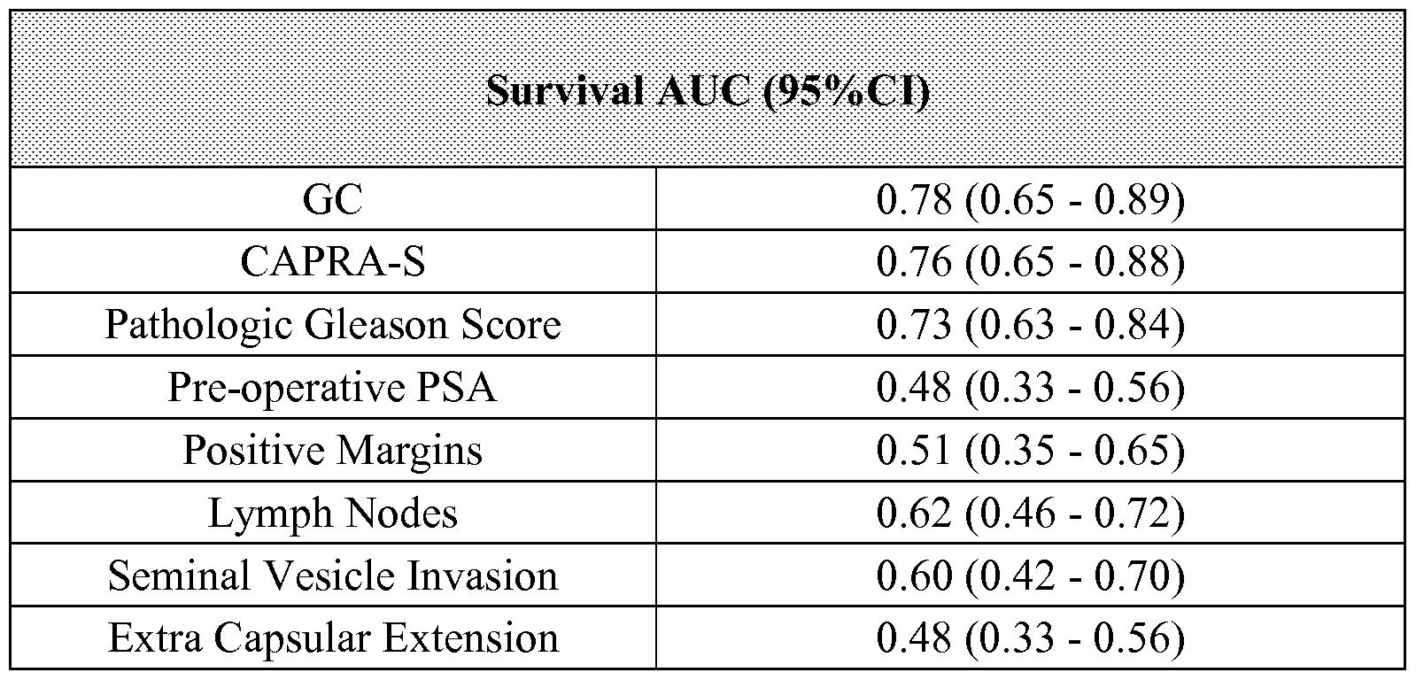

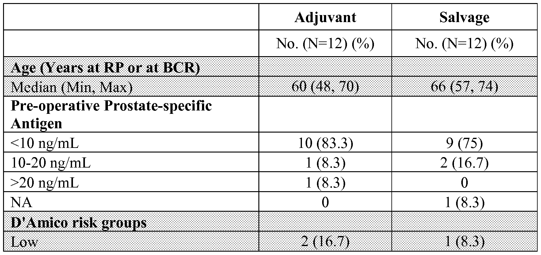


Claims
Priority Applications (8)
| Application Number | Priority Date | Filing Date | Title |
|---|---|---|---|
| EP18179289.6A EP3435084B1 (en) | 2012-08-16 | 2013-08-16 | Prostate cancer prognostics using biomarkers |
| EP13829137.2A EP2885640B1 (en) | 2012-08-16 | 2013-08-16 | Prostate cancer prognostics using biomarkers |
| CA2881627A CA2881627A1 (en) | 2012-08-16 | 2013-08-16 | Cancer diagnostics using biomarkers |
| AU2013302365A AU2013302365B2 (en) | 2012-08-16 | 2013-08-16 | Cancer diagnostics using biomarkers |
| EP23156159.8A EP4219765A3 (en) | 2012-08-16 | 2013-08-16 | Prostate cancer prognosis using biomarkers |
| HK15111421.9A HK1210832A1 (en) | 2012-08-16 | 2015-11-19 | Cancer diagnostics using biomarkers |
| AU2019201577A AU2019201577B2 (en) | 2012-08-16 | 2019-03-07 | Cancer diagnostics using biomarkers |
| AU2021273545A AU2021273545A1 (en) | 2012-08-16 | 2021-11-23 | Cancer diagnostics using biomarkers |
Applications Claiming Priority (6)
| Application Number | Priority Date | Filing Date | Title |
|---|---|---|---|
| US201261684066P | 2012-08-16 | 2012-08-16 | |
| US61/684,066 | 2012-08-16 | ||
| US201361764365P | 2013-02-13 | 2013-02-13 | |
| US61/764,365 | 2013-02-13 | ||
| US201361783124P | 2013-03-14 | 2013-03-14 | |
| US61/783,124 | 2013-03-14 |
Publications (2)
| Publication Number | Publication Date |
|---|---|
| WO2014028884A2 true WO2014028884A2 (en) | 2014-02-20 |
| WO2014028884A3 WO2014028884A3 (en) | 2014-05-08 |
Family
ID=50101621
Family Applications (1)
| Application Number | Title | Priority Date | Filing Date |
|---|---|---|---|
| PCT/US2013/055429 WO2014028884A2 (en) | 2012-08-16 | 2013-08-16 | Cancer diagnostics using biomarkers |
Country Status (8)
| Country | Link |
|---|---|
| US (2) | US11035005B2 (en) |
| EP (3) | EP3435084B1 (en) |
| AU (3) | AU2013302365B2 (en) |
| CA (1) | CA2881627A1 (en) |
| DK (1) | DK3435084T3 (en) |
| ES (1) | ES2945036T3 (en) |
| HK (1) | HK1210832A1 (en) |
| WO (1) | WO2014028884A2 (en) |
Cited By (18)
| Publication number | Priority date | Publication date | Assignee | Title |
|---|---|---|---|---|
| US9057109B2 (en) | 2008-05-14 | 2015-06-16 | Dermtech International | Diagnosis of melanoma and solar lentigo by nucleic acid analysis |
| WO2017059549A1 (en) * | 2015-10-08 | 2017-04-13 | Genomedx Biosciences, Inc. | Use of a genetic signature diagnostically to evaluate treatment strategies for prostate cancer |
| WO2017070582A1 (en) * | 2015-10-23 | 2017-04-27 | Novartis Ag | Computer method and system for deriving cell-to-cell spatial proximities |
| WO2018039490A1 (en) * | 2016-08-24 | 2018-03-01 | Genomedx Biosciences, Inc. | Use of genomic signatures to predict responsiveness of patients with prostate cancer to post-operative radiation therapy |
| US10407731B2 (en) | 2008-05-30 | 2019-09-10 | Mayo Foundation For Medical Education And Research | Biomarker panels for predicting prostate cancer outcomes |
| US10494677B2 (en) | 2006-11-02 | 2019-12-03 | Mayo Foundation For Medical Education And Research | Predicting cancer outcome |
| US10513737B2 (en) | 2011-12-13 | 2019-12-24 | Decipher Biosciences, Inc. | Cancer diagnostics using non-coding transcripts |
| EP3636779A1 (en) * | 2018-10-11 | 2020-04-15 | Koninklijke Philips N.V. | Pre-surgical risk stratification based on pde4d7 and dhx9 expression |
| WO2020076897A1 (en) * | 2018-10-09 | 2020-04-16 | Genecentric Therapeutics, Inc. | Detecting cancer cell of origin |
| CN111139298A (en) * | 2018-11-05 | 2020-05-12 | 王辉云 | Application of 4-LncRNA molecular label in lung cancer prognosis evaluation |
| US10865452B2 (en) | 2008-05-28 | 2020-12-15 | Decipher Biosciences, Inc. | Systems and methods for expression-based discrimination of distinct clinical disease states in prostate cancer |
| WO2021048445A1 (en) * | 2019-09-12 | 2021-03-18 | Uea Enterprises Limited | Novel biomarkers and diagnostic profiles for prostate cancer integrating clinical variables and gene expression data |
| US11035005B2 (en) | 2012-08-16 | 2021-06-15 | Decipher Biosciences, Inc. | Cancer diagnostics using biomarkers |
| US11078542B2 (en) | 2017-05-12 | 2021-08-03 | Decipher Biosciences, Inc. | Genetic signatures to predict prostate cancer metastasis and identify tumor aggressiveness |
| US11208697B2 (en) | 2017-01-20 | 2021-12-28 | Decipher Biosciences, Inc. | Molecular subtyping, prognosis, and treatment of bladder cancer |
| WO2022109125A1 (en) * | 2020-11-20 | 2022-05-27 | Decipher Biosciences, Inc. | Methods and genomic classifiers for identifying homologous recombination deficiency prostate cancer |
| US11578373B2 (en) | 2019-03-26 | 2023-02-14 | Dermtech, Inc. | Gene classifiers and uses thereof in skin cancers |
| US11873532B2 (en) | 2017-03-09 | 2024-01-16 | Decipher Biosciences, Inc. | Subtyping prostate cancer to predict response to hormone therapy |
Families Citing this family (6)
| Publication number | Priority date | Publication date | Assignee | Title |
|---|---|---|---|---|
| WO2016183414A1 (en) * | 2015-05-14 | 2016-11-17 | The Arizona Board Of Regents On Behalf Of The University Of Arizona | Systems and methods for characterizing granulomatous diseases |
| GB201616912D0 (en) | 2016-10-05 | 2016-11-16 | University Of East Anglia | Classification of cancer |
| EP3625357A4 (en) * | 2017-05-16 | 2021-02-24 | The Chinese University Of Hong Kong | Integrative single-cell and cell-free plasma rna analysis |
| US20180357361A1 (en) * | 2017-06-13 | 2018-12-13 | Feliks Frenkel | Systems and methods for identifying responders and non-responders to immune checkpoint blockade therapy |
| CN108830039A (en) * | 2018-03-19 | 2018-11-16 | 青岛理工大学 | A kind of PCR control system control node calculation method |
| WO2024026103A1 (en) * | 2022-07-28 | 2024-02-01 | Univeristy Of Pittsburgh - Of The Commonwealth System Of Higher Education | Systems and methods for predicting prostate cancer recurrence |
Citations (11)
| Publication number | Priority date | Publication date | Assignee | Title |
|---|---|---|---|---|
| US3687808A (en) | 1969-08-14 | 1972-08-29 | Univ Leland Stanford Junior | Synthetic polynucleotides |
| WO1990015070A1 (en) | 1989-06-07 | 1990-12-13 | Affymax Technologies N.V. | Very large scale immobilized peptide synthesis |
| WO1992010092A1 (en) | 1990-12-06 | 1992-06-25 | Affymax Technologies N.V. | Very large scale immobilized polymer synthesis |
| WO1993009668A1 (en) | 1991-11-22 | 1993-05-27 | Affymax Technology N.V. | Combinatorial strategies for polymer synthesis |
| US5288514A (en) | 1992-09-14 | 1994-02-22 | The Regents Of The University Of California | Solid phase and combinatorial synthesis of benzodiazepine compounds on a solid support |
| US5384261A (en) | 1991-11-22 | 1995-01-24 | Affymax Technologies N.V. | Very large scale immobilized polymer synthesis using mechanically directed flow paths |
| US5538848A (en) | 1994-11-16 | 1996-07-23 | Applied Biosystems Division, Perkin-Elmer Corp. | Method for detecting nucleic acid amplification using self-quenching fluorescence probe |
| US5846717A (en) | 1996-01-24 | 1998-12-08 | Third Wave Technologies, Inc. | Detection of nucleic acid sequences by invader-directed cleavage |
| US5985557A (en) | 1996-01-24 | 1999-11-16 | Third Wave Technologies, Inc. | Invasive cleavage of nucleic acids |
| US5994069A (en) | 1996-01-24 | 1999-11-30 | Third Wave Technologies, Inc. | Detection of nucleic acids by multiple sequential invasive cleavages |
| US9304145B2 (en) | 2014-07-30 | 2016-04-05 | Hitachi High-Tech Fine Systems Corporation | Inspection method and its apparatus for thermal assist type magnetic head element |
Family Cites Families (284)
| Publication number | Priority date | Publication date | Assignee | Title |
|---|---|---|---|---|
| AT277936B (en) | 1968-02-27 | 1970-01-12 | Guenter Dipl Ing Knapp | Method for the quantitative determination of iodine and thyroid hormones and device for carrying out the method |
| US4323546A (en) | 1978-05-22 | 1982-04-06 | Nuc Med Inc. | Method and composition for cancer detection in humans |
| US4683202A (en) | 1985-03-28 | 1987-07-28 | Cetus Corporation | Process for amplifying nucleic acid sequences |
| US4965188A (en) | 1986-08-22 | 1990-10-23 | Cetus Corporation | Process for amplifying, detecting, and/or cloning nucleic acid sequences using a thermostable enzyme |
| US4683195A (en) | 1986-01-30 | 1987-07-28 | Cetus Corporation | Process for amplifying, detecting, and/or-cloning nucleic acid sequences |
| EP0232967B1 (en) | 1986-01-10 | 1993-04-28 | Amoco Corporation | Competitive homogeneous assay |
| US4800159A (en) | 1986-02-07 | 1989-01-24 | Cetus Corporation | Process for amplifying, detecting, and/or cloning nucleic acid sequences |
| US5283174A (en) | 1987-09-21 | 1994-02-01 | Gen-Probe, Incorporated | Homogenous protection assay |
| US5130238A (en) | 1988-06-24 | 1992-07-14 | Cangene Corporation | Enhanced nucleic acid amplification process |
| US5225326A (en) | 1988-08-31 | 1993-07-06 | Research Development Foundation | One step in situ hybridization assay |
| US5744101A (en) | 1989-06-07 | 1998-04-28 | Affymax Technologies N.V. | Photolabile nucleoside protecting groups |
| EP0731175A3 (en) | 1989-07-11 | 2004-05-26 | Gen-Probe Incorporated | Oligonucleotide probes for HIV nucleic acid |
| CA2020958C (en) | 1989-07-11 | 2005-01-11 | Daniel L. Kacian | Nucleic acid sequence amplification methods |
| US5494810A (en) | 1990-05-03 | 1996-02-27 | Cornell Research Foundation, Inc. | Thermostable ligase-mediated DNA amplifications system for the detection of genetic disease |
| US5798257A (en) | 1990-07-09 | 1998-08-25 | Research Corporation Technologies, Inc. | Nucleic acid encoding human MTS-1 protein |
| US5455166A (en) | 1991-01-31 | 1995-10-03 | Becton, Dickinson And Company | Strand displacement amplification |
| US5270184A (en) | 1991-11-19 | 1993-12-14 | Becton, Dickinson And Company | Nucleic acid target generation |
| US5545524A (en) | 1991-12-04 | 1996-08-13 | The Regents Of The University Of Michigan | Compositions and methods for chromosome region-specific probes |
| US6204370B1 (en) | 1992-03-11 | 2001-03-20 | Institute Of Virology, Slovak Academy Of Sciences | MN gene and protein |
| US6027887A (en) | 1992-03-11 | 2000-02-22 | Institute Of Virology, Solvak Academy Of Sciences | MN gene and protein |
| US5541061A (en) | 1992-04-29 | 1996-07-30 | Affymax Technologies N.V. | Methods for screening factorial chemical libraries |
| US5925517A (en) | 1993-11-12 | 1999-07-20 | The Public Health Research Institute Of The City Of New York, Inc. | Detectably labeled dual conformation oligonucleotide probes, assays and kits |
| US5989815A (en) | 1994-03-18 | 1999-11-23 | University Of Utah Research Foundation | Methods for detecting predisposition to cancer at the MTS gene |
| US5648211A (en) | 1994-04-18 | 1997-07-15 | Becton, Dickinson And Company | Strand displacement amplification using thermophilic enzymes |
| US7625697B2 (en) | 1994-06-17 | 2009-12-01 | The Board Of Trustees Of The Leland Stanford Junior University | Methods for constructing subarrays and subarrays made thereby |
| US5830753A (en) | 1994-09-30 | 1998-11-03 | Ludwig Institute For Cancer Research | Isolated nucleic acid molecules coding for tumor rejection antigen precursor dage and uses thereof. |
| JP3189000B2 (en) | 1994-12-01 | 2001-07-16 | 東ソー株式会社 | Specific nucleic acid sequence detection method |
| US5710029A (en) | 1995-06-07 | 1998-01-20 | Gen-Probe Incorporated | Methods for determining pre-amplification levels of a nucleic acid target sequence from post-amplification levels of product |
| US5705365A (en) | 1995-06-07 | 1998-01-06 | Gen-Probe Incorporated | Kits for determining pre-amplification levels of a nucleic acid target sequence from post-amplification levels of product |
| US5854206A (en) | 1995-08-25 | 1998-12-29 | Corixa Corporation | Compounds and methods for treatment and diagnosis of prostate cancer |
| US5854033A (en) | 1995-11-21 | 1998-12-29 | Yale University | Rolling circle replication reporter systems |
| US6121489A (en) | 1996-03-05 | 2000-09-19 | Trega Biosciences, Inc. | Selectively N-alkylated peptidomimetic combinatorial libraries and compounds therein |
| CA2249742A1 (en) | 1996-03-15 | 1997-09-18 | Steven G. Reed | Compounds and methods for immunotherapy and immunodiagnosis of prostate cancer |
| AU713667B2 (en) | 1996-04-12 | 1999-12-09 | Phri Properties, Inc. | Detection probes, kits and assays |
| JP3122771B2 (en) | 1996-04-16 | 2001-01-09 | 岸本 忠三 | Method for detecting solid cancer cells and tissue atypia, and method for testing tissues for bone marrow transplantation and peripheral blood stem cell transplantation |
| US6136182A (en) | 1996-06-07 | 2000-10-24 | Immunivest Corporation | Magnetic devices and sample chambers for examination and manipulation of cells |
| US5711029A (en) | 1996-06-21 | 1998-01-27 | Visco; Raymond D. | Protective apparatus for dispersing pressure applied at a joint |
| HUP0002095A3 (en) | 1997-02-25 | 2002-02-28 | Corixa Corp Seattle | Compounds for immunotherapy of prostate cancer and methods for their use |
| US20030185830A1 (en) | 1997-02-25 | 2003-10-02 | Corixa Corporation | Compositions and methods for the therapy and diagnosis of prostate cancer |
| EP0965034B1 (en) | 1997-03-07 | 2007-05-30 | Clare Chemical Research, Inc. | Fluorometric detection using visible light |
| ATE438719T1 (en) | 1997-03-07 | 2009-08-15 | Siemens Healthcare Diagnostics | SPECIFIC MARKER FOR PROSTATE CANCER |
| AU7019498A (en) | 1997-04-10 | 1998-10-30 | Diagnocure Inc. | Pca3, pca3 genes, and methods of use |
| GB9711040D0 (en) | 1997-05-29 | 1997-07-23 | Duff Gordon W | Prediction of inflammatory disease |
| US8101349B2 (en) | 1997-12-23 | 2012-01-24 | Novartis Vaccines And Diagnostics, Inc. | Gene products differentially expressed in cancerous cells and their methods of use II |
| US20020076735A1 (en) | 1998-09-25 | 2002-06-20 | Williams Lewis T. | Diagnostic and therapeutic methods using molecules differentially expressed in cancer cells |
| GB9805918D0 (en) * | 1998-03-19 | 1998-05-13 | Nycomed Amersham Plc | Sequencing by hybridisation |
| JP3991327B2 (en) | 1998-06-08 | 2007-10-17 | 扶桑薬品工業株式会社 | Method for detecting thyroid cancer cells in thyroid tissue sample and composition for histological diagnosis of thyroid cancer |
| ES2330812T3 (en) | 1998-07-02 | 2009-12-15 | Gen-Probe Incorporated | MOLECULAR TORCHES. |
| US6406921B1 (en) | 1998-07-14 | 2002-06-18 | Zyomyx, Incorporated | Protein arrays for high-throughput screening |
| US20060204989A1 (en) | 1998-09-22 | 2006-09-14 | Kopreski Michael S | Comparative analysis of extracellular RNA species |
| US6573043B1 (en) | 1998-10-07 | 2003-06-03 | Genentech, Inc. | Tissue analysis and kits therefor |
| US6468476B1 (en) | 1998-10-27 | 2002-10-22 | Rosetta Inpharmatics, Inc. | Methods for using-co-regulated genesets to enhance detection and classification of gene expression patterns |
| EP1873260B1 (en) | 1998-11-09 | 2009-03-18 | Eiken Kagaku Kabushiki Kaisha | Process for synthesizing nucleic acid |
| US6828429B1 (en) | 1999-03-26 | 2004-12-07 | Henry M. Jackson Foundation For The Advancement Of Military Medicine | Prostate-specific gene, PCGEM1, and the methods of using PCGEM1 to detect, treat, and prevent prostate cancer |
| US6303305B1 (en) | 1999-03-30 | 2001-10-16 | Roche Diagnostics, Gmbh | Method for quantification of an analyte |
| US6436642B1 (en) | 1999-04-20 | 2002-08-20 | Curagen Corporation | Method of classifying a thyroid carcinoma using differential gene expression |
| EP1055734B1 (en) | 1999-05-24 | 2004-10-13 | Tosoh Corporation | Method for assaying ribonucleic acid |
| NZ517121A (en) | 1999-09-13 | 2004-05-28 | Nugen Technologies Inc | Methods and compositions for linear isothermal amplification of polynucleotide sequences |
| US7244559B2 (en) | 1999-09-16 | 2007-07-17 | 454 Life Sciences Corporation | Method of sequencing a nucleic acid |
| US7211390B2 (en) | 1999-09-16 | 2007-05-01 | 454 Life Sciences Corporation | Method of sequencing a nucleic acid |
| US6723506B2 (en) | 2000-01-20 | 2004-04-20 | Brigham And Women's Hospital | Method of identifying PAX8-PPAR gamma-nucleic acid molecules |
| US20020168638A1 (en) | 2000-01-24 | 2002-11-14 | Robert Schlegel | Compositions, kits, and methods for identification, assessment, prevention, and therapy of human prostate cancer |
| US7700341B2 (en) | 2000-02-03 | 2010-04-20 | Dendreon Corporation | Nucleic acid molecules encoding transmembrane serine proteases, the encoded proteins and methods based thereon |
| US7361488B2 (en) | 2000-02-07 | 2008-04-22 | Illumina, Inc. | Nucleic acid detection methods using universal priming |
| AU2001241541A1 (en) | 2000-02-17 | 2001-08-27 | Millennium Predictive Medicine, Inc. | Novel genes, compositions, kits, and methods for identification, assessment, prevention, and therapy of human prostate cancer |
| EP1263956A2 (en) | 2000-03-09 | 2002-12-11 | Chiron Corporation | Human genes and gene expression products |
| US7700359B2 (en) | 2000-06-02 | 2010-04-20 | Novartis Vaccines And Diagnostics, Inc. | Gene products differentially expressed in cancerous cells |
| FR2810677B1 (en) | 2000-06-27 | 2004-10-01 | Urogene | PROCESS FOR IN VITRO DIAGNOSIS OF PROSTATE CANCER AND IMPLEMENTATION KIT |
| US6673545B2 (en) | 2000-07-28 | 2004-01-06 | Incyte Corporation | Prostate cancer markers |
| US20070037165A1 (en) | 2000-09-08 | 2007-02-15 | Applera Corporation | Polymorphisms in known genes associated with human disease, methods of detection and uses thereof |
| US7892730B2 (en) | 2000-12-22 | 2011-02-22 | Sagres Discovery, Inc. | Compositions and methods for cancer |
| US6988040B2 (en) | 2001-01-11 | 2006-01-17 | Affymetrix, Inc. | System, method, and computer software for genotyping analysis and identification of allelic imbalance |
| US20040029114A1 (en) | 2001-01-24 | 2004-02-12 | Eos Technology, Inc. | Methods of diagnosis of breast cancer, compositions and methods of screening for modulators of breast cancer |
| WO2002064781A2 (en) | 2001-02-09 | 2002-08-22 | Active Pass Pharmaceuticals, Inc. | Regulation of amyloid precursor protein expression by modification of abc transporter expression or activity |
| US7026163B1 (en) | 2001-02-23 | 2006-04-11 | Mayo Foundation For Medical Education And Research | Sulfotransferase sequence variants |
| WO2002068677A2 (en) | 2001-02-27 | 2002-09-06 | Eos Biotechnology, Inc. | Novel methods of diagnosis of metastatic colorectal cancer, compositions and methods of screening for modulators of metastatic colorectal cancer |
| US20030190602A1 (en) | 2001-03-12 | 2003-10-09 | Monogen, Inc. | Cell-based detection and differentiation of disease states |
| US20040009481A1 (en) | 2001-06-11 | 2004-01-15 | Millennium Pharmaceuticals, Inc. | Compositions, kits, and methods for identification, assessment, prevention, and therapy of human prostate cancer |
| US7229774B2 (en) | 2001-08-02 | 2007-06-12 | Regents Of The University Of Michigan | Expression profile of prostate cancer |
| JP4291143B2 (en) | 2001-08-31 | 2009-07-08 | ジェン−プロウブ インコーポレイテッド | Affinity-shifted probe for quantification of analyte polynucleotide |
| ATE309393T1 (en) | 2001-09-06 | 2005-11-15 | Adnagen Ag | METHOD FOR THE QUALITATIVE AND/OR QUANTITATIVE DETECTION OF CELLS |
| US20040142325A1 (en) | 2001-09-14 | 2004-07-22 | Liat Mintz | Methods and systems for annotating biomolecular sequences |
| US20050202442A1 (en) | 2003-12-15 | 2005-09-15 | Morris David W. | Novel therapeutic targets in cancer |
| DE60235413D1 (en) | 2001-12-07 | 2010-04-01 | Novartis Vaccines & Diagnostic | IN PROSTATE CANCER HIGHLY REGULATED ENDOGENIC RETROVIRUS |
| US6949342B2 (en) | 2001-12-21 | 2005-09-27 | Whitehead Institute For Biomedical Research | Prostate cancer diagnosis and outcome prediction by expression analysis |
| US7704700B2 (en) | 2002-02-12 | 2010-04-27 | Burnham Institute For Medical Research | Methods for determining the prognosis for patients with a prostate neoplastic condition using inhibitor of apoptosis polypeptides |
| US20030194734A1 (en) | 2002-03-29 | 2003-10-16 | Tim Jatkoe | Selection of markers |
| US20030186248A1 (en) | 2002-03-29 | 2003-10-02 | Erlander Mark G. | Interpreting cytological specimens via molecular histological signatures |
| US20050143933A1 (en) | 2002-04-23 | 2005-06-30 | James Minor | Analyzing and correcting biological assay data using a signal allocation model |
| US20050032065A1 (en) | 2002-06-24 | 2005-02-10 | Afar Daniel E. H. | Methods of prognosis of prostate cancer |
| US7662553B2 (en) | 2002-07-31 | 2010-02-16 | University Of Southern California | Polymorphisms in the ERCC1 gene for predicting treatment outcome |
| US7282328B2 (en) | 2002-09-20 | 2007-10-16 | New England Biolabs, Inc. | Helicase dependent amplification of nucleic acids |
| US7300788B2 (en) | 2002-10-08 | 2007-11-27 | Affymetrix, Inc. | Method for genotyping polymorphisms in humans |
| US20050266443A1 (en) | 2002-10-11 | 2005-12-01 | Thomas Jefferson University | Novel tumor suppressor gene and compositions and methods for making and using the same |
| KR101078977B1 (en) | 2002-11-07 | 2011-11-01 | 세키스이 메디칼 가부시키가이샤 | Method of detecting gene mutation |
| US7244567B2 (en) | 2003-01-29 | 2007-07-17 | 454 Life Sciences Corporation | Double ended sequencing |
| JP4568716B2 (en) | 2003-02-20 | 2010-10-27 | ジェノミック ヘルス, インコーポレイテッド | Use of intron RNA to measure gene expression |
| US7378233B2 (en) | 2003-04-12 | 2008-05-27 | The Johns Hopkins University | BRAF mutation T1796A in thyroid cancers |
| US20070275915A1 (en) | 2003-04-15 | 2007-11-29 | Cell Genesys, Inc. | Tmprss2 Regulatory Sequences and Uses Thereof |
| AU2004235747B2 (en) | 2003-05-01 | 2009-05-28 | Gen-Probe Incorporated | Oligonucleotides comprising a molecular switch |
| JP2007507222A (en) | 2003-05-28 | 2007-03-29 | ゲノミック ヘルス, インコーポレイテッド | Gene expression markers for predicting response to chemotherapy |
| US7638275B2 (en) | 2003-05-30 | 2009-12-29 | The Board Of Trustees Of The University Of Illinois | Gene expression profiles that identify genetically elite cattle |
| CA2528669A1 (en) | 2003-06-09 | 2005-01-20 | The Regents Of The University Of Michigan | Compositions and methods for treating and diagnosing cancer |
| EP1644858B1 (en) | 2003-07-10 | 2017-12-06 | Genomic Health, Inc. | Expression profile algorithm and test for cancer prognosis |
| US20070048738A1 (en) | 2003-07-14 | 2007-03-01 | Mayo Foundation For Medical Education And Research | Methods and compositions for diagnosis, staging and prognosis of prostate cancer |
| US20050118625A1 (en) | 2003-10-02 | 2005-06-02 | Mounts William M. | Nucleic acid arrays for detecting gene expression associated with human osteoarthritis and human proteases |
| CA2542656A1 (en) | 2003-10-16 | 2005-05-06 | Genomic Health, Inc. | Qrt-pcr assay system for gene expression profiling |
| US20050130177A1 (en) | 2003-12-12 | 2005-06-16 | 3M Innovative Properties Company | Variable valve apparatus and methods |
| CA2549324A1 (en) | 2003-12-12 | 2005-06-30 | Bayer Pharmaceuticals Corporation | Gene expression profiles and methods of use |
| WO2005062770A2 (en) | 2003-12-19 | 2005-07-14 | Novakoff James L | Method for conducting pharmacogenomics-based studies |
| US20090020433A1 (en) | 2003-12-31 | 2009-01-22 | Microfabrica Inc. | Electrochemical Fabrication Methods for Producing Multilayer Structures Including the use of Diamond Machining in the Planarization of Deposits of Material |
| US7407755B2 (en) | 2004-01-15 | 2008-08-05 | Lubinski Jan | Determining a predisposition to cancer |
| WO2005078139A2 (en) | 2004-02-09 | 2005-08-25 | Thomas Jefferson University | DIAGNOSIS AND TREATMENT OF CANCERS WITH MicroRNA LOCATED IN OR NEAR CANCER-ASSOCIATED CHROMOSOMAL FEATURES |
| WO2005085471A2 (en) | 2004-03-03 | 2005-09-15 | Universität Leipzig | Method and means for differential diagnosis of thyroid tumours |
| US8426126B2 (en) | 2004-03-18 | 2013-04-23 | Applied Biosystems, Llc | Modified surfaces as solid supports for nucleic acid purification |
| WO2005095654A1 (en) | 2004-03-25 | 2005-10-13 | Biohelix Corporation | Helicase-dependent amplification of circular nucleic acids |
| US7319011B2 (en) | 2004-04-08 | 2008-01-15 | Duke University | Method for distinguishing follicular thyroid adenoma (FTA) from follicular thyroid carcinoma (FTC) |
| JP5813908B2 (en) | 2004-04-09 | 2015-11-17 | ジェノミック ヘルス, インコーポレイテッド | Gene expression markers to predict response to chemotherapeutic agents |
| DE602005025211D1 (en) | 2004-04-09 | 2011-01-20 | Us Gov Health & Human Serv | DIAGNOSTIC DEVICE FOR THE DIAGNOSIS OF GOOD-TO-BROKEN THYROID LESIONS |
| US20050240357A1 (en) | 2004-04-26 | 2005-10-27 | Minor James M | Methods and systems for differential clustering |
| JP2007535966A (en) | 2004-05-04 | 2007-12-13 | ダコ デンマーク アクティーゼルスカブ | Method for detecting chromosomal abnormalities |
| EP2290067B1 (en) | 2004-05-28 | 2014-12-10 | Asuragen, Inc. | Methods and compositions involving microRNA |
| US20060088851A1 (en) | 2004-06-04 | 2006-04-27 | Arcturus Bioscience, Inc. | Invasion/migration gene |
| US20060019615A1 (en) | 2004-07-24 | 2006-01-26 | Ditmer Larry R | Wireless keying for a continuous wave (CW) amateur radio transmitter |
| EP2311953A1 (en) | 2004-08-04 | 2011-04-20 | Riken | Bone/joint disease susceptibility gene and use thereof |
| WO2006020837A2 (en) | 2004-08-11 | 2006-02-23 | The Cleveland Clinic Foundation | Method of detecting thyroid cancer |
| EP1970454B1 (en) | 2004-08-27 | 2012-09-19 | Gen-Probe Incorporated | Single-primer nucleic acid amplification methods |
| US20060046253A1 (en) | 2004-09-02 | 2006-03-02 | Suntory Limited | Method for analyzing genes of industrial yeasts |
| US7642348B2 (en) | 2004-10-04 | 2010-01-05 | Rosetta Genomics Ltd | Prostate cancer-related nucleic acids |
| ITBZ20040048A1 (en) | 2004-10-06 | 2005-01-06 | Genprofiler | METHOD FOR THE IDENTIFICATION OF NEOPLASTIC TRANSFORMATION, WITH PARTICULAR REFERENCE TO PROSTATIC CANCER |
| EP1810026B1 (en) | 2004-10-06 | 2018-04-18 | Mayo Foundation For Medical Education And Research | B7-h1 and pd-1 in treatment of renal cell carcinoma |
| US7629325B2 (en) | 2004-10-11 | 2009-12-08 | Technion Research & Development Foundation Ltd. | Human Sef isoforms and methods of using same for cancer diagnosis and gene therapy |
| US7485468B2 (en) | 2004-10-15 | 2009-02-03 | Galapagos Bv | Molecular targets and compounds, and methods to identify the same, useful in the treatment of joint degenerative and inflammatory diseases |
| US20060088870A1 (en) | 2004-10-22 | 2006-04-27 | Finkelstein Sydney D | Topographic genotyping for determining the diagnosis, malignant potential, and biologic behavior of pancreatic cysts and related conditions |
| JP2008518610A (en) * | 2004-11-03 | 2008-06-05 | アルマック ダイアグノスティックス リミテッド | Transcriptome microarray technique and method of using the same |
| US7666595B2 (en) * | 2005-02-25 | 2010-02-23 | The Brigham And Women's Hospital, Inc. | Biomarkers for predicting prostate cancer progression |
| US20120122718A1 (en) | 2005-03-01 | 2012-05-17 | Reisman David N | BRM Expression and Related Diagnostics |
| WO2006110264A2 (en) * | 2005-03-16 | 2006-10-19 | Sidney Kimmel Cancer Center | Methods and compositions for predicting death from cancer and prostate cancer survival using gene expression signatures |
| US20070020657A1 (en) | 2005-05-20 | 2007-01-25 | Grebe Stefan K | Methods for detecting circulating tumor cells |
| US20070037186A1 (en) | 2005-05-20 | 2007-02-15 | Yuqiu Jiang | Thyroid fine needle aspiration molecular assay |
| US20060292610A1 (en) | 2005-06-06 | 2006-12-28 | Regents Of The University Of Michigan | Prognostic meta signatures and uses thereof |
| WO2006135886A2 (en) | 2005-06-13 | 2006-12-21 | The Regents Of The University Of Michigan | Compositions and methods for treating and diagnosing cancer |
| JP2008546387A (en) | 2005-06-13 | 2008-12-25 | ザ リージェンツ オブ ザ ユニバーシティ オブ ミシガン | Compositions and methods for treating and diagnosing cancer |
| IL177006A0 (en) | 2005-08-02 | 2006-12-10 | Veridex Llc | Predicting bone relapse of breast cancer |
| EP1995321A2 (en) | 2005-08-15 | 2008-11-26 | Genentech, Inc. | Gene disruptions, compositions and methods relating thereto |
| US9957569B2 (en) | 2005-09-12 | 2018-05-01 | The Regents Of The University Of Michigan | Recurrent gene fusions in prostate cancer |
| CA2814598A1 (en) | 2005-09-12 | 2007-03-22 | The Regents Of The University Of Michigan | Recurrent gene fusions in prostate cancer |
| CN102346816B (en) | 2005-09-22 | 2014-12-03 | 中国合成橡胶股份有限公司 | Gene expression profiling for identification of prognostic subclasses in nasopharyngeal carcinomas |
| WO2007038792A2 (en) | 2005-09-28 | 2007-04-05 | H. Lee Moffitt Cancer Center | Individualized cancer treatments |
| US7962291B2 (en) | 2005-09-30 | 2011-06-14 | Affymetrix, Inc. | Methods and computer software for detecting splice variants |
| EP1943356B1 (en) | 2005-10-03 | 2014-05-14 | Epigenomics AG | Methods and nucleic acids for the analysis of gene expression associated with the prognosis of cell proliferative disorders |
| US7598052B2 (en) | 2005-10-11 | 2009-10-06 | The Regents Of The University Of Michigan | Expression profile of thyroid cancer |
| US20070220621A1 (en) | 2005-10-31 | 2007-09-20 | Clarke Michael F | Genetic characterization and prognostic significance of cancer stem cells in cancer |
| EP1945817A4 (en) * | 2005-11-02 | 2008-12-10 | Univ Michigan Technology Man W | Molecular profiling of cancer |
| WO2007070621A2 (en) | 2005-12-13 | 2007-06-21 | Children's Medical Center Corporation | Prognosis indicators for solid human tumors |
| EP2487258B1 (en) | 2006-01-05 | 2014-10-01 | The Ohio State University Research Foundation | MicroRNA-based methods for the diagnosis of colon, pancreas and stomach cancer |
| ES2553442T3 (en) | 2006-01-05 | 2015-12-09 | The Ohio State University Research Foundation | Procedures based on microRNAs for the diagnosis, prognosis and treatment of lung cancer |
| US20070172841A1 (en) | 2006-01-25 | 2007-07-26 | Hui Wang | Probe/target stabilization with add-in oligo |
| FR2896881B1 (en) | 2006-01-31 | 2008-04-18 | Biomerieux Sa | METHOD FOR DETERMINING PRONGF FOR IN VITRO DIAGNOSIS OF BREAST CANCER AND USE OF PRONGF IN THERAPY |
| US7670775B2 (en) | 2006-02-15 | 2010-03-02 | The Ohio State University Research Foundation | Method for differentiating malignant from benign thyroid tissue |
| JP4867018B2 (en) | 2006-03-22 | 2012-02-01 | 富士フイルム株式会社 | Cancer detection method and suppression method |
| US7914988B1 (en) * | 2006-03-31 | 2011-03-29 | Illumina, Inc. | Gene expression profiles to predict relapse of prostate cancer |
| US20100279327A1 (en) | 2006-06-12 | 2010-11-04 | Bipar Sciences, Inc. | Method of treating diseases with parp inhibitors |
| US20080076674A1 (en) | 2006-07-06 | 2008-03-27 | Thomas Litman | Novel oligonucleotide compositions and probe sequences useful for detection and analysis of non coding RNAs associated with cancer |
| US20080028302A1 (en) | 2006-07-31 | 2008-01-31 | Steffen Meschkat | Method and apparatus for incrementally updating a web page |
| FI20060751A0 (en) | 2006-08-23 | 2006-08-23 | Valtion Teknillinen | Method of treating prostate cancer and screening of patients who benefit from said method |
| US8273539B2 (en) | 2006-09-25 | 2012-09-25 | Mayo Foundation For Medical Education And Research | Extracellular and membrane-associated prostate cancer markers |
| US8541169B2 (en) | 2006-10-10 | 2013-09-24 | The Henry M. Jackson Foundation For The Advancement Of Military Medicine, Inc. | Prostate cancer-specific alternations in ERG gene expression and detection and treatment methods based on those alterations |
| WO2008046911A2 (en) | 2006-10-20 | 2008-04-24 | Exiqon A/S | Novel human micrornas associated with cancer |
| WO2008058018A2 (en) | 2006-11-02 | 2008-05-15 | Mayo Foundation For Medical Education And Research | Predicting cancer outcome |
| WO2008069906A2 (en) | 2006-11-14 | 2008-06-12 | The Regents Of The University Of California | Digital expression of gene analysis |
| US8470534B2 (en) | 2006-12-01 | 2013-06-25 | Erik S. Knudsen | Methods of predicting resistance or sensitivity to therapies for cancer |
| WO2008086478A2 (en) | 2007-01-10 | 2008-07-17 | Henry Ford Health System | Methods and compositions for identification of prostate cancer markers |
| WO2008112283A2 (en) | 2007-03-12 | 2008-09-18 | Government Of The United States Of America, As Represented By The Secretary, Department Of Health And Human Services | Microrna profiling of androgen responsiveness for predicting the appropriate prostate cancer treatment |
| US8802599B2 (en) | 2007-03-27 | 2014-08-12 | Rosetta Genomics, Ltd. | Gene expression signature for classification of tissue of origin of tumor samples |
| AU2008231393A1 (en) | 2007-03-27 | 2008-10-02 | Rosetta Genomics Ltd. | Gene expression signature for classification of cancers |
| US9096906B2 (en) | 2007-03-27 | 2015-08-04 | Rosetta Genomics Ltd. | Gene expression signature for classification of tissue of origin of tumor samples |
| US20100273172A1 (en) | 2007-03-27 | 2010-10-28 | Rosetta Genomics Ltd. | Micrornas expression signature for determination of tumors origin |
| EP1975252A1 (en) | 2007-03-29 | 2008-10-01 | INSERM (Institut National de la Santé et de la Recherche Medicale) | Methods for the prognosis or for the diagnosis of a thyroid disease |
| US20090062144A1 (en) | 2007-04-03 | 2009-03-05 | Nancy Lan Guo | Gene signature for prognosis and diagnosis of lung cancer |
| US7901888B2 (en) | 2007-05-09 | 2011-03-08 | The Regents Of The University Of California | Multigene diagnostic assay for malignant thyroid neoplasm |
| EP3018216B1 (en) | 2007-07-06 | 2018-09-12 | The Regents Of The University Of Michigan | Recurrent gene fusions in prostate cancer |
| US8465918B2 (en) | 2007-08-03 | 2013-06-18 | The Ohio State University Research Foundation | Ultraconserved regions encoding ncRNAs |
| WO2009020521A2 (en) | 2007-08-03 | 2009-02-12 | The Brigham And Women's Hospital, Inc. | Identification and treatment of estrogen responsive prostate tumors |
| WO2009029266A2 (en) | 2007-08-27 | 2009-03-05 | The Government Of The United States Of America, As Represented By The Secretary, Department Of Health Of Human Services, National Institutes Of Health | Diagnostic tool for diagnosing benign versus malignant thyroid lesions |
| AT505726A2 (en) | 2007-08-30 | 2009-03-15 | Arc Austrian Res Centers Gmbh | SET OF TUMOR MARKERS |
| NZ562237A (en) | 2007-10-05 | 2011-02-25 | Pacific Edge Biotechnology Ltd | Proliferation signature and prognosis for gastrointestinal cancer |
| WO2009074968A2 (en) | 2007-12-12 | 2009-06-18 | Ecole Polytechnique Federale De Lausanne (Epfl) | Method for predicting the efficacy of cancer therapy |
| US20090191535A1 (en) | 2007-12-22 | 2009-07-30 | Mark Carle Connelly | Method of assessing metastatic carcinomas from circulating endothelial cells and disseminated tumor cells |
| WO2009087139A1 (en) | 2008-01-04 | 2009-07-16 | Centre National De La Recherche Scientifique | Molecular in vitro diagnosis of breast cancer |
| US20100021538A1 (en) | 2008-02-29 | 2010-01-28 | Youngro Byun | Pharmaceutical compositions containing heparin derivatives |
| US8293880B2 (en) | 2008-03-25 | 2012-10-23 | University Of Southern California | Prognostic panel for urinary bladder cancer |
| JP2011523049A (en) | 2008-05-14 | 2011-08-04 | ドナー, インコーポレイテッド | Biomarkers for head and neck cancer identification, monitoring and treatment |
| EP2806054A1 (en) | 2008-05-28 | 2014-11-26 | Genomedx Biosciences Inc. | Systems and methods for expression-based discrimination of distinct clinical disease states in prostate cancer |
| US10407731B2 (en) | 2008-05-30 | 2019-09-10 | Mayo Foundation For Medical Education And Research | Biomarker panels for predicting prostate cancer outcomes |
| US20090298082A1 (en) * | 2008-05-30 | 2009-12-03 | Klee George G | Biomarker panels for predicting prostate cancer outcomes |
| US20110077168A1 (en) | 2008-06-17 | 2011-03-31 | Nitzan Rosenfeld | Methods for distinguishing between specific types of lung cancers |
| US20120122698A1 (en) | 2008-07-07 | 2012-05-17 | Decode Genetics Ehf. | Genetic Variants Predictive of Cancer Risk in Humans |
| CA2734123A1 (en) | 2008-08-15 | 2010-02-18 | Decode Genetics Ehf | Genetic variants predictive of cancer risk |
| WO2010030365A2 (en) | 2008-09-12 | 2010-03-18 | Cornell Research Foundation, Inc. Wmc | Thyroid tumors identified |
| CA2739140A1 (en) | 2008-10-01 | 2010-04-08 | Noviogendix Research B.V. | Molecular markers in prostate cancer |
| CN102232117A (en) | 2008-10-14 | 2011-11-02 | 卡里斯Mpi公司 | Gene and gene expressed protein targets depicting biomarker patterns and signature sets by tumor type |
| WO2010056351A2 (en) | 2008-11-14 | 2010-05-20 | Stc.Unm | Gene expression classifiers for relapse free survival and minimal residual disease improve risk classification and out come prediction in pedeatric b-precursor acute lymphoblastic leukemia |
| WO2010056931A1 (en) | 2008-11-14 | 2010-05-20 | Intelligent Oncotherapeutics, Inc. | Methods for identification of tumor phenotype and treatment |
| US9495515B1 (en) | 2009-12-09 | 2016-11-15 | Veracyte, Inc. | Algorithms for disease diagnostics |
| AU2009314502B2 (en) | 2008-11-17 | 2015-01-22 | Veracyte, Inc. | Methods and compositions of molecular profiling for disease diagnostics |
| US10236078B2 (en) | 2008-11-17 | 2019-03-19 | Veracyte, Inc. | Methods for processing or analyzing a sample of thyroid tissue |
| WO2010058572A1 (en) | 2008-11-20 | 2010-05-27 | Oncotherapy Science, Inc. | Methods for diagnosing or treating prostate cancer |
| CA2777638A1 (en) | 2008-11-26 | 2010-06-03 | Decode Genetics Ehf. | Genetic variants useful for risk assessment of thyroid cancer |
| WO2010061396A1 (en) | 2008-11-30 | 2010-06-03 | Rosetta Genomics Ltd. | Methods for detecting an increased susceptibility to cancer |
| CA2745961A1 (en) | 2008-12-04 | 2010-06-10 | The Regents Of The University Of California | Materials and methods for determining diagnosis and prognosis of prostate cancer |
| CN102333888B (en) | 2008-12-24 | 2013-07-10 | 姜桥 | Gene expression signature for classification of tissue of origin of tumor samples |
| US20120041274A1 (en) | 2010-01-07 | 2012-02-16 | Myriad Genetics, Incorporated | Cancer biomarkers |
| JP2012514475A (en) | 2009-01-09 | 2012-06-28 | ザ リージェンツ オブ ザ ユニバーシティ オブ ミシガン | Reproducible gene fusions in cancer |
| WO2010091049A2 (en) | 2009-02-03 | 2010-08-12 | Children's Medical Center Corporation | Diagnosis and treatment of cancer |
| US9074258B2 (en) | 2009-03-04 | 2015-07-07 | Genomedx Biosciences Inc. | Compositions and methods for classifying thyroid nodule disease |
| US8697275B2 (en) | 2009-03-04 | 2014-04-15 | Samsung Sdi Co., Ltd. | Rechargeable battery having an extendable case region |
| WO2010123626A1 (en) | 2009-04-24 | 2010-10-28 | University Of Southern California | Cd133 polymorphisms and expression predict clinical outcome in patients with cancer |
| US9206482B2 (en) | 2009-04-29 | 2015-12-08 | Genomedx Biosciences Inc. | Systems and methods for expression-based classification of thyroid tissue |
| JP6078339B2 (en) | 2009-05-07 | 2017-02-08 | ベラサイト インコーポレイテッド | Methods and compositions for diagnosis of thyroid status |
| EP2473628A1 (en) | 2009-08-31 | 2012-07-11 | Alcedo Biotech GmbH | Microrna-based methods and compositions for the diagnosis, prognosis and treatment of tumor involving chromosomal rearrangements |
| US9110065B2 (en) | 2009-09-21 | 2015-08-18 | Paul Walfish | Methods and compositions for the diagnosis and treatment of thyroid cancer |
| EP2483813A1 (en) | 2009-10-01 | 2012-08-08 | Chipdx LLC | System and method for classification of patients |
| US8975019B2 (en) | 2009-10-19 | 2015-03-10 | University Of Massachusetts | Deducing exon connectivity by RNA-templated DNA ligation/sequencing |
| US20130231258A1 (en) | 2011-12-09 | 2013-09-05 | Veracyte, Inc. | Methods and Compositions for Classification of Samples |
| US10446272B2 (en) | 2009-12-09 | 2019-10-15 | Veracyte, Inc. | Methods and compositions for classification of samples |
| GB0922085D0 (en) | 2009-12-17 | 2010-02-03 | Cambridge Entpr Ltd | Cancer diagnosis and treatment |
| US9642821B2 (en) | 2010-01-14 | 2017-05-09 | Dana-Farber Cancer Institute, Inc. | MiR-182 in the diagnosis and treatment of cancer |
| EP2366800A1 (en) | 2010-03-01 | 2011-09-21 | Centrum Onkologii-Instytut im M. Sklodowskiej-Curie Oddzial w Gliwicach | Kit, method and use for the diagnosis of papillary thyroid cancer using a gene expression profile |
| WO2011116380A2 (en) | 2010-03-19 | 2011-09-22 | H. Lee Moffitt Cancer Center And Research Institute, Inc. | Hybrid model for the classification of carcinoma subtypes |
| US9157123B2 (en) | 2010-04-20 | 2015-10-13 | The Johns Hopkins University | Genetic amplification of IQGAP1 in cancer |
| CN103038635B (en) | 2010-05-11 | 2016-12-28 | 威拉赛特公司 | For diagnosing the method and composition of condition of illness |
| WO2011150453A1 (en) | 2010-06-01 | 2011-12-08 | The University Of Queensland | Diagnostic, prognostic and therapeutic use of a long non-coding rna |
| WO2012012225A2 (en) | 2010-07-22 | 2012-01-26 | Mayo Foundation For Medical Education And Research | Predicting cancer outcome |
| US20140045915A1 (en) | 2010-08-31 | 2014-02-13 | The General Hospital Corporation | Cancer-related biological materials in microvesicles |
| ES2629890T3 (en) | 2010-11-17 | 2017-08-16 | Interpace Diagnostics, Llc | miRNA as biomarkers to distinguish between benign and malignant thyroid neoplasms |
| WO2012068383A2 (en) | 2010-11-19 | 2012-05-24 | The Regents Of The University Of Michigan | ncRNA AND USES THEREOF |
| US20130267443A1 (en) | 2010-11-19 | 2013-10-10 | The Regents Of The University Of Michigan | ncRNA AND USES THEREOF |
| WO2012085948A1 (en) | 2010-12-21 | 2012-06-28 | Decode Genetics Ehf | Genetic variants useful for risk assessment of thyroid cancer |
| WO2012094744A1 (en) | 2011-01-11 | 2012-07-19 | University Health Network | Prognostic signature for oral squamous cell carcinoma |
| EP2686443A4 (en) | 2011-03-17 | 2014-12-17 | Decode Genetics Ehf | Genetic variants useful for risk assessment of thyroid cancer |
| US8945829B2 (en) | 2011-03-22 | 2015-02-03 | Cornell University | Distinguishing benign and malignant indeterminate thyroid lesions |
| CA2831074A1 (en) | 2011-03-26 | 2012-10-04 | Oregon Health And Science University | Gene expression predictors of cancer prognosis |
| EP2505664B1 (en) | 2011-03-30 | 2014-12-03 | Universität Leipzig | Method and means for distinguishing malignant from benign tumor samples, in particular in routine air dried fine needle aspiration biopsy (FNAB) |
| WO2013006495A2 (en) | 2011-07-01 | 2013-01-10 | Dana-Farber Cancer Institute, Inc. | Methods of predicting prognosis in cancer |
| US9435812B2 (en) | 2011-08-31 | 2016-09-06 | Ventana Medical Systems, Inc. | Expression of ETS related gene (ERG) and phosphatase and tensin homolog (PTEN) correlates with prostate cancer capsular penetration |
| WO2013066678A1 (en) | 2011-10-26 | 2013-05-10 | Georgetown University | Microrna expression profiling of thyroid cancer |
| US20130142728A1 (en) | 2011-10-27 | 2013-06-06 | Asuragen, Inc. | Mirnas as diagnostic biomarkers to distinguish benign from malignant thyroid tumors |
| WO2013086524A1 (en) | 2011-12-09 | 2013-06-13 | The Johns Hopkins University | Compositions and methods for characterizing thyroid neoplasia |
| GB2513732B (en) | 2011-12-10 | 2020-12-02 | Veracyte Inc | Methods and compositions for sample identification |
| CA2858581A1 (en) | 2011-12-13 | 2013-06-20 | Genomedx Biosciences, Inc. | Cancer diagnostics using non-coding transcripts |
| WO2013088457A1 (en) | 2011-12-13 | 2013-06-20 | Decode Genetics Ehf | Genetic variants useful for risk assessment of thyroid cancer |
| US20130184999A1 (en) | 2012-01-05 | 2013-07-18 | Yan Ding | Systems and methods for cancer-specific drug targets and biomarkers discovery |
| WO2013116742A1 (en) | 2012-02-01 | 2013-08-08 | Mayo Foundation For Medical Education And Research | Predicting responses to androgen deprivation therapy and methods for treating prostate cancer |
| US20150038376A1 (en) | 2012-03-15 | 2015-02-05 | Qiagen Sciences Llc | Thyroid cancer biomarker |
| US20150141470A1 (en) | 2012-05-08 | 2015-05-21 | The Broad Institute, Inc. | Diagnostic and treatment methods in patients having or at risk of developing resistance to cancer therapy |
| EP3435084B1 (en) | 2012-08-16 | 2023-02-22 | Decipher Biosciences, Inc. | Prostate cancer prognostics using biomarkers |
| CA2885202A1 (en) | 2012-09-20 | 2014-03-27 | Genomedx Biosciences, Inc. | Thyroid cancer diagnostics |
| WO2014058317A1 (en) | 2012-10-10 | 2014-04-17 | Stichting Het Nederlands Kanker Instituut-Antoni van Leeuwenhoek Ziekenhuis | Methods and means for predicting resistance to anti-cancer treatment |
| BR102013027577A2 (en) | 2012-10-25 | 2016-03-29 | Memorialsloan Kettering Cancer Ct | method of detecting an increased axl or gas6 level and method of identifying an egfr inhibitor resistant cancer patient |
| US20140143188A1 (en) | 2012-11-16 | 2014-05-22 | Genformatic, Llc | Method of machine learning, employing bayesian latent class inference: combining multiple genomic feature detection algorithms to produce an integrated genomic feature set with specificity, sensitivity and accuracy |
| CN104937111B (en) | 2012-11-27 | 2018-05-11 | 智利天主教教皇大学 | For diagnosing the composition and method of thyroid tumors |
| WO2014085666A1 (en) | 2012-11-27 | 2014-06-05 | Board Of Regents, The University Of Texas System | Methods of characterizing and treating molecular subset of muscle-invasive bladder cancer |
| CA2893745A1 (en) | 2012-12-04 | 2014-06-12 | Caris Mpi, Inc. | Molecular profiling for cancer |
| US20160024586A1 (en) | 2013-03-12 | 2016-01-28 | Cepheid | Methods of detecting cancer |
| US20160032395A1 (en) | 2013-03-14 | 2016-02-04 | Elai Davicioni | Cancer biomarkers and classifiers and uses thereof |
| CA2905620A1 (en) | 2013-03-15 | 2014-10-02 | Biotheranostics, Inc. | Neuroendocrine tumors |
| US20160032400A1 (en) | 2013-03-15 | 2016-02-04 | Fundació Institut De Recerca Biomèdica (Irb Barcelona) | Method for the prognosis and treatment of cancer metastasis |
| EP2971164B1 (en) | 2013-03-15 | 2023-07-26 | Veracyte, Inc. | Methods and compositions for classification of samples |
| CN105378110A (en) | 2013-04-17 | 2016-03-02 | 生命技术公司 | Gene fusions and gene variants associated with cancer |
| EP2993980B1 (en) | 2013-05-10 | 2023-11-01 | Memorial Sloan Kettering Cancer Center | Lipid scavenging in ras cancers |
| PL406033A1 (en) | 2013-11-14 | 2015-05-25 | Warszawski Uniwersytet Medyczny | Method for diagnosing of thyroid papillous carcinoma, application of microRNA marker for diagnosing of the thyroid carcinoma, assessment of the disease progress and evaluation of patient and/or disease susceptibility to the proposed treatment and diagnostic set containing such markers |
| WO2015073949A1 (en) | 2013-11-15 | 2015-05-21 | The University Of North Carolina At Chapel Hill | Method of subtyping high-grade bladder cancer and uses thereof |
| US9708667B2 (en) | 2014-05-13 | 2017-07-18 | Rosetta Genomics, Ltd. | MiRNA expression signature in the classification of thyroid tumors |
| JP2018514187A (en) | 2015-03-04 | 2018-06-07 | ベラサイト インコーポレイテッド | Method for assessing risk of disease onset or recurrence using expression level and sequence variant information |
| US20190204322A1 (en) | 2015-09-09 | 2019-07-04 | Genomedx Biosciences, Inc. | Molecular subtyping, prognosis and treatment of prostate cancer |
| WO2017062505A1 (en) | 2015-10-05 | 2017-04-13 | Cedars-Sinai Medical Center | Method of classifying and diagnosing cancer |
| WO2017059549A1 (en) | 2015-10-08 | 2017-04-13 | Genomedx Biosciences, Inc. | Use of a genetic signature diagnostically to evaluate treatment strategies for prostate cancer |
| JP2019528482A (en) | 2016-08-22 | 2019-10-10 | オストロー、マイケル | Laser LED hybrid light source for projection display |
| US11414708B2 (en) | 2016-08-24 | 2022-08-16 | Decipher Biosciences, Inc. | Use of genomic signatures to predict responsiveness of patients with prostate cancer to post-operative radiation therapy |
| EP3571322B9 (en) | 2017-01-20 | 2023-10-04 | VERACYTE SD, Inc. | Molecular subtyping, prognosis, and treatment of bladder cancer |
| AU2018230784A1 (en) | 2017-03-09 | 2019-10-10 | Decipher Biosciences, Inc. | Subtyping prostate cancer to predict response to hormone therapy |
| AU2018266733A1 (en) | 2017-05-12 | 2020-01-16 | Veracyte, Inc. | Genetic signatures to predict prostate cancer metastasis and identify tumor aggressiveness |
| WO2019023517A2 (en) | 2017-07-27 | 2019-01-31 | Veracyte, Inc. | Genomic sequencing classifier |
-
2013
- 2013-08-16 EP EP18179289.6A patent/EP3435084B1/en active Active
- 2013-08-16 EP EP23156159.8A patent/EP4219765A3/en active Pending
- 2013-08-16 AU AU2013302365A patent/AU2013302365B2/en active Active
- 2013-08-16 US US13/968,838 patent/US11035005B2/en active Active
- 2013-08-16 WO PCT/US2013/055429 patent/WO2014028884A2/en active Application Filing
- 2013-08-16 CA CA2881627A patent/CA2881627A1/en active Pending
- 2013-08-16 EP EP13829137.2A patent/EP2885640B1/en active Active
- 2013-08-16 DK DK18179289.6T patent/DK3435084T3/en active
- 2013-08-16 ES ES18179289T patent/ES2945036T3/en active Active
-
2015
- 2015-11-19 HK HK15111421.9A patent/HK1210832A1/en unknown
-
2019
- 2019-03-07 AU AU2019201577A patent/AU2019201577B2/en active Active
-
2021
- 2021-06-11 US US17/346,106 patent/US20220349011A1/en active Pending
- 2021-11-23 AU AU2021273545A patent/AU2021273545A1/en active Pending
Patent Citations (14)
| Publication number | Priority date | Publication date | Assignee | Title |
|---|---|---|---|---|
| US3687808A (en) | 1969-08-14 | 1972-08-29 | Univ Leland Stanford Junior | Synthetic polynucleotides |
| WO1990015070A1 (en) | 1989-06-07 | 1990-12-13 | Affymax Technologies N.V. | Very large scale immobilized peptide synthesis |
| US5143854A (en) | 1989-06-07 | 1992-09-01 | Affymax Technologies N.V. | Large scale photolithographic solid phase synthesis of polypeptides and receptor binding screening thereof |
| WO1992010092A1 (en) | 1990-12-06 | 1992-06-25 | Affymax Technologies N.V. | Very large scale immobilized polymer synthesis |
| US5384261A (en) | 1991-11-22 | 1995-01-24 | Affymax Technologies N.V. | Very large scale immobilized polymer synthesis using mechanically directed flow paths |
| WO1993009668A1 (en) | 1991-11-22 | 1993-05-27 | Affymax Technology N.V. | Combinatorial strategies for polymer synthesis |
| US5288514A (en) | 1992-09-14 | 1994-02-22 | The Regents Of The University Of California | Solid phase and combinatorial synthesis of benzodiazepine compounds on a solid support |
| US5538848A (en) | 1994-11-16 | 1996-07-23 | Applied Biosystems Division, Perkin-Elmer Corp. | Method for detecting nucleic acid amplification using self-quenching fluorescence probe |
| US5846717A (en) | 1996-01-24 | 1998-12-08 | Third Wave Technologies, Inc. | Detection of nucleic acid sequences by invader-directed cleavage |
| US5985557A (en) | 1996-01-24 | 1999-11-16 | Third Wave Technologies, Inc. | Invasive cleavage of nucleic acids |
| US5994069A (en) | 1996-01-24 | 1999-11-30 | Third Wave Technologies, Inc. | Detection of nucleic acids by multiple sequential invasive cleavages |
| US6001567A (en) | 1996-01-24 | 1999-12-14 | Third Wave Technologies, Inc. | Detection of nucleic acid sequences by invader-directed cleavage |
| US6090543A (en) | 1996-01-24 | 2000-07-18 | Third Wave Technologies, Inc. | Cleavage of nucleic acids |
| US9304145B2 (en) | 2014-07-30 | 2016-04-05 | Hitachi High-Tech Fine Systems Corporation | Inspection method and its apparatus for thermal assist type magnetic head element |
Non-Patent Citations (29)
| Title |
|---|
| AGELL ET AL., AM J PATHOL, vol. 181, no. 5, 2012, pages 1585 - 94 |
| AUSUBEL ET AL.: "Current Protocols in Molecular Biology", 1997, WILEY & SONS |
| BARLOW ET AL., J CLIN EPIDEMIOL, vol. 52, no. 12, 1999, pages 1165 - 72 |
| BARLOW ET AL., JOURNAL OF CLINICAL EPIDEMIOLOGY, vol. 52, no. 12, 1999, pages 1165,72 |
| BARLOW WE., ROBUST VARIANCE ESTIMATION FOR THE CASE-COHORT DESIGN, vol. 50, no. 4, 2013, pages 1064 - 1072 |
| BOORMANS ET AL., INT J CANCER, vol. 133, no. 2, 15 July 2013 (2013-07-15), pages 335 - 45 |
| COOPERBERG ET AL., CANCER, vol. 117, no. 22, 2011, pages 5039 - 46 |
| COOPERBERG, M. R. ET AL.: "The CAPRA-S score: A straightforward tool for improved prediction of outcomes after radical prostatectomy", CANCER, vol. 117, no. 22, pages 5039 - 46 |
| ENGLISCH ET AL.: "Angewandte Chemie, Int. Ed.", vol. 30, 1991, JOHN WILEY & SONS, pages: 613 |
| FINE ET AL., JOURNAL OF THE AMERICAN STATISTICAL ASSOCIATION, vol. 94, no. 446, pages 496 - 509 |
| FODOR ET AL., SCIENCE, vol. 251, 1991, pages 767 - 777 |
| GRAMBSCH PM; THERNEAU TM, PROPORTIONAL HAZARDS TESTS AND DIAGNOSTICS BASED ON WEIGHTED RESIDUALS, vol. 81, no. 3, 2013, pages 515 - 526 |
| HEAGERTY ET AL., BIOMETRICS, vol. 56, no. 2, 2000, pages 337 - 44 |
| HORNBERGER ET AL., MOLE GEN, 2012, CDC, 2007 |
| JHAVAR ET AL., BJU INT, vol. 103, no. 9, 2009, pages 1256 - 69 |
| LIN; YING, JOURNAL OF THE AMERICAN STATISTICAL ASSOCIATION, vol. 88, no. 424, pages 1341 - 1349 |
| LOCKSTONE, BRIEFINGS IN BIOINFORMATICS, vol. 12, no. 6, 2011, pages 634 - 44 |
| MCCALL ET AL., BIOSTATISTICS (OXFORD, ENGLAND), vol. 11, no. 2, 2010, pages 242 - 53 |
| POUND ET AL., JAMA, vol. 281, no. 17, 1999, pages 1591 - 7 |
| ROSENFELD S.: "NCCN Clinical Practice Guidelines in Oncology (NCCN Guidelines®", PROSTATE CANCER, 2013 |
| SANGHVI, Y. S.: "Antisense Research and Applications", 1993, CRC PRESS, pages: 289 - 302 |
| SOLO ET AL.: "ASCO Annual Meeting", JOURNAL OF CLINICAL ONCOLOGY, vol. 29, pages 4637 |
| STEPHENSON ET AL., POSTOPERATIVE NOMOGRAM PREDICTING THE 10-YEAR PROBABILITY OF PROSTATE CANCER RECURRENCE AFTER RADICAL PROSTATECTOMY, vol. 23, no. 28, 2008, pages 7005 - 7012 |
| TAYLOR ET AL., CANCER CELL, vol. 18, no. 1, 22 November 2010 (2010-11-22) |
| THE CONCISE ENCYCLOPEDIA OF POLYMER SCIENCE AND ENGINEERING, 1990, pages 858 - 859 |
| THOMPSON ET AL., THE JOURNAL OF UROLOGY, vol. 178, no. 2, 2007, pages 459 - 63 |
| THOMPSON ET AL.: "Adjuvant and Salvage Radiotherapy After Prostatectomy: AUA/ASTRO Guideline", J UROL. |
| VICKERS ET AL., BMC MEDICAL INFORMATICS AND DECISION MAKING, vol. 8, 2008, pages 53 |
| YATES ET AL.: "X:Map: Annotation and visualization of genome structure for Affymetrix exon array analysis", NUCLEIC ACIDS RES., 2008 |
Cited By (33)
| Publication number | Priority date | Publication date | Assignee | Title |
|---|---|---|---|---|
| US10494677B2 (en) | 2006-11-02 | 2019-12-03 | Mayo Foundation For Medical Education And Research | Predicting cancer outcome |
| US11753687B2 (en) | 2008-05-14 | 2023-09-12 | Dermtech, Inc. | Diagnosis of melanoma and solar lentigo by nucleic acid analysis |
| US11332795B2 (en) | 2008-05-14 | 2022-05-17 | Dermtech, Inc. | Diagnosis of melanoma and solar lentigo by nucleic acid analysis |
| US9057109B2 (en) | 2008-05-14 | 2015-06-16 | Dermtech International | Diagnosis of melanoma and solar lentigo by nucleic acid analysis |
| US10407729B2 (en) | 2008-05-14 | 2019-09-10 | Dermtech, Inc. | Diagnosis of melanoma by nucleic acid analysis |
| US10865452B2 (en) | 2008-05-28 | 2020-12-15 | Decipher Biosciences, Inc. | Systems and methods for expression-based discrimination of distinct clinical disease states in prostate cancer |
| US10407731B2 (en) | 2008-05-30 | 2019-09-10 | Mayo Foundation For Medical Education And Research | Biomarker panels for predicting prostate cancer outcomes |
| US10513737B2 (en) | 2011-12-13 | 2019-12-24 | Decipher Biosciences, Inc. | Cancer diagnostics using non-coding transcripts |
| US11035005B2 (en) | 2012-08-16 | 2021-06-15 | Decipher Biosciences, Inc. | Cancer diagnostics using biomarkers |
| WO2017059549A1 (en) * | 2015-10-08 | 2017-04-13 | Genomedx Biosciences, Inc. | Use of a genetic signature diagnostically to evaluate treatment strategies for prostate cancer |
| US11747319B2 (en) | 2015-10-23 | 2023-09-05 | Novartis Ag | Computer method and system for deriving cell-to-cell spatial proximities |
| US10712331B2 (en) | 2015-10-23 | 2020-07-14 | Novartis Ag | Computer method and system for deriving cell-to-cell spatial proximities |
| CN108463727A (en) * | 2015-10-23 | 2018-08-28 | 诺华股份有限公司 | Computer approach for obtaining the intercellular space degree of approach and system |
| US11249066B2 (en) | 2015-10-23 | 2022-02-15 | Novartis Ag | Computer method and system for deriving cell-to-cell spatial proximities |
| CN108463727B (en) * | 2015-10-23 | 2021-06-18 | 诺华股份有限公司 | Computer method and system for obtaining spatial proximity between cells |
| WO2017070582A1 (en) * | 2015-10-23 | 2017-04-27 | Novartis Ag | Computer method and system for deriving cell-to-cell spatial proximities |
| US11414708B2 (en) | 2016-08-24 | 2022-08-16 | Decipher Biosciences, Inc. | Use of genomic signatures to predict responsiveness of patients with prostate cancer to post-operative radiation therapy |
| WO2018039490A1 (en) * | 2016-08-24 | 2018-03-01 | Genomedx Biosciences, Inc. | Use of genomic signatures to predict responsiveness of patients with prostate cancer to post-operative radiation therapy |
| US11208697B2 (en) | 2017-01-20 | 2021-12-28 | Decipher Biosciences, Inc. | Molecular subtyping, prognosis, and treatment of bladder cancer |
| US11873532B2 (en) | 2017-03-09 | 2024-01-16 | Decipher Biosciences, Inc. | Subtyping prostate cancer to predict response to hormone therapy |
| US11078542B2 (en) | 2017-05-12 | 2021-08-03 | Decipher Biosciences, Inc. | Genetic signatures to predict prostate cancer metastasis and identify tumor aggressiveness |
| US11851715B2 (en) | 2018-10-09 | 2023-12-26 | Genecentric Therapeutics, Inc. | Detecting cancer cell of origin |
| WO2020076897A1 (en) * | 2018-10-09 | 2020-04-16 | Genecentric Therapeutics, Inc. | Detecting cancer cell of origin |
| JP7463357B2 (en) | 2018-10-11 | 2024-04-08 | コーニンクレッカ フィリップス エヌ ヴェ | Preoperative risk stratification based on PDE4D7 and DHX9 expression |
| EP3636779A1 (en) * | 2018-10-11 | 2020-04-15 | Koninklijke Philips N.V. | Pre-surgical risk stratification based on pde4d7 and dhx9 expression |
| WO2020074679A1 (en) | 2018-10-11 | 2020-04-16 | Koninklijke Philips N.V. | Pre-surgical risk stratification based on pde4d7 and dhx9 expression |
| CN113166812A (en) * | 2018-10-11 | 2021-07-23 | 皇家飞利浦有限公司 | Pre-operative risk stratification based on PDE4D7 and DHX9 expression |
| CN111139298B (en) * | 2018-11-05 | 2023-04-11 | 中山大学肿瘤防治中心(中山大学附属肿瘤医院、中山大学肿瘤研究所) | Application of 4-LncRNA molecular label in lung cancer prognosis evaluation |
| CN111139298A (en) * | 2018-11-05 | 2020-05-12 | 王辉云 | Application of 4-LncRNA molecular label in lung cancer prognosis evaluation |
| US11578373B2 (en) | 2019-03-26 | 2023-02-14 | Dermtech, Inc. | Gene classifiers and uses thereof in skin cancers |
| WO2021048445A1 (en) * | 2019-09-12 | 2021-03-18 | Uea Enterprises Limited | Novel biomarkers and diagnostic profiles for prostate cancer integrating clinical variables and gene expression data |
| WO2022109125A1 (en) * | 2020-11-20 | 2022-05-27 | Decipher Biosciences, Inc. | Methods and genomic classifiers for identifying homologous recombination deficiency prostate cancer |
| GB2616359A (en) * | 2020-11-20 | 2023-09-06 | Decipher Biosciences Inc | Methods and genomic classifiers for identifying homologous recombination deficiency prostate cancer |
Also Published As
| Publication number | Publication date |
|---|---|
| US20140066323A1 (en) | 2014-03-06 |
| US20220349011A1 (en) | 2022-11-03 |
| EP4219765A2 (en) | 2023-08-02 |
| US11035005B2 (en) | 2021-06-15 |
| ES2945036T3 (en) | 2023-06-28 |
| AU2013302365B2 (en) | 2019-03-21 |
| HK1210832A1 (en) | 2016-05-06 |
| WO2014028884A3 (en) | 2014-05-08 |
| EP3435084A1 (en) | 2019-01-30 |
| EP2885640A2 (en) | 2015-06-24 |
| CA2881627A1 (en) | 2014-02-20 |
| AU2021273545A1 (en) | 2021-12-16 |
| EP2885640A4 (en) | 2016-05-04 |
| AU2019201577B2 (en) | 2021-08-26 |
| EP3435084B1 (en) | 2023-02-22 |
| EP2885640B1 (en) | 2018-07-18 |
| DK3435084T3 (en) | 2023-05-30 |
| EP4219765A3 (en) | 2023-09-20 |
| AU2013302365A1 (en) | 2015-02-26 |
| AU2019201577A1 (en) | 2019-03-28 |
Similar Documents
| Publication | Publication Date | Title |
|---|---|---|
| US20220349011A1 (en) | Cancer diagnostics using biomarkers | |
| US20230220485A1 (en) | Cancer biomarkers and classifiers and uses thereof | |
| US20190100805A1 (en) | Thyroid cancer diagnostics | |
| EP2791359B1 (en) | Cancer diagnostics using non-coding transcripts | |
| US11873532B2 (en) | Subtyping prostate cancer to predict response to hormone therapy | |
| US20180291459A1 (en) | Use of a genetic signature diagnostically to evaluate treatment strategies for prostate cancer | |
| US11078542B2 (en) | Genetic signatures to predict prostate cancer metastasis and identify tumor aggressiveness | |
| US20160258026A1 (en) | Cancer biomarkers and classifiers and uses thereof | |
| WO2017042625A2 (en) | Molecular subtyping, prognosis and treatment of prostate cancer | |
| US20240002944A1 (en) | Methods and genomic classifiers for identifying homologous recombination deficiency prostate cancer | |
| WO2017205972A1 (en) | Systems methods and compositions for predicting metastasis in bladder cancer | |
| WO2023220314A1 (en) | Prognosis and treatment of molecular subtypes of prostate cancer |
Legal Events
| Date | Code | Title | Description |
|---|---|---|---|
| 121 | Ep: the epo has been informed by wipo that ep was designated in this application |
Ref document number: 13829137 Country of ref document: EP Kind code of ref document: A2 |
|
| ENP | Entry into the national phase |
Ref document number: 2881627 Country of ref document: CA |
|
| NENP | Non-entry into the national phase |
Ref country code: DE |
|
| ENP | Entry into the national phase |
Ref document number: 2013302365 Country of ref document: AU Date of ref document: 20130816 Kind code of ref document: A |
|
| WWE | Wipo information: entry into national phase |
Ref document number: 2013829137 Country of ref document: EP |
|
| 121 | Ep: the epo has been informed by wipo that ep was designated in this application |
Ref document number: 13829137 Country of ref document: EP Kind code of ref document: A2 |